. 2022 Sep 20;94(37):12683-12690.
doi: 10.1021/acs.analchem.2c01993. Epub 2022 Aug 16.

SCORe: SARS-CoV-2 Omicron Variant RBD-Binding DNA Aptamer for Multiplexed Rapid Detection and Pseudovirus Neutralization

Affiliations

SCORe: SARS-CoV-2 Omicron Variant RBD-Binding DNA Aptamer for Multiplexed Rapid Detection and Pseudovirus Neutralization

Lucy F Yang et al. Anal Chem. .

Abstract

During the COVID-19 (coronavirus disease 2019) pandemic, several SARS-CoV-2 variants of concern emerged, including the Omicron variant, which has enhanced infectivity and immune invasion. Many antibodies and aptamers that bind the spike (S) of previous strains of SARS-CoV-2 either do not bind or bind with low affinity to Omicron S. In this study, we report a high-affinity SARS-CoV-2 Omicron RBD-binding aptamer (SCORe) that binds Omicron BA.1 and BA.2 RBD with nanomolar KD1. We employ aptamers SCORe.50 and SNAP4.74 in a multiplexed lateral flow assay (LFA) to distinguish between Omicron and wild-type S at concentrations as low as 100 pM. Finally, we show that SCORe.50 and its dimerized form SCOReD can neutralize Omicron S-pseudotyped virus infection of ACE2-overexpressing cells by >70%. SCORe therefore has potential applications in COVID-19 rapid diagnostics as well as in viral neutralization.


Figures

Figure 1.
Figure 1.
Schematic of SELEX process. A single-stranded DNA (ssDNA) aptamer library is subjected to SARS-CoV-2 Omicron (BA.1) S1 protein for seven rounds of positive selection with increasing stringency, and SARS-CoV-2 Omicron (BA.1) NTD negative selection in round 6 and 7 only. Binding of aptamer round pool is evaluated by protein binding assay, and aptamer round pools with positive binding are sequenced and analyzed.
Figure 2.
Figure 2.
A) Secondary structure prediction with the following conditions: 22°C, 137 mM Na+, 5.5 mM Mg++. Arrows represent the 3’ end of the aptamer. B, D) Proteins (B) or inactivated virus (D) adsorbed onto surface are incubated with 100 nM biotinylated aptamer and stained with streptavidin-HRP with detection of the HRP substrate TMB by UV absorbance. Bars indicate mean, brackets indicate S.D., n = 3. C) Biolayer interferometry assessment of binding. His-tagged RBD was loaded on Ni-NTA biosensors and associated with SCORe-FITC or SCORe.50-biotin. The gray line indicates the switch from analyte association to dissociation after 600 s. KD values (mean ± S.D., n = 3–4) were determined from a global fit (dotted line) of the kinetic data at various concentrations of proteins for a 2:1 binding model.
Figure 3.
Figure 3.
A) LFA schematic. B) Detection and differentiation between Omicron (BA.1) and wild-type SARS-CoV-2 S protein. “None” and “0 pM” indicate negative controls, which were treated identically as other samples except with no S protein added. Bars indicate mean. Brackets indicate S.D. Statistical significance determined by one-way ANOVA with Dunnett correction; ** indicates p < 0.01, **** indicates p < 0.0001 when compared to no protein; n = 3 per experimental group. Representative images are shown.
Figure 4.
Figure 4.
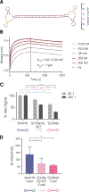
A) Secondary structure prediction as described in Fig 2. B) BLI characterization of SCOReD binding to immobilized RBD with experimental details and KD determination as described in Figure 2. C) ACE2-overexpressing HEK293 cells were incubated with 25 nM Omicron S protein and 250 nM or 2.5 μM of indicated dimer or 500 nM or 5 μM of monomeric SCORe.50-30T. S binding was measured by flow cytometry. Ratios (20:1, 200:1) indicate molar ratio of monometic aptamer units to S protein. Three technical replicates shown. Bars represent mean, brackets represent S.D. **** denotes p < 0.0001 as determined by 2-way ANOVA with Tukey’s multiple comparison test. D) Omicron S pseudotyped virus infection of ACE2-overexpressing HEK293 cells in the presence of various aptamer constructs. Three biological replicates performed with four technical replicates each shown. Bars represent mean, brackets represent S.D. * denotes p < 0.05 and ** denotes p < 0.01 as determined by 2-way ANOVA with Dunnett’s multiple comparison test.

Similar articles

Cited by

References

    1. Singh J; Pandit P; McArthur AG; Banerjee A; Mossman K Evolutionary trajectory of SARS-CoV-2 and emerging variants. Virology journal 2021, 18 (1), 1–21. - PMC - PubMed
    1. Tao K; Tzou PL; Nouhin J; Gupta RK; de Oliveira T; Kosakovsky Pond SL; Fera D; Shafer RW The biological and clinical significance of emerging SARS-CoV-2 variants. Nature Reviews Genetics 2021, 22 (12), 757–773. DOI: 10.1038/s41576-021-00408-x. - DOI - PMC - PubMed
    1. Cameroni E; Bowen JE; Rosen LE; Saliba C; Zepeda SK; Culap K; Pinto D; VanBlargan LA; De Marco A; di Iulio J Broadly neutralizing antibodies overcome SARS-CoV-2 Omicron antigenic shift. Nature 2022, 602 (7898), 664–670. - PMC - PubMed
    1. Cele S; Jackson L; Khoury DS; Khan K; Moyo-Gwete T; Tegally H; San JE; Cromer D; Scheepers C; Amoako DG Omicron extensively but incompletely escapes Pfizer BNT162b2 neutralization. Nature 2022, 602 (7898), 654–656. - PMC - PubMed
    1. Walls AC; Park Y-J; Tortorici MA; Wall A; McGuire AT; Veesler D Structure, function, and antigenicity of the SARS-CoV-2 spike glycoprotein. Cell 2020, 181 (2), 281–292. e286. - PMC - PubMed

Publication types

Substances

Supplementary concepts