. 2022 Sep 1;276(3):450-462.
doi: 10.1097/SLA.0000000000005558. Epub 2022 Jun 27.

Pancreatic Cancer Patient-derived Organoids Can Predict Response to Neoadjuvant Chemotherapy

Affiliations

Pancreatic Cancer Patient-derived Organoids Can Predict Response to Neoadjuvant Chemotherapy

Lyudmyla Demyan et al. Ann Surg. .

Abstract

Objective: To evaluate if patient-derived organoids (PDOs) may predict response to neoadjuvant (NAT) chemotherapy in patients with pancreatic adenocarcinoma.

Background: PDOs have been explored as a biomarker of therapy response and for personalized therapeutics in patients with pancreatic cancer.

Methods: During 2017-2021, patients were enrolled into an IRB-approved protocol and PDO cultures were established. PDOs of interest were analyzed through a translational pipeline incorporating molecular profiling and drug sensitivity testing.

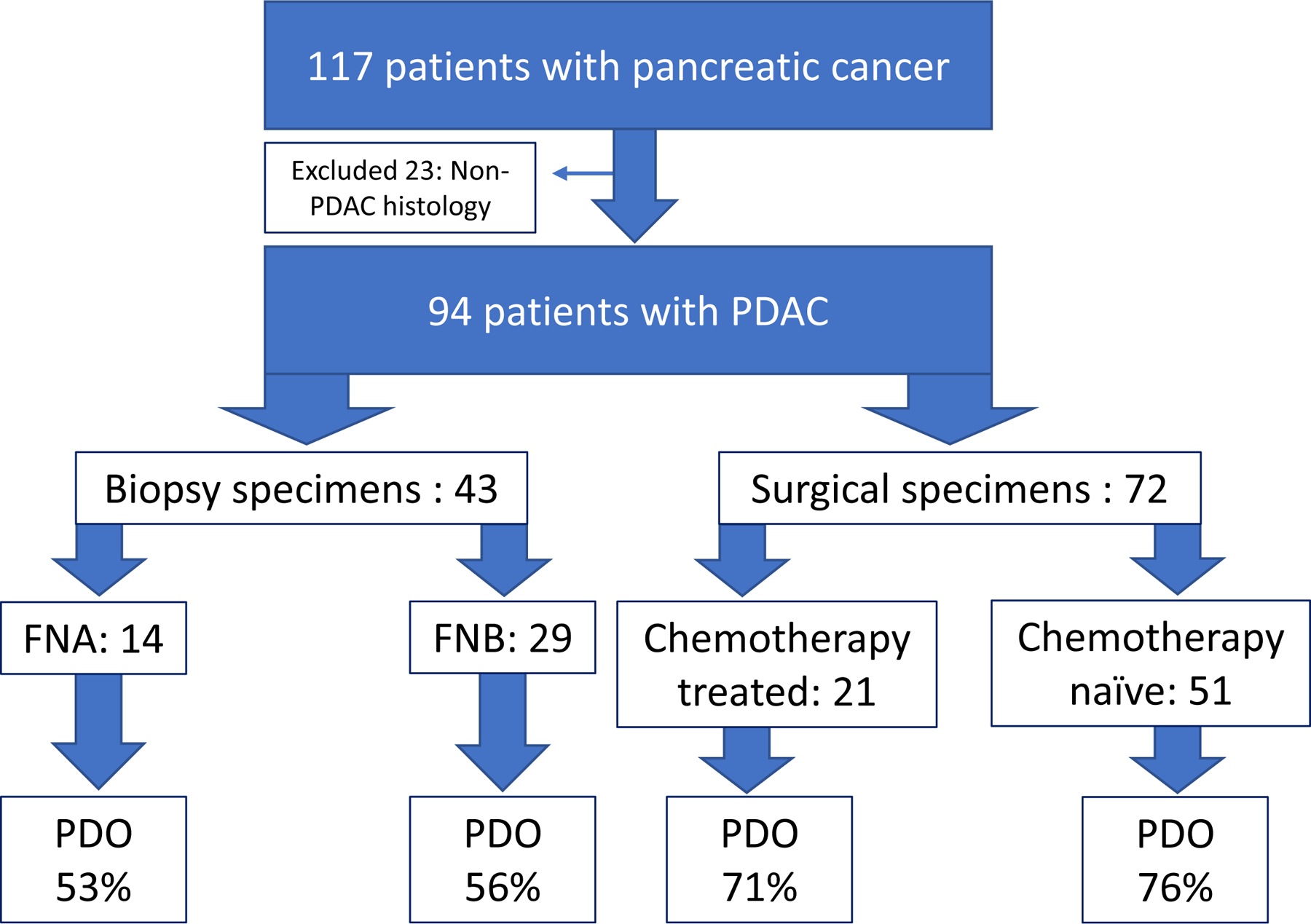
Results: One hundred thirty-six samples, including both surgical resections and fine needle aspiration/biopsy from 117 patients with pancreatic cancer were collected. This biobank included diversity in stage, sex, age, and race, with minority populations representing 1/3 of collected cases (16% Black, 9% Asian, 7% Hispanic/Latino). Among surgical specimens, PDO generation was successful in 71% (15 of 21) of patients who had received NAT prior to sample collection and in 76% (39 of 51) of patients who were untreated with chemotherapy or radiation at the time of collection. Pathological response to NAT correlated with PDO chemotherapy response, particularly oxaliplatin. We demonstrated the feasibility of a rapid PDO drug screen and generated data within 7 days of tissue resection.

Conclusion: Herein we report a large single-institution organoid biobank, including ethnic minority samples. The ability to establish PDOs from chemotherapy-naive and post-NAT tissue enables longitudinal PDO generation to assess dynamic chemotherapy sensitivity profiling. PDOs can be rapidly screened and further development of rapid screening may aid in the initial stratification of patients to the most active NAT regimen.


Conflict of interest statement

The authors report no conflicts of interest.

Figures

Figure 1:
Figure 1:. PDAC PDOs were generated with acceptable success rates regardless of tissue acquisition and chemotherapy treatment status.
Schematic diagram shows patient inclusion and exclusion criteria: only patients with confirmed PDAC histology were included in further analysis. Early (<5 passages) PDO establishment rate is reported as percent (%) for each respective tissue acquisition modality and systemic chemotherapy treatment status.
Figure 2:
Figure 2:. Neoadjuvant cohort timeline of clinical course and longitudinal specimen collection.
21 patients were enrolled into the NAT cohort. Four patients (20%) had a pre-treatment biopsy sent for PDO establishment with 75% success rate. 14% of patients (n=3) had progression of the disease while on NAT and did not undergo surgical resection. 86% of patients (n=18) completed NAT pathway, underwent oncological resection and 100% of samples were sent for PDO with 61% establishment rate.
Figure 3:
Figure 3:. Histological comparison of radiation effects.
Representative H&E slides from patients with comparable tumor cellularity but differing radiation exposure. A) Radiation exposed tumor with 84% cellularity, 100x compared with B) tumor without radiation exposure with 86% cellularity, 100x.
Figure 4:
Figure 4:. Organoids can be successfully established from FNB prior to NAT and from resections post-NAT.
A) Microscopic images of PDO line hF161, which was established from an FNB. Following establishment this line was frozen and then thawed at passage 6. PDOs are shown recovering well post thaw and continuing to expand shortly after undergoing an additional passage. B) hT220 was established from resected tissue from the same patient collected after NAT. Following establishment PDOs were frozen and then thawed at passage 4. There was a successful recovery from freezing and continued growth when expanded in passage 5. All images taken at 2x and scale bar is 1000 µm.
Figure 5:
Figure 5:. Pharmacotyping of PDOs reveals sensitivities and resistance.
Five standard-of-care chemotherapies (5-FU, oxaliplatin, SN-38 (Irinotecan), gemcitabine, and paclitaxel) were tested and dose response curves are shown in (A) for 7 PDOs collected from NAT patients and 2 pre-NAT PDOs. B) Screening results for all 9 PDOs across the five chemotherapies was merged with the biobank generated in Tiriac et al. Each dot in the violin plot represents a single PDO and sensitive PDOs have a lower normalized AUC whereas more resistant PDOs have a higher normalized AUC. Violin plots are broken down into tertiles with the bottom representing predicted drug sensitivity and top drug resistance. Black dots are biobank PDOs. Blue dots are PDOs derived from GnP treated patients, orange dots are PDOs from FFX treated patients, and purple are PDOs derived from patients prior to receiving NAT.
Figure 6:
Figure 6:. PDOs derived from matched pre- and post-NAT reveal emergence of resistance.
Schematic describing collection of matched PDOs: hF161 and hT220. B) Dose response curves for hF161 (orange) and hT220 (red) for NAT the patient received: gemcitabine and paclitaxel. Flattening of paclitaxel curve is driven by emergence of resistant cells as highlighted in the graph. C) Table displaying the predicted sensitivity and resistance which was determined for the 5 chemotherapies based on the PDO biobank Tiriac et al.
Figure 7:
Figure 7:. Rapid drug screening is feasible for organoids derived from pre- and post-NAT treated patients.
A) Table summarizing the 6 PDO lines that underwent rapid drug screening including 2 NAT PDOs. The number of days each PDO line was cultured prior to screening along with the total duration until data was generated, which includes the 5-day long screen, is reported. Several of the PDOs were screened repeatedly on consecutive passages. B) Microscopic image of hT268 PDO at passage 1 taken simultaneously to ongoing drug screen. Image taken at 4x, scale bar is 500 µm. C) Dose response curve from five standard-of-care chemotherapies from a rapid screen of hT268 during passage 2. D) Dose response curve for 5-FU in three consecutive passages of hT268. E) Microscope image of hT270 NAT PDO at passage 1 prior to rapid drug screening. Image taken at 4x, scale bar is 500 µm. F) Dose response curve from five standard-of-care chemotherapies from a rapid screen of hT270 passage 2.

References

    1. Saad AM, Turk T, Al-Husseini MJ, Abdel-Rahman O. Trends in pancreatic adenocarcinoma incidence and mortality in the United States in the last four decades; a SEER-based study. BMC Cancer 2018;18(1):688. - PMC - PubMed
    1. Tempero MA, Malafa MP, Al-Hawary M, et al. Pancreatic Adenocarcinoma, Version 2.2021, NCCN Clinical Practice Guidelines in Oncology. J Natl Compr Canc Netw 2021;19(4):439–457. - PubMed
    1. Fathi A, Christians KK, George B, et al. Neoadjuvant therapy for localized pancreatic cancer: guiding principles. J Gastrointest Oncol 2015;6(4):418–429. - PMC - PubMed
    1. Aung KL, Fischer SE, Denroche RE, et al. Genomics-Driven Precision Medicine for Advanced Pancreatic Cancer: Early Results from the COMPASS Trial. Clin Cancer Res 2018;24(6):1344–1354. - PMC - PubMed
    1. Law HC, Lagundzin D, Clement EJ, et al. The Proteomic Landscape of Pancreatic Ductal Adenocarcinoma Liver Metastases Identifies Molecular Subtypes and Associations with Clinical Response. Clin Cancer Res 2020;26(5):1065–1076. - PMC - PubMed

Publication types

Substances