. 2022 Sep 26;32(18):3939-3951.e6.
doi: 10.1016/j.cub.2022.07.037. Epub 2022 Aug 15.

Molecular conflicts disrupting centromere maintenance contribute to Xenopus hybrid inviability

Affiliations

Molecular conflicts disrupting centromere maintenance contribute to Xenopus hybrid inviability

Maiko Kitaoka et al. Curr Biol. .

Abstract

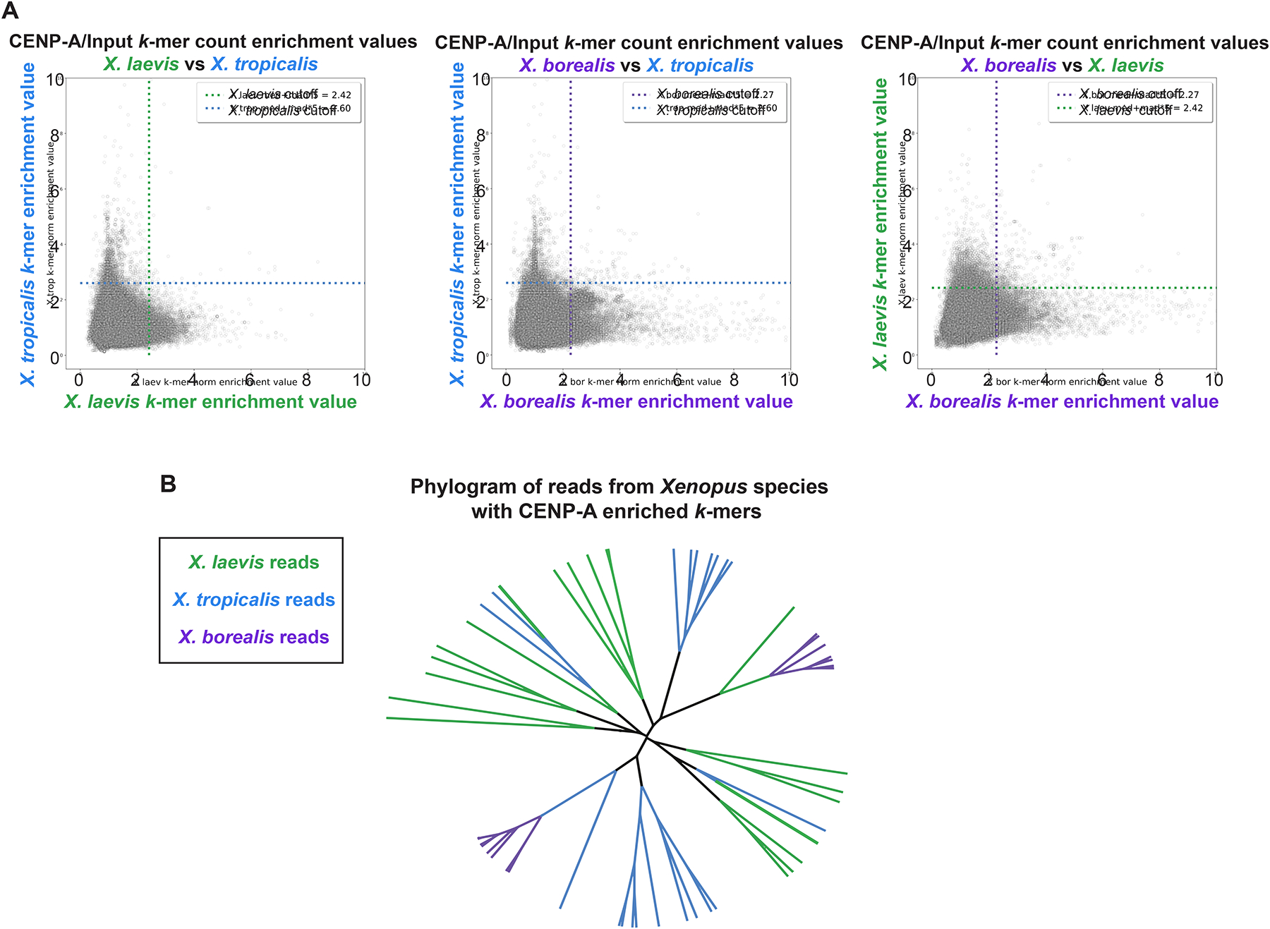
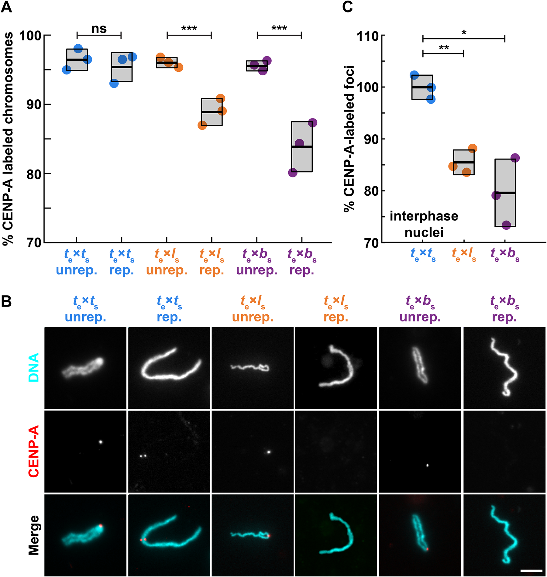
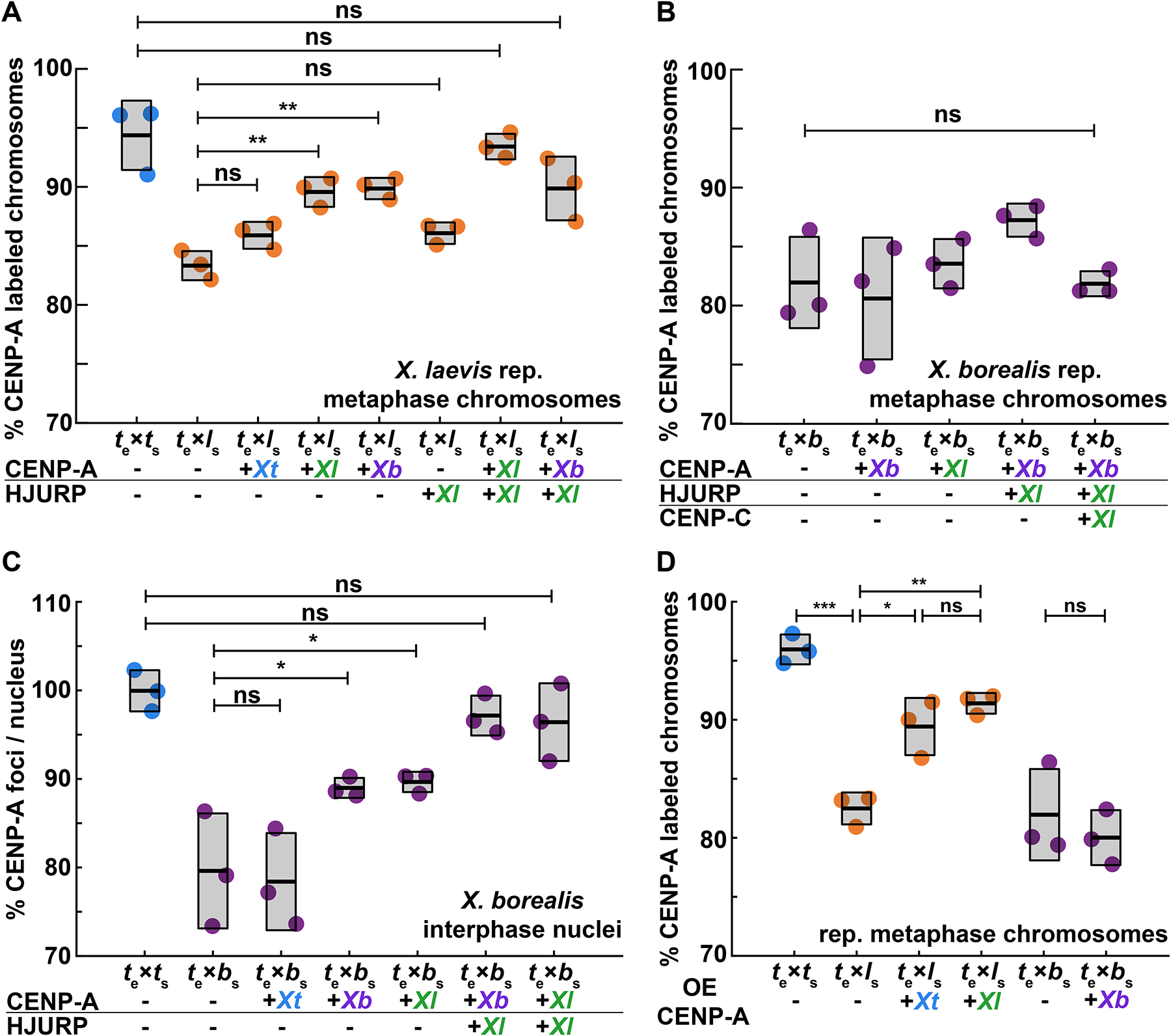
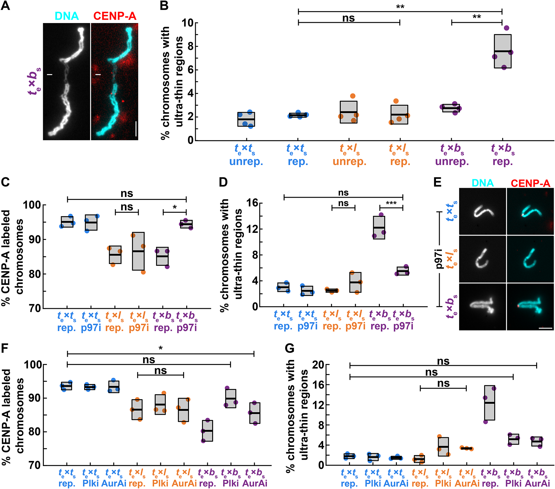
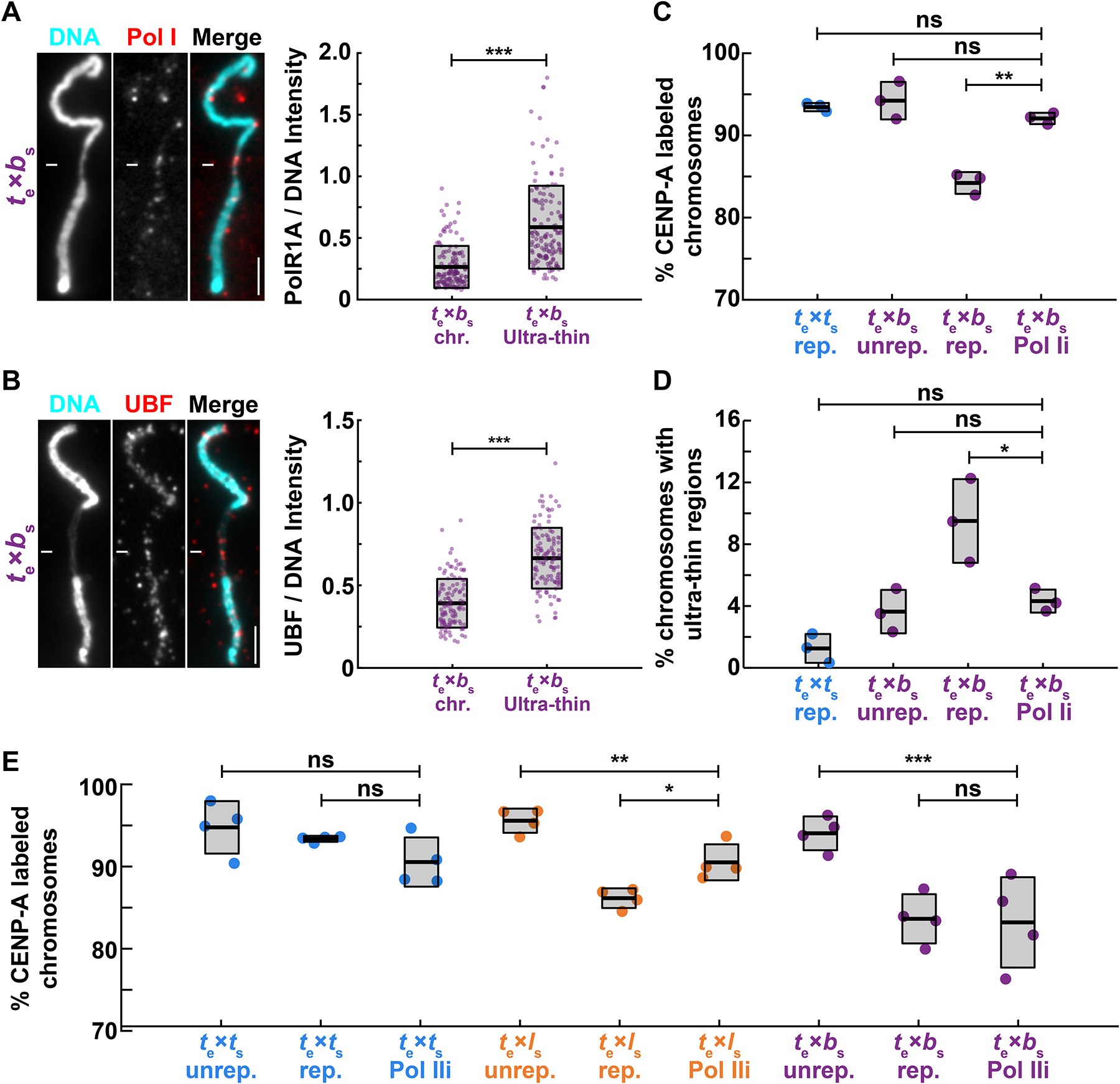
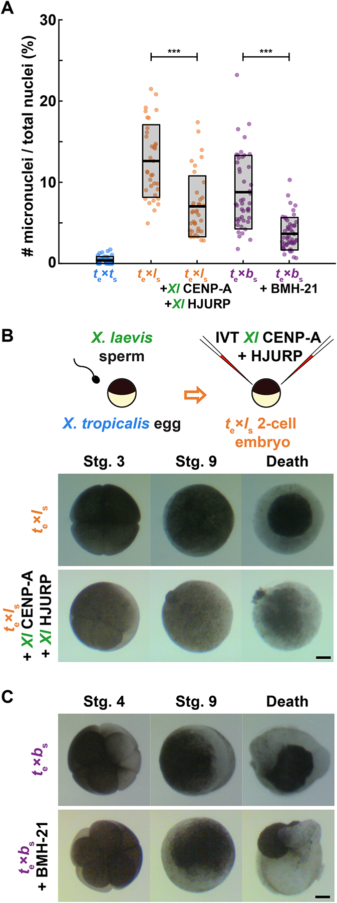
Although central to evolution, the causes of hybrid inviability that drive reproductive isolation are poorly understood. Embryonic lethality occurs when the eggs of the frog X. tropicalis are fertilized with either X. laevis or X. borealis sperm. We observed that distinct subsets of paternal chromosomes failed to assemble functional centromeres, causing their mis-segregation during embryonic cell divisions. Core centromere DNA sequence analysis revealed little conservation among the three species, indicating that epigenetic mechanisms that normally operate to maintain centromere integrity are disrupted on specific paternal chromosomes in hybrids. In vitro reactions combining X. tropicalis egg extract with either X. laevis or X. borealis sperm chromosomes revealed that paternally matched or overexpressed centromeric histone CENP-A and its chaperone HJURP could rescue centromere assembly on affected chromosomes in interphase nuclei. However, although the X. laevis chromosomes maintained centromeric CENP-A in metaphase, X. borealis chromosomes did not and also displayed ultra-thin regions containing ribosomal DNA. Both centromere assembly and morphology of X. borealis mitotic chromosomes could be rescued by inhibiting RNA polymerase I or preventing the collapse of stalled DNA replication forks. These results indicate that specific paternal centromeres are inactivated in hybrids due to the disruption of associated chromatin regions that interfere with CENP-A incorporation, at least in some cases due to conflicts between replication and transcription machineries. Thus, our findings highlight the dynamic nature of centromere maintenance and its susceptibility to disruption in vertebrate interspecies hybrids.

Keywords: CENP-A; Xenopus; centromere; chromosome segregation; hybrid incompatibility; speciation.


Conflict of interest statement

Declaration of interests The authors declare no competing interests.

Figures

Figure 1:
Figure 1:. Comparison of X. laevis, X. tropicalis, and X. borealis core centromere sequences
(A) Scatter plots of k-mer enrichment values (normalized CENP-A counts / normalized input counts) compared between species. Only k-mers found in both species are plotted. Dotted lines indicate enrichment value for each species that is five median absolute deviations above the median enrichment value to denote highly enriched k-mers, which are not well conserved across species. (B) Phylogram of full-length sequencing reads from each Xenopus species. Branches are colored according to species of origin. Sequencing reads were selected first by the presence of at least 80 CENP-A enriched 25bp k-mers and then by hierarchical clustering. The phylogram illustrates a striking divergence of core centromere sequences. See also Figure S1.
Figure 2:
Figure 2:. Loss of centromeric CENP-A is cell cycle-dependent
(A) Percentage of mitotic chromosomes with centromeric CENP-A staining in X. tropicalis egg extract. Over 95% of X. tropicalis, X. laevis, and X. borealis unreplicated sperm chromosomes added directly to metaphase-arrested X. tropicalis egg extracts possess centromeres, as indicated by immunofluorescence of the centromeric histone CENP-A. Following progression through the cell cycle, a fraction of replicated X. laevis and X. borealis mitotic chromosomes completely lose centromeric CENP-A foci. Unrep., unreplicated chromosomes; rep, replicated chromosomes. N = 3 extracts, N > 275 chromosomes per extract. p-values (left to right) by two-tailed two-sample unequal variance t-tests: 0.3356, 0.0008, 0.0004; ns, not significant. (B) Representative images of mitotic unreplicated and replicated X. tropicalis, X. laevis, and X. borealis chromosomes formed in X. tropicalis egg extracts. The chromosomes shown here are not identified, but selected from a population of paternal chromosomes. DNA in cyan, CENP-A in red. Scale bar is 10 μm. (C) Percentage of total expected CENP-A foci observed in nuclei formed in interphase X. tropicalis egg extract. X. laevis and X. borealis interphase nuclei both lose centromere foci during interphase, prior to entry into metaphase, whereas X. tropicalis nuclei do not. From N = 3 extracts, N > 64 nuclei per extract. p-values (top to bottom) by one-way ANOVA with Tukey post-hoc analysis: 0.0025, 0.0133. Species nomenclature throughout figures denotes egg extract as subscript e and chromosomes as subscript s, for example te × ls indicates X. tropicalis egg extract combined with X. laevis sperm chromosomes. X. tropicalis is color-coded blue, while X. laevis and X. borealis hybrid combinations are orange and purple, respectively. See also Figure S2.
Figure 3:
Figure 3:. Driving CENP-A assembly rescues centromere localization in interphase, which persists on mitotic X. laevis, but not on X. borealis chromosomes
(A) Percentage of replicated X. laevis chromosomes with centromeric CENP-A staining in X. tropicalis extract supplemented with in vitro translated CENP-A and HJURP proteins from different Xenopus species. X. laevis chromosomes are fully rescued with species-matched centromere proteins. Quantification with N = 3 extracts, N > 315 chromosomes per extract. p-values (top to bottom) by one-way ANOVA with Tukey post-hoc analysis: 0.1734, 0.9999, 0.5522, 0.0057, 0.0086, 0.6281. (B) Percentage of replicated X. borealis chromosomes with centromeric CENP-A staining in X. tropicalis extract supplemented with in vitro translated centromere proteins from different Xenopus species. No combination or increased amounts of centromeric proteins CENP-A (CA), HJURP (HJ), and CENP-C (CC) restored CENP-A localization on X. borealis mitotic chromosomes. Quantification with N = 3 extracts, N > 216 chromosomes per extract. p-value by one-way ANOVA = 0.0786. (C) Percentage of CENP-A-labeled centromeric foci in X. borealis nuclei assembled in X. tropicalis extract supplemented with in vitro translated centromere proteins from different Xenopus species. Driving centromere assembly with species-matched proteins fully restores formation of centromere foci in interphase, but CENP-A staining is subsequently lost in metaphase (panel B). Quantification with N = 3 extracts, N > 67 nuclei per extract. p-values (top to bottom) by one-way ANOVA: 0.9996, 0.0562, 0.0433, 0.9690, 0.9109. (D) Percentage of replicated X. laevis or X. borealis chromosomes with centromeric CENP-A staining in X. tropicalis extract supplemented with excess (~80X endogenous levels) of in vitro translated X. laevis or X. tropicalis CENP-A. Whereas centromere staining is fully rescued on X. laevis mitotic chromosomes by CENP-A from either species, X. borealis centromere staining is not affected. Quantification with N = 3 extracts, N > 204 chromosomes per extract. p-values (top to bottom, then left to right) by one-way ANOVA with Tukey post-hoc analysis: 0.0042, 0.0001, 0.0249, 0.8845, 0.88946. A-C: Centromere proteins were added at ~8X endogenous levels. A-D: ns, not significant. See also Figure S3.
Figure 4:
Figure 4:. Mitotic replication stress leads to X. borealis centromere and chromosome morphology defects
(A) Representative image showing an ultra-thin region of a mitotic X. borealis chromosome formed in X. tropicalis egg extract. Note that the chromosome has an intact centromere. DNA in cyan, CENP-A in red. Scale bar is 5 μm. (B) Percentage of unreplicated and replicated mitotic chromosomes with ultrathin morphology defects in X. tropicalis extract. A low percentage of X. tropicalis, X. laevis or X. borealis unreplicated chromosomes display ultra-thin regions. After cycling through interphase, only X. borealis chromosomes exhibit a significant increase in this defect. Quantification with N = 3 extracts, N > 310 chromosomes per extract. p-values (top to bottom, then left to right) by one-way ANOVA with Tukey post-hoc analysis: 2.9352e-7, 0.9999, 1.6475e-6. (C) Percentage of replicated chromosomes with centromeric CENP-A staining in X. tropicalis extracts treated with solvent control or 10 μM p97 ATPase inhibitor NMS-873 (p97i). Inhibition of p97 restores CENP-A staining on X. borealis mitotic chromosomes, but does not affect X. tropicalis or X. laevis chromosomes. p-values (top to bottom, then left to right) by one-way ANOVA with Tukey post-hoc analysis: 0.9997, 0.9978, 0.0204. (D) Percentage of chromosomes with ultrathin regions in X. tropicalis extracts treated with solvent control or 10 μM p97 ATPase inhibitor NMS-873 (p97i). Inhibition of p97 rescues X. borealis chromosome morphology defects, but does not affect X. tropicalis or X. laevis chromosomes. p-values (top to bottom, then left to right) by one-way ANOVA with Tukey post-hoc analysis: 0.1114, 0.6903, 6.2572e-5. (E) Representative images of mitotic replicated X. tropicalis, X. laevis, and X. borealis chromosomes following treatment with 10 μM p97 ATPase inhibitor NMS-873 (p97i). X. borealis chromosome morphology and centromere localization are rescued (bottom panels, compare to Fig. 4A, 2B images), similar to X. tropicalis, while X. laevis chromosomes have lost CENP-A staining (middle panels). DNA in cyan, CENP-A in red. Scale bar is 5 μm. (F) Percentage of replicated chromosomes with centromeric CENP-A staining in X. tropicalis extracts treated with solvent control, 1 μM Polo-like kinase 1 inhibitor BI-2536 (Plk1i), or 1 μM Aurora A kinase inhibitor MLN-8237 (AurAi). CENP-A localization is fully or partially rescued on X. borealis mitotic chromosomes, whereas X. tropicalis or X. laevis chromosomes are not affected. p-values (top to bottom) by one-way ANOVA with Tukey post-hoc analysis: 0.0276, 0.7003, 0.9999. (G) Percentage of chromosomes with ultrathin regions in X. tropicalis extracts treated with solvent control, 1 μM Polo-like kinase 1 inhibitor BI-2536 (Plk1i), or 1 μM Aurora A kinase inhibitor MLN-8237 (AurAi). Inhibition of Plk1 and AurA rescued X. borealis mitotic chromosome morphology defects, but did not affect X. tropicalis or X. laevis chromosomes. p-values (top to bottom) by one-way ANOVA with Tukey post-hoc analysis: 0.2882, 0.1525, 0.5887. C, D: N = 3 extracts, N > 179 chromosomes per extract. E, F: N = 3 extracts, N > 155 chromosomes per extract. B-F: ns, not significant. See also Figure S4.
Figure 5:
Figure 5:. Replication-transcription conflicts at rDNA on X. borealis chromosomes can be rescued by inhibiting RNA Pol I
(A) Representative images and fluorescence intensity quantification of RNA Pol I staining relative to DNA on ultrathin and normal regions of X. borealis mitotic chromosomes, revealing enrichment of Pol I on ultra-thin regions. Quantification with N = 3 extracts, N = 140 chromosomes. p-value = 9.4793e-20 by two-tailed two-sample unequal variance t-tests. (B) Representative images and fluorescence intensity quantification of UBF staining relative to DNA on ultrathin and normal regions of X. borealis mitotic chromosomes, revealing enrichment of UBF on ultra-thin regions. Quantification with N = 3 extracts, N = 62 chromosomes. p-value = 4.5004e-13 by two-tailed two-sample unequal variance t-tests. (C) Percentage of mitotic chromosomes with centromeric CENP-A staining in X. tropicalis extracts treated with solvent control or 1 μM BMH-21 to inhibit RNA Pol I (Pol Ii), which fully rescues CENP-A localization on replicated X. borealis chromosomes. p-values (top to bottom) by one-way ANOVA with Tukey post-hoc analysis: 0.9794, 0.7979, 0.0005. (D) Percentage of mitotic chromosomes with ultrathin regions in X. tropicalis extracts treated with solvent control or 1 μM BMH-21 (Pol Ii). Pol I inhibition also rescues X. borealis chromosome morphology defects. p-values (top to bottom) by one-way ANOVA with Tukey post-hoc analysis: 0.5078, 0.9999, 0.0469. (E) Percentage of chromosomes with centromeric CENP-A staining in X. tropicalis extracts treated with solvent control or 25 μM triptolide to inhibit RNA Pol II (Pol IIi). X. laevis chromosomes are partially rescued, while X. tropicalis and X. borealis chromosomes are not affected. Quantification with N = 3 extracts, N > 322 chromosomes per extract. p-values (top to bottom, then left to right) by one-way ANOVA with Tukey post-hoc analysis: 0.4785, 0.8797, 0.0052, 0.0125, 0.0003, 0.9999. A, B: DNA in cyan, Pol I in red. Scale bar is 5 μm. C, D: N = 3 extracts, N > 172 chromosomes per extract. C-E: ns, not significant. See also Figure S5.
Figure 6:
Figure 6:. Treatments that rescue CENP-A localization in egg extracts reduce micronuclei formation in hybrid embryos, but inviability persists
(A) Quantification of chromosome mis-segregation events as measured by the number of micronuclei compared to total nuclei in treated hybrid embryos. X. tropicalis eggs fertilized with X. laevis sperm were microinjected with X. laevis CENP-A/HJURP, while X. tropicalis eggs fertilized with X. borealis sperm were treated with Pol 1 inhibitor BMH-21. Embryos were fixed at stage 9 (7 hpf) just before gastrulation and hybrid death. The number of micronuclei was significantly reduced in both cases, but not to control levels measured in X. tropicalis eggs fertilized with X. tropicalis sperm. N = 3 clutches for each hybrid, N > 15 embryos and > 200 cells per embryo. p-values (left to right) by two-tailed two-sample unequal variance t-tests: 2.111e-7, 2.651e-9; ns, not significant. (B) Schematic of experiment and video frames of X. tropicalis eggs fertilized with X. laevis sperm microinjected at the two-cell stage with X. laevis CENP-A/HJURP, increasing centromeric protein concentration by ~44.5%. Microinjected hybrid embryos die at the same time and in the same manner as uninjected hybrid controls. N = 10 embryos across 4 clutches. Scale bar is 200 μm. See also Video S1. (C) Video frames of X. tropicalis eggs fertilized with X. borealis sperm that were incubated from the two-cell stage with 1 μM RNA Pol I inhibitor, BMH-21. Treated hybrid embryos die at the same time and in the same manner as untreated hybrid controls. N = 12 embryos across 2 clutches. Scale bar is 200 μm. See also Video S2. See also Figure S6.

Comment in

References

    1. Sanei M, Pickering R, Kumke K, Nasuda S, and Houben A (2011). Loss of centromeric histone H3 (CENH3) from centromeres precedes uniparental chromosome elimination in interspecific barley hybirds. PNAS 108, E498–E505. - PMC - PubMed
    1. Maheshwari S, and Barbash DA (2011). The Genetics of Hybrid Incompatibilities. Annu. Rev. Genet 45, 331–355. - PubMed
    1. Fujiwara A, Abe S, Yamaha E, Yamazaki F, and Yoshida MC (1997). Uniparental chromosome elimination in the early embryogenesis of the inviable salmonid hybrids between masu salmon female and rainbow trout male. Chromosoma 106, 44–52. - PubMed
    1. Gernand D, Rutten T, Varshney A, Rubtsova M, Prodanovic S, and Bru C (2005). Uniparental Chromosome Elimination at Mitosis and Interphase in Wheat and Pearl Millet Crosses Involves Micronucleus Formation, Progressive Heterochromatinization, and DNA Fragmentation. Plant Cell 17, 2431–2438. - PMC - PubMed
    1. Malik HS, and Henikoff S (2001). Adaptive evolution of Cid, a centromere-specific histone in Drosophila. Genetics 157, 1293–1298. - PMC - PubMed

Publication types