Review
. 2022 Aug 16;8(1):363.
doi: 10.1038/s41420-022-01154-7.

Role of necroptosis in airflow limitation in chronic obstructive pulmonary disease: focus on small-airway disease and emphysema

Affiliations
Review

Role of necroptosis in airflow limitation in chronic obstructive pulmonary disease: focus on small-airway disease and emphysema

Chanjing Liu et al. Cell Death Discov. .

Abstract

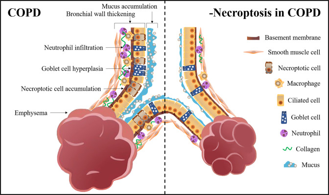
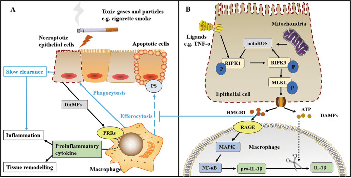
Airflow limitation with intractable progressive mechanisms is the main disease feature of chronic obstructive pulmonary disease (COPD). The pathological process of airflow limitation in COPD involves necroptosis, a form of programmed necrotic cell death with pro-inflammatory properties. In this paper, the correlations of small-airway disease and emphysema with airflow limitation in COPD were firstly reviewed; then, based on this, the effects of necroptosis on small-airway disease and emphysema were analysed, and the possible mechanisms of necroptosis causing airflow limitation in COPD were explored. The results showed that airflow limitation is caused by a combination of small-airway disease and emphysema. In addition, toxic particulate matter stimulates epithelial cells to trigger necroptosis, and necroptosis promotes the expulsion of cell contents, the abnormal hyperplasia of pro-inflammatory mediators and the insufficient clearance of dead cells by macrophages; these processes, coupled with the interaction of necroptosis and oxidative stress, collectively result in small-airway disease and emphysema in COPD.


Conflict of interest statement

The authors declare no competing interests.

Figures

Fig. 1
Fig. 1. Main detection methods of necroptosis.
The left figure is a schematic diagram of the morphological changes in the necroptotic cell. The figure shows cellular swelling, organelle swelling, plasma membrane rupture, and cell lysis causing the release of the contents. Generally, it can be observed directly by an electron microscope. The right figure is a schematic diagram of the necroptotic signalling pathway. The figure shows RIPK1 phosphorylation upon receiving a signal from ligand binding and then RIPK3 activation, which interacts with RIPK1 to form a necrosome. Phosphorylated MLKL mediated by RIPK3 induces the formation of plasma membrane pores. In addition, DAI and TLRs can directly activate RIPK3 leading to necroptosis after detecting exogenous viral DNA or RNA. Key proteins in the necroptotic signalling pathway, such as RIPK3 and MLKL, can be detected by Western Blot, Immunohistochemistry or Enzyme-Linked Immunosorbent Assay. The expression levels of the genes corresponding to key proteins can be detected by Polymerase Chain Reaction. The above methods are suitable for detecting necroptosis. DAI DNA-dependent activator of interferon regulatory factors, MLKL mixed-lineage kinase domain-like, P phosphorylates, RIPK receptor-interacting protein kinase, TLRs toll-like receptors, TNF tumour necrosis factor.
Fig. 2
Fig. 2. Pathological changes after inhibiting necroptosis in COPD.
The left side of the dashed line shows the pathological manifestations of airflow limitation in COPD, including small-airway disease and emphysema; the right side shows the pathological changes after inhibiting necroptosis in COPD, the specific as follows: in the small airway, the increased number of ciliated cells, the reduced thickness of the bronchial wall, and the improvements of neutrophilic airway inflammation, mucus accumulation and collagen deposition; in the alveoli, the improvements of alveolar lumen enlargement and alveolar wall destruction.
Fig. 3
Fig. 3. The mechanism of necroptosis leads to airflow limitation in COPD.
A Lung parenchymal epithelial cells undergo necroptosis due to direct exposure to toxic gases and particles (e.g. cigarette smoke). Excessive DAMPs are released and linked to PRRs on neighbouring macrophages after plasma membrane rupture, leading to pro-inflammatory cytokine production through multiple pathways and consequently causing inflammation and tissue remodelling in the lung parenchyma. In addition, the impaired ability of macrophages to phagocytose apoptotic epithelial cells in COPD and the slower clearance of necroptosis cells by macrophages can cause inadequate phagocytosis of dead cells, which triggers inflammation. B Epithelial cell membrane receptors bind to the corresponding ligands (e.g. TNF-α) and send signals to cause RIPK1 activation, followed by RIPK3 recruitment and phosphorylation. Mitochondrial ROS contributes to RIPK1 autophosphorylation, but the production is dependent on RIPK3. RIPK3 can also mediate MLKL phosphorylation, causing MLKL oligomerisation and translocation to the plasma membrane for forming plasma membrane pores and therefore releasing excessive DAMPs (including HMGB1 and ATP) to the extracellular environment. HMGB1 activates RAGE receptors on macrophages and promotes pro–IL-1β production via the MAPK/NF-κB pathway. Pro–IL-1β is cleaved by ATP-induced pathway products to form IL-1β. In addition, HMGB1 interacts with RAGE on the surface of macrophages and PS on the surface of apoptotic cells to participate in the inhibition of the macrophage uptake of apoptotic epithelial cells. ATP adenosine 5′-triphosphate, DAMPs damage-associated molecular patterns, HMGB1 high-mobility group box-1, IL interleukin, MAPK mitogen-activated protein kinase, MitoROS mitochondrial reactive oxygen species, MLKL mixed-lineage kinase domain-like, NF-κB nuclear factor kappa-B, P phosphorylates, PRRs pattern recognition receptors, PS phosphatidylserine, RAGE receptor for advanced glycation end-products, RIPK receptor-interacting protein kinase, TNF tumour necrosis factor.

References

    1. WHO. The top 10 causes of death. https://www.who.int/news-room/fact-sheets/detail/the-top-10-causes-of-death (2020).
    1. Global Initiative for Chronic Obstructive Lung Disease. Global strategy for the diagnosis, management, and prevention of chronic obstructive pulmonary disease. https://goldcopd.org/wp-content/uploads/2021/11/GOLD-REPORT-2022-v1.1-22... (2022).
    1. Gamble E, Grootendorst DC, Hattotuwa K, O’Shaughnessy T, Ram FS, Qiu Y, et al. Airway mucosal inflammation in COPD is similar in smokers and ex-smokers: a pooled analysis. Eur Respir J. 2007;30:467–71. doi: 10.1183/09031936.00013006. - DOI - PubMed
    1. Miller M, Cho JY, Pham A, Friedman PJ, Ramsdell J, Broide DH. Persistent airway inflammation and emphysema progression on CT scan in ex-smokers observed for 4 years. Chest. 2011;139:1380–7. doi: 10.1378/chest.10-0705. - DOI - PMC - PubMed
    1. Wen Y, Reid DW, Zhang D, Ward C, Wood-Baker R, Walters EH. Assessment of airway inflammation using sputum, BAL, and endobronchial biopsies in current and ex-smokers with established COPD. Int J Chronic Obstr Pulm Dis. 2010;5:327–34. - PMC - PubMed