. 2022 Aug 17;7(1):275.
doi: 10.1038/s41392-022-01096-7.

DDX39B drives colorectal cancer progression by promoting the stability and nuclear translocation of PKM2

Affiliations

DDX39B drives colorectal cancer progression by promoting the stability and nuclear translocation of PKM2

Gang Zhao et al. Signal Transduct Target Ther. .

Abstract

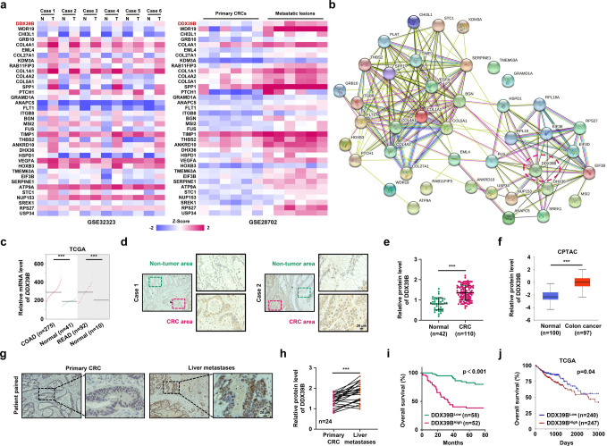
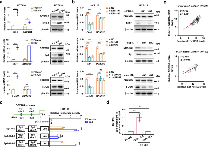
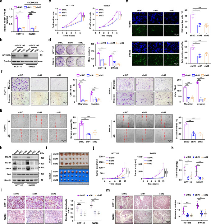
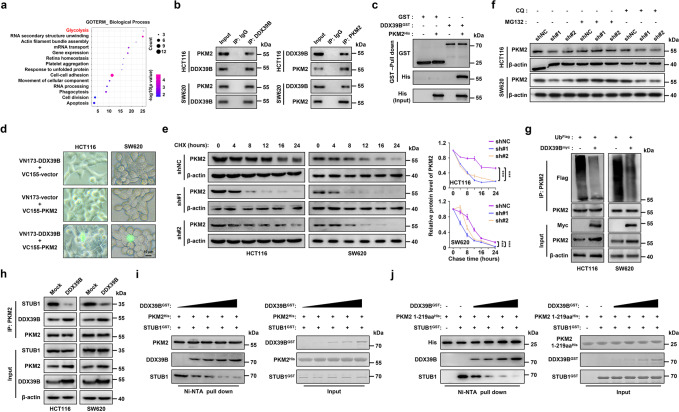
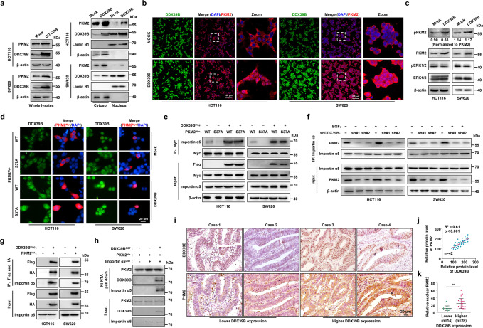
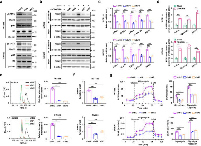
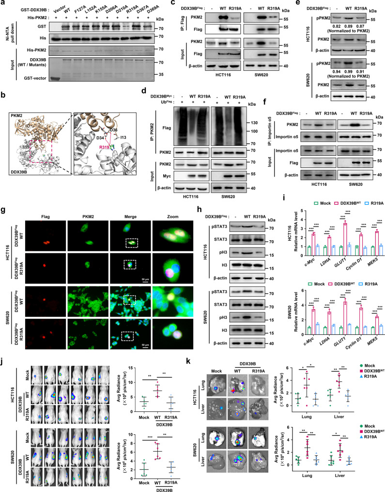
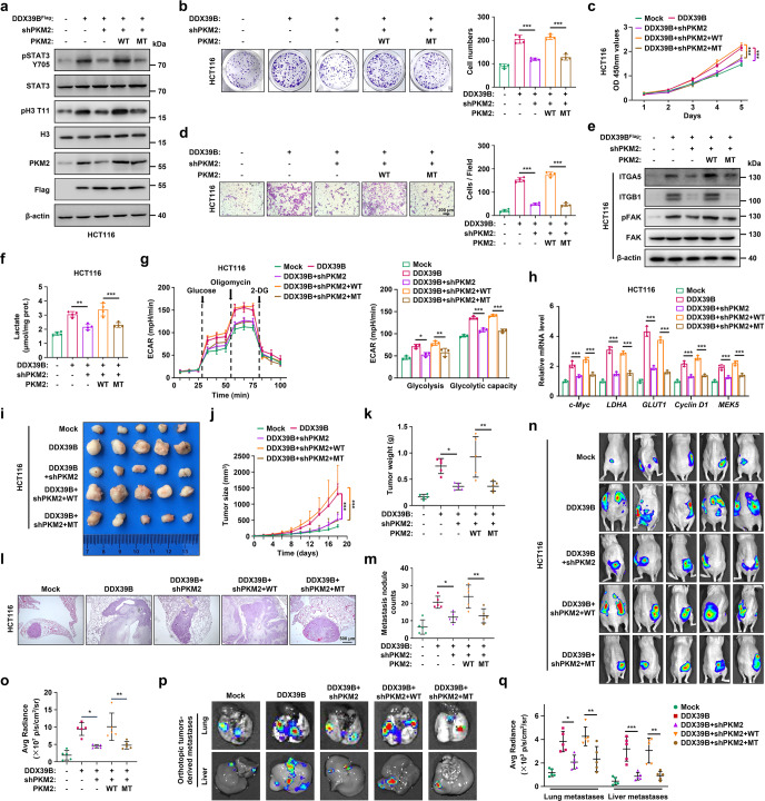
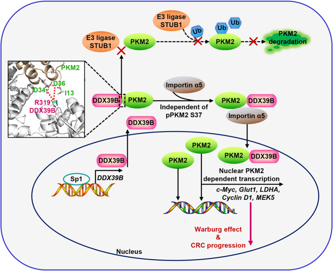
Metastasis is a major cause of colorectal cancer (CRC) mortality, but its molecular mechanisms are still not fully understood. Here, we show that upregulated DDX39B correlates with liver metastases and aggressive phenotypes in CRC. DDX39B is an independent prognostic factor associated with poor clinical outcome in CRC patients. We demonstrate that Sp1 potently activates DDX39B transcription by directly binding to the GC box of the DDX39B promoter in CRC cells. DDX39B overexpression augments the proliferation, migration, and invasion of CRC cells, while the opposite results are obtained in DDX39B-deficient CRC cells. Mechanistically, DDX39B interacts directly with and stabilizes PKM2 by competitively suppressing STUB1-mediated PKM2 ubiquitination and degradation. Importantly, DDX39B recruits importin α5 to accelerate the nuclear translocation of PKM2 independent of ERK1/2-mediated phosphorylation of PKM2, leading to the transactivation of oncogenes and glycolysis-related genes. Consequently, DDX39B enhances glucose uptake and lactate production to activate Warburg effect in CRC. We identify that Arg319 of DDX39B is required for PKM2 binding as well as PKM2 nuclear accumulation and for DDX39B to promote CRC growth and metastasis. In addition, blocking PKM2 nuclear translocation or treatment with glycolytic inhibitor 2-deoxy-D-glucose efficiently abolishes DDX39B-triggered malignant development in CRC. Taken together, our findings uncover a key role for DDX39B in modulating glycolytic reprogramming and aggressive progression, and implicate DDX39B as a potential therapeutic target in CRC.


Conflict of interest statement

The authors declare no competing interests.

Figures

Fig. 1
Fig. 1
Upregulation of DDX39B correlates with aggressive phenotypes and poor prognosis in human CRC. A total of 37 up-regulated genes both in primary and metastatic CRC tumors were displayed as a heatmap (a) and by STRING network (b) analysis. N normal tissues; T: CRC tumors. c DDX39B transcripts in colon adenocarcinoma (COAD), rectal adenocarcinoma (READ) and normal tissues from the TCGA database were analyzed through an online tool (http://gepia2.cancer-pku.cn/). d, e DDX39B protein levels in a microarray of CRC and normal mucosal tissues were detected by immunohistochemistry (IHC) staining. d Representative images are shown, and e the relative staining intensities were analyzed by two-tailed unpaired t test. f DDX39B protein level in normal and colon cancer tissues from the Clinical Proteomic Tumor Analysis Consortium (CPTAC) database was analyzed through an online tool (http://ualcan.path.uab.edu/index.html). g, h DDX39B protein expression in primary CRCs and patient-paired liver metastases were detected by IHC. g Representative images are shown, and h the relative staining intensities were analyzed by two-tailed paired t test. i The correlation between DDX39B protein level and the overall survival of CRC patients was tested by Kaplan-Meier analysis. j Kaplan-Meier survival analysis for DDX39B transcripts in colon adenocarcinoma patients was carried out using an online tool (https://xenabrowser.net/). ***p < 0.001
Fig. 2
Fig. 2
Sp1 activates DDX39B transcription in CRC cells. a HCT116 cells were transiently transfected with the plasmids of ETS-1, Sp1, or c-JUN, respectively. The mRNA and protein levels of DDX39B were measured by qPCR and western blotting. b HCT116 cells were transiently transfected with siRNAs targeting ETS-1, Sp1, or c-JUN, respectively. The mRNA and protein expressions of DDX39B were determined by qRT-PCR and western blotting. c The relative luciferase activity was detected in HCT116 cells transfected with the indicated DDX39B promoter in the presence or absence of Sp1. d DNA fragments from HCT116 cells were immunoprecipitated with the Sp1-specific antibody and analyzed by qPCR using the indicated primers. e The correlations of transcript levels between DDX39B and Sp1 in human colon and rectal adenocarcinoma tissues were analyzed through an online tool (https://xenabrowser.net/). Data are presented as mean ± SD. The p values were obtained by two-tailed unpaired t test (a) or one-way ANOVA (b–d). *p < 0.05, ***p < 0.001, ns not significant
Fig. 3
Fig. 3
DDX39B knockdown inhibits CRC growth and metastasis in vitro and in vivo. Knockdown efficiency mediated by two lentivirus-delivered DDX39B-specific shRNA (sh#1 and sh#2) was verified by qRT-PCR (a) and western blotting (b) in HCT116 and SW620 cells. The cell viability and proliferation of DDX39B-knockdown CRC cells were measured by CCK8 assays (c), colony formation assays (d), and EdU assays (e). The motility of DDX39B-knockdown CRC cells was examined by transwell migration/invasion assays (f) and wound healing assays (g). h The protein levels of ITGA5, ITGB1, pFAKY397, and FAK in DDX39B-knockdown CRC cells were detected by western blotting. The growth and metastatic abilities of DDX39B-knockdown CRC cells in vivo were assessed in nude mice by subcutaneous and lung metastasis tumor models (n = 8), respectively. The images (i), tumor sizes (j), tumor weights (k), and Ki-67 expression (l) of subcutaneous xenografts are presented. Representative pulmonary metastases detected by H&E staining are shown, along with the number of metastatic nodules (m). Data are presented as mean ± SD. The p values were obtained by two-way ANOVA (c, j) or one-way ANOVA (others). **p < 0.01, ***p < 0.001
Fig. 4
Fig. 4
DDX39B interacts with PKM2 and reduces its degradation. a The Gene Ontology analysis of DDX39B interactome. b The endogenous interaction between DDX39B and PKM2 was examined by immunoprecipitation assay in CRC cells. c GST pull-down assays were carried out with bacterially expressed GST-fused DDX39B and His-fused PKM2. d HCT116 and SW620 cells were transfected with pBiFC-VN173-DDX39B and/or pBiFC-VC155-PKM2, and the fluorescence signals were imaged. e The degradation of PKM2 protein in DDX39B knockdown CRC cells was measured by cycloheximide (CHX) chase analysis. f DDX39B-deficient CRC cells were treated with MG132 or chloroquine (CQ), and PKM2 protein levels were determined by western blotting. g The indicated CRC cells were transfected with plasmids expressing UbFlag and DDX39Bmyc, and cell lysates were immunoprecipitated with anti-PKM2 antibody followed by western blotting. h The binding affinity of PKM2 and STUB1 in CRC cells stably expressing mock or DDX39B was measured by immunoprecipitation assay. In vitro competitive binding analysis was executed with the indicated purified proteins, and the effect of DDX39B on PKM2WT-STUB1 (i) or PKM21–218,219aa-STUB1 (j) binding was determined by western blotting. Data represent as mean ± SD. The p values were determined by two-way ANOVA (e). ***p < 0.001
Fig. 5
Fig. 5
DDX39B promotes ERK-independent PKM2 nuclear translocation. a Nuclear and cytosolic protein lysates prepared from HCT116 and SW620 cells stably expressing mock or DDX39B were assayed by western blotting. b The subcellular localization of PKM2 in HCT116 and SW620 cells stably expressing mock or DDX39B was visualized by immunofluorescence assay. c Phosphorylation of PKM2S37 and ERK1T202/Y204, ERK2T185/Y187 in CRC cells stably expressing mock or DDX39B was detected by western blotting. d The indicated CRC cells were cotransfected with Flag-DDX39B and Myc-PKM2WT or Myc-PKM2S37A, and immunofluorescence assay was performed. e The CRC cells were cotransfected as indicated, and the binding of importin α5 to PKM2WT or PKM2S37A was evaluated by immunoprecipitation assay. f DDX39B-knockdown CRC cells were treated with or without 150 ng/ml EGF, and the association of importin α5 with PKM2 was detected by immunoprecipitation assay. g Lysates prepared from CRC cells cotransfected with Flag-DDX39B and HA-PKM2 were subjected to tandem affinity purification using anti-Flag and anti-HA magnetic beads. h In vitro binding analysis was performed using the indicated purified proteins. i DDX39B and PKM2 protein levels in serial sections of CRC tissues (n = 42) were measured by immunohistochemistry staining. j The correlation of relative staining intensities between DDX39B and PKM2. k The association of DDX39B and nuclear PKM2 in CRC tissues. The p values were determined by Pearson correlation coefficient analysis (j) or Student’s t test (k). **p < 0.01
Fig. 6
Fig. 6
DDX39B enhances nuclear PKM2 function and aerobic glycolysis in CRC cells. a Phosphorylation of STAT3Y705 and histone H3T11 in DDX39B-knockdown CRC cells was detected by western blotting. b DDX39B-deficient CRC cells were treated with or without 150 ng/ml EGF, and the association of PKM2 with β-catenin was detected by immunoprecipitation assay. c The relative mRNA levels of c-Myc, GLUT1, LDHA, Cyclin D1, and MEK5 were measured by qPCR in CRC cells stably expressing shNC or shDDX39B. d Binding of PKM2 to the promoters of c-Myc, Cyclin D1, and MEK5 was detected by chromatin immunoprecipitation analysis. e Glucose uptake in DDX39B-knockdown CRC cells was detected by flow cytometry using the fluorescent glucose analog 2-NBDG. f The lactate levels in DDX39B-deficient CRC cells were quantified. g The extracellular acidification rate (ECAR) of DDX39B-knockdown CRC cells was monitored, and the levels of glycolysis and glycolytic capacity were calculated. Data are presented as mean ± SD. The p values were determined by Student’s t test (d) or one-way ANOVA (others). **p < 0.01, ***p < 0.001
Fig. 7
Fig. 7
Arg319 of DDX39B is required for PKM2 binding and the promotion of CRC carcinogenesis and metastasis. a A Ni-NTA pull-down assay was carried out using bacterially-expressed wild type and the indicated mutant forms of GST-tagged DDX39B and His-tagged PKM2 in vitro. b The binding interface between DDX39B and PKM2 was based on the molecular docking model. c–f, h–k The indicated CRC cells were divided into three groups: stably expressing mock, DDX39BWT, and DDX39BR319A. c Interaction between PKM2 with DDX39BWT or DDX39BR319A was determined by immunoprecipitation assay. d Cell lysates were immunoprecipitated with anti-PKM2 antibody, and the ubiquitination of PKM2 was detected. e The phosphorylation of PKM2S37 was determined by western blotting. f Binding of PKM2 with importin α5 was examined by immunoprecipitation assay. g HCT116 and SW620 cells were transfected with the indicated plasmids, and subcellular localization of DDX39B and PKM2 signals were observed by immunofluorescence assay. h The phosphorylation of STAT3Y705 and histone H3T11 were detected by western blotting. (i) The relative transcription of c-Myc, GLUT1, LDHA, Cyclin D1 and MEK5 genes was measured by qPCR. The indicated CRC cells were orthotopically inoculated into the cecum of mice (n = 6). At day 60 after inoculation, the bioluminescent images and light emission of orthotopic tumors were captured and quantified (j). The representative bioluminescent images of the isolated lungs and livers were obtained, and the metastases were quantified (k). Data are presented as mean ± SD. The p values were obtained by one-way ANOVA. *p < 0.05, **p < 0.01, ***p < 0.001
Fig. 8
Fig. 8
Blocking PKM2 nuclear accumulation impairs DDX39B-triggered Warburg effect and tumorigenicity in CRC. a–q HCT116 cells simultaneously expressing DDX39B cDNA and PKM2-shRNA were re-introduced into RNAi-resistant PKM2WT or PKM2R399/400A mutant (MT), respectively. a Phosphorylation of STAT3Y705 and histone H3T11 was detected by western blotting. b The cell proliferation was measured by colony formation assay. c Cell viability was measured by CCK8 assay. d Cell motility was determined by transwell migration assays. e The indicated protein levels were detected by western blotting. f The lactate production in indicated HCT116 cells was quantified. g The extracellular acidification rate (ECAR) of indicated HCT116 cells was monitored, and the levels of glycolysis and glycolytic capacity were calculated. h The relative transcriptions of c-Myc, GLUT1, LDHA, Cyclin D1 and MEK5 were measured by qPCR. The growth and metastatic abilities of indicated HCT116 cells in vivo were assessed in nude mice by subcutaneous and lung metastasis tumor models (n = 5), respectively. The images (i), tumor sizes (j), and tumor weights (k) of subcutaneous xenografts are presented. Representative pulmonary metastases detected by H&E staining are shown (l), along with the number of metastatic nodules (m). n–q Indicated HCT116 cells were orthotopically inoculated into the cecum of mice (n = 5). At day 60 after inoculation, the bioluminescent images of orthotopic tumors were captured (n) and light emissions were quantified (o). The representative bioluminescent images of the isolated lungs and livers were obtained (p), and the metastases were quantified (q). Data are presented as mean ± SD. The p values were obtained by two-way ANOVA (c, j) or one-way ANOVA (others). *p < 0.05, **p < 0.01, ***p < 0.001
Fig. 9
Fig. 9
Schematic diagram of the Sp1/DDX39B/PKM2 axis in regulation of the Warburg effect and CRC progression

References

    1. Siegel RL, et al. Colorectal cancer statistics. CA Cancer J. Clin. 2020;70:145–164. doi: 10.3322/caac.21601. - DOI - PubMed
    1. Keum N, Giovannucci E. Global burden of colorectal cancer: emerging trends, risk factors and prevention strategies. Nat. Rev. Gastroenterol. Hepatol. 2019;16:713–732. doi: 10.1038/s41575-019-0189-8. - DOI - PubMed
    1. Arnold M, et al. Global patterns and trends in colorectal cancer incidence and mortality. Gut. 2017;66:683–691. doi: 10.1136/gutjnl-2015-310912. - DOI - PubMed
    1. Sveen A, Kopetz S, Lothe RA. Biomarker-guided therapy for colorectal cancer: strength in complexity. Nat. Rev. Clin. Oncol. 2020;17:11–32. doi: 10.1038/s41571-019-0241-1. - DOI - PMC - PubMed
    1. Piawah S, Venook AP. Targeted therapy for colorectal cancer metastases: a review of current methods of molecularly targeted therapy and the use of tumor biomarkers in the treatment of metastatic colorectal cancer. Cancer. 2019;125:4139–4147. doi: 10.1002/cncr.32163. - DOI - PubMed

Publication types