Review
. 2022 Nov;21(11):799-820.
doi: 10.1038/s41573-022-00520-5. Epub 2022 Aug 16.

Macrophages as tools and targets in cancer therapy

Affiliations
Review

Macrophages as tools and targets in cancer therapy

Alberto Mantovani et al. Nat Rev Drug Discov. 2022 Nov.

Abstract

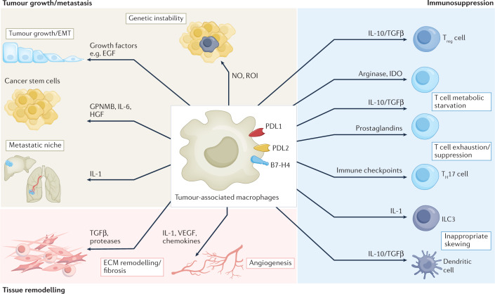
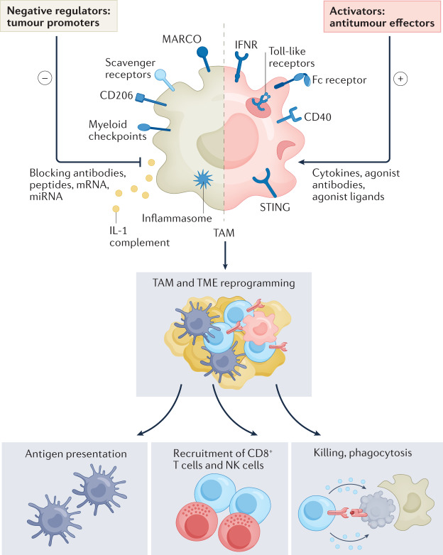
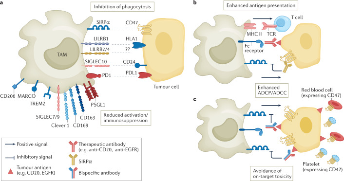
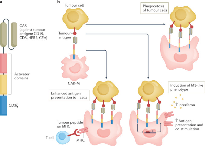
Tumour-associated macrophages are an essential component of the tumour microenvironment and have a role in the orchestration of angiogenesis, extracellular matrix remodelling, cancer cell proliferation, metastasis and immunosuppression, as well as in resistance to chemotherapeutic agents and checkpoint blockade immunotherapy. Conversely, when appropriately activated, macrophages can mediate phagocytosis of cancer cells and cytotoxic tumour killing, and engage in effective bidirectional interactions with components of the innate and adaptive immune system. Therefore, they have emerged as therapeutic targets in cancer therapy. Macrophage-targeting strategies include inhibitors of cytokines and chemokines involved in the recruitment and polarization of tumour-promoting myeloid cells as well as activators of their antitumorigenic and immunostimulating functions. Early clinical trials suggest that targeting negative regulators (checkpoints) of myeloid cell function indeed has antitumor potential. Finally, given the continuous recruitment of myelomonocytic cells into tumour tissues, macrophages are candidates for cell therapy with the development of chimeric antigen receptor effector cells. Macrophage-centred therapeutic strategies have the potential to complement, and synergize with, currently available tools in the oncology armamentarium.


Conflict of interest statement

A.M. has been a recipient of commercial research grants from Sigma Tau, Roche, Novartis, Compugen and Efranat, and has been a consultant/advisory board member/lecturer for Novartis, Roche, Ventana, Pierre Fabre, Verily, Abbvie, BMS, J&J, Compugen, Imcheck, Macrophage Therapeutics, AstraZeneca, Biovelocita, BG Fund, Third Rock, Verseau Therapeutics and Olatec Therapeutics. C.G. and P.A. are recipients of research grants from Imcheck and Macrophage Therapeutics. A.M. and C.G. are inventors of patents related to PTX3 and other innate immunity molecules. A.M., C.G. and P.A. receive royalties for reagents related to innate immunity.

Figures

Fig. 1
Fig. 1. Pro-tumoural functions and immunosuppressive activity of TAMs.
The pro-tumour functions of tumour-associated macrophages (TAMs) are diverse and act at different phases of tumour development. TAMs release nitric oxide (NO) and reactive oxygen intermediates (ROI), which cause DNA damage and genetic instability during the initiation phase. TAMs produce epidermal growth factor (EGF) and several mediators such as IL-6, hepatocyte growth factor (HGF) and GPNMB, which support cancer stem cell expansion. At later stages, TAMs contribute to metastatic spread by releasing IL-1 and transforming growth factor-β (TGFβ), which are also involved — together with several proteases — in extracellular matrix (ECM) remodelling and pathological fibrosis. TAMs are a critical source of angiogenic factors: vascular endothelial growth factor (VEGF) and pro-angiogenic chemokines. TAMs are drivers of immunosuppression in the tumour microenvironment. Secretion of IL-10, TGFβ, prostaglandins and indoleamine 2,3-dioxygenase (IDO) promote the expansion of regulatory T (Treg) cells, inappropriate skewing of dendritic cells towards an immature and tolerogenic state, and T cell metabolic starvation. Immunosuppressive TAMs are characterized by a high expression of immune-checkpoint molecules (PDL1, PDL2, B7-H4) causing T cell exhaustion. EMT, epithelial–mesenchymal transition; ILC3, type 3 innate lymphoid cell; TH17, T helper 17.
Fig. 2
Fig. 2. Macrophage reprogramming and activation of innate and adaptive immune responses.
Cytokines (such as interferons), Toll-like receptors, stimulator of interferon genes (STING) agonists, and monoclonal antibodies activate macrophage-mediated tumour cell killing (red shading). A number of signals, including complement, inflammasome activators, ligands for scavenger receptors MARCO and CD206, and myeloid checkpoints (Fig. 3) can set macrophages in a pro-tumour mode (brown shading). MicroRNAs (miRNAs) and mRNA represent strategies to reprogramme macrophage functions. Macrophage reprogramming induces macrophage-mediated killing of cancer cells, recruitment and activation of innate and adaptive lymphoid cells, and reshaping of the tumour microenvironment. IFNR, interferon receptor; NK, natural killer; TAM, tumour-associated macrophages.
Fig. 3
Fig. 3. Myeloid checkpoints and other inhibitory receptors expressed by macrophages.
a | Overview of myeloid checkpoints and inhibitory receptors expressed by tumour-associated macrophages (TAMs) and their ligands expressed on tumour cells or cell debris. These include the receptor/ligand pairs signal regulatory protein-α (SIRPα)–CD47, LILRB1–HLA1, sialic acid-binding immunoglobulin-like lectin 10 (SIGLEC10)–CD24, and PD1–PDL1, which inhibit phagocytosis, and macrophage receptor with collagenous structure (MARCO), CD169 and mannose receptor scavenger receptors. Clever 1, triggering receptor expressed on myeloid cells 2 (TREM2) and P-selectin glycoprotein ligand 1 (PSGL1) are also depicted. Targeting of Clever 1 and TREM2 does not specifically interfere with phagocytosis but with immunosuppressive activation. b | In strategies featuring the use of therapeutic antibodies, such as anti-CD20 or anti-epithelial growth factor receptor (EGFR), combinatorial use of anti-CD47 enhances antibody-dependent cellular phagocytosis (ADCP) and increases antigen presentation to T cells. c | Bispecific CD47 antibodies are designed to recognize CD47 and tumour-associated antigens (such as CD20 or PDL1), which enhances selective blocking of CD47 on tumour cells, avoiding on-target toxicity due to recognition of CD47 on red blood cells and platelets. ADCC, antibody-dependent cellular cytotoxicity; MHC II, MHC class II.
Fig. 4
Fig. 4. Cell therapy based on CAR-M cells.
a | Schematic of chimeric antigen receptor (CAR) molecule designed to be expressed by macrophages (CAR-M). Antibody specificity is provided by the extracellular module recognizing tumour antigens. Transmembrane and intracellular modules allow downstream activation signalling. Common CAR molecules have a T cell-activating module, which is preserved in one of the CAR-M developed so far because it is shown to retain activator functions on macrophages. b | Upon binding of CAR-M to tumour cells expressing the antigen, phagocytosis is increased and the pro-inflammatory M1 programme is induced, featuring release of interferon and upregulation of antigen-presentation molecules as well as enhanced presentation to T cells.

References

    1. Balkwill F, Mantovani A. Inflammation and cancer: back to Virchow? Lancet. 2001;357:539–545. doi: 10.1016/S0140-6736(00)04046-0. - DOI - PubMed
    1. Mantovani A, Allavena P, Sica A, Balkwill F. Cancer-related inflammation. Nature. 2008;454:436–444. doi: 10.1038/nature07205. - DOI - PubMed
    1. Engblom C, Pfirschke C, Pittet MJ. The role of myeloid cells in cancer therapies. Nat. Rev. Cancer. 2016;16:447–462. doi: 10.1038/nrc.2016.54. - DOI - PubMed
    1. Mantovani A, Marchesi F, Malesci A, Laghi L, Allavena P. Tumour-associated macrophages as treatment targets in oncology. Nat. Rev. Clin. Oncol. 2017;14:399–416. doi: 10.1038/nrclinonc.2016.217. - DOI - PMC - PubMed
    1. Cassetta L, Pollard JW. Targeting macrophages: therapeutic approaches in cancer. Nat. Rev. Drug Discov. 2018;17:887–904. doi: 10.1038/nrd.2018.169. - DOI - PubMed

Substances