. 2022 Aug 16;12(1):13865.
doi: 10.1038/s41598-022-17921-6.

Application of immune enhanced organoids in modeling personalized Merkel cell carcinoma research

Affiliations

Application of immune enhanced organoids in modeling personalized Merkel cell carcinoma research

Steven D Forsythe et al. Sci Rep. .

Abstract

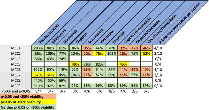
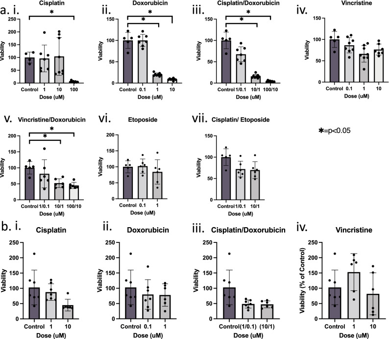
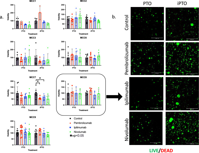
Merkel cell carcinoma (MCC) is a rare neuroendocrine cutaneous cancer, with incidence of less than 1/100,000, low survival rates and variable response to chemotherapy or immunotherapy. Herein we explore the application of patient tumor organoids (PTOs) in modeling personalized research in this rare malignancy. Unsorted and non-expanded MCC tumor cells were isolated from surgical specimens and suspended in an ECM based hydrogel, along with patient matched blood and lymph node tissue to generate immune enhanced organoids (iPTOs). Organoids were treated with chemotherapy or immunotherapy agents and efficacy was determined by post-treatment viability. Nine specimens from seven patients were recruited from December 2018-January 2022. Establishment rate was 88.8% (8/9) for PTOs and 77.8% (7/9) for iPTOs. Histology on matched patient tissues and PTOs demonstrated expression of MCC markers. Chemotherapy response was exhibited in 4/6 (66.6%) specimens with cisplatin and doxorubicin as the most effective agents (4/6 PTO sets) while immunotherapy was not effective in tested iPTO sets. Four specimens from two patients demonstrated resistance to pembrolizumab, correlating with the corresponding patient's treatment response. Routine establishment and immune enhancement of MCC PTOs is feasible directly from resected surgical specimens allowing for personalized research and exploration of treatment regimens in the preclinical setting.


Conflict of interest statement

The authors declare no competing interests.

Figures

Figure 1
Figure 1
Flow chart of Merkel cell carcinoma organoid fabrication. Tumor cells were isolated from digested tumor tissue and fabricated into PTOs, while immune cells were isolated from patient blood or from lymph tissue and combined with tumor cells to create iPTOs. These organoids were cultured for 1 week, after which they were characterized and screened for therapeutic efficacy. Created with BioRender.com.
Figure 2
Figure 2
Immunohistochemistry of matched patient tissue (Bx) and Merkel cell carcinoma PTO set (a) MCC5 and (b) MCC7. Comparison of tissue and PTOs shows high concordance of identification markers including (i) cellular morphology (H&E), (ii) iPTO cellular morphology (H&E), (iii) pan cytokeratin (Pan CK), (iv) cytokeratin 20 (CK20), (v) neurofilament (NFH), (vi) chromogranin A (ChgA), (vii) synaptophysin (SYP and (viii) Merkel cell polyomavirus (MCPyV). Additionally, sets MCC5 (a) and MCC7 (b) demonstrated variable expression of markers related to disease identification and potential outcomes, including MCPyV. All images taken at 40 × magnification with scale bar 20 μM.
Figure 3
Figure 3
Chemotherapy screening of PTO demonstrates intertumor heterogeneity in tumor treatment response. Percentages are viability compared to patient matched control organoids. Doses for treatments in table are as follows: cisplatin (10 uM), doxorubicin (1 μM), vincristine (1 μM), etoposide (1 μM), cisplatin/doxorubicin (10 μM/1 μM), cisplatin/etoposide (10 μM/1 μM), and vincristine/doxorubicin (1 μM/1 μM). Green boxes represent treatments resulting in < 50% viability decrease and p > 0.05, yellow boxes > 50% or p < 0.05, red boxes represent treatments with > 50% viability decrease and p < 0.05. The bottom line is number of PTO sets in which the treatment is effective (< 50% viable and p < 0.05). The column on the far right represents the number of effective treatments in the respective patient organoid set.
Figure 4
Figure 4
Application of personalized chemotherapy treatment platform for (a) MCC2 and (b) MCC5 with (i) Cisplatin. (ii) Doxorubicin. (iii) Vincristine. (iv) Etoposide. (v) Cisplatin/Doxorubicin. (vi) Cisplatin/Etoposide and (vii) Vincristine/Doxorubicin. Summary of CellTiter-Glo 3D assay results for (a) MCC2 demonstrate dose-dependent treatment responses for (i) Cisplatin (ii) Doxorubicin (iii) Cisplatin/Doxorubicin and (vii) Vincristine/Doxorubicin while (b) MCC5 illustrated these results for (i) cisplatin and (iii) cisplatin/doxorubicin. Under each dose, replicates listed as data points. *p < 0.05.
Figure 5
Figure 5
Demonstration of iPTOs in immunotherapy testing applications. (a) iPTO sets were treated with immunotherapies and viability was recorded with CellTiter-Glo 3D. No iPTOs were determined to have significant response of < 50% viability compared to control, statistical significance of p < 0.05 compared to iPTO control and to PTO counter treatment to any immunotherapies tested. (b) LIVE/DEAD Panel for MCC8. Scale bars represent 250 microns. *p < 0.05.
Figure 6
Figure 6
Comparative analysis of tumors temporally separated in the same patient. (a) Immunohistochemistry of iPTOs demonstrates robust CD8+ t-cell populations but low granzyme B mediated killing of CK20+ cells. (b) LDH cellular stress analysis determines an increase in pembrolizumab MCC7 iPTO set but not in MCC9, suggesting acquisition of resistance between the two tumors in this patient. All images taken at 40X magnification with scale bar 20 μM. *p < 0.05.

References

    1. Paulson, K.G., et al. Merkel cell carcinoma: Current US incidence and projected increases based on changing demographics. J. Am. Acad. Dermatol.78(3), 457–463 e2 (2018). - PMC - PubMed
    1. Woo SH, et al. Identification of epidermal progenitors for the Merkel cell lineage. Development. 2010;137(23):3965–3971. doi: 10.1242/dev.055970. - DOI - PMC - PubMed
    1. Harms PW, et al. The biology and treatment of Merkel cell carcinoma: Current understanding and research priorities. Nat. Rev. Clin. Oncol. 2018;15(12):763–776. doi: 10.1038/s41571-018-0103-2. - DOI - PMC - PubMed
    1. Stachyra, K., et al., Merkel cell carcinoma from molecular pathology to novel therapies. Int. J. Mol. Sci.22(12) (2021). - PMC - PubMed
    1. Ricco, G., et al. Multimodal strategy in localized merkel cell carcinoma: Where are we and where are we heading? Int. J. Mol. Sci.22(19) (2021). - PMC - PubMed

Publication types

MeSH terms