Review
. 2023 Mar;23(3):174-188.
doi: 10.1038/s41577-022-00761-w. Epub 2022 Aug 16.

Tuning immunity through tissue mechanotransduction

Affiliations
Review

Tuning immunity through tissue mechanotransduction

Huixun Du et al. Nat Rev Immunol. 2023 Mar.

Abstract

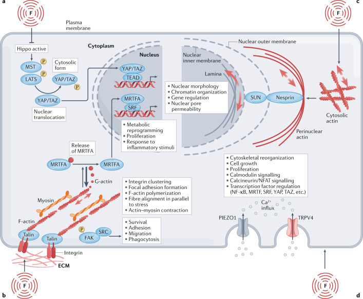
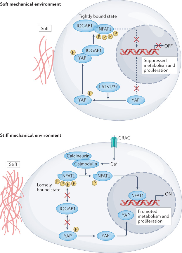
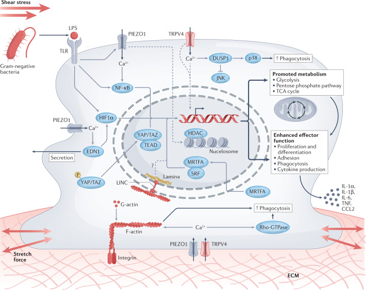
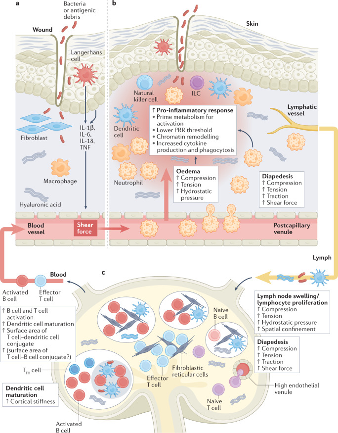
Immune responses are governed by signals from the tissue microenvironment, and in addition to biochemical signals, mechanical cues and forces arising from the tissue, its extracellular matrix and its constituent cells shape immune cell function. Indeed, changes in biophysical properties of tissue alter the mechanical signals experienced by cells in many disease conditions, in inflammatory states and in the context of ageing. These mechanical cues are converted into biochemical signals through the process of mechanotransduction, and multiple pathways of mechanotransduction have been identified in immune cells. Such pathways impact important cellular functions including cell activation, cytokine production, metabolism, proliferation and trafficking. Changes in tissue mechanics may also represent a new form of 'danger signal' that alerts the innate and adaptive immune systems to the possibility of injury or infection. Tissue mechanics can change temporally during an infection or inflammatory response, offering a novel layer of dynamic immune regulation. Here, we review the emerging field of mechanoimmunology, focusing on how mechanical cues at the scale of the tissue environment regulate immune cell behaviours to initiate, propagate and resolve the immune response.


Conflict of interest statement

The authors declare no competing interests.

Figures

Fig. 1
Fig. 1. An overview of mechanotransduction pathways in cells.
a | Classical Hippo pathway activation induces phosphorylation of kinases MST1 and/or MST2, as well as LATS1 and/or LATS2, which in turn phosphorylate yes-associated protein (YAP) and transcriptional co-activator with PDZ-binding motif (TAZ). Phosphorylated YAP and TAZ are sequestered in the cytoplasm. Upon Hippo pathway inhibition, such as in response to mechanical force (denoted by F), YAP and TAZ translocate to the nucleus and drive gene expression that is key to immune function, including genes involved in cellular metabolic reprogramming, proliferation and responsiveness to inflammatory stimuli, through the TEA domain (TEAD) transcription factor family. b | Integrins anchor cells to the extracellular matrix (ECM), cluster and form focal adhesions, which act as multiprotein signalling hubs. Focal adhesion kinase (FAK) and SRC are key downstream effectors of focal adhesions, orchestrating various immune cell functions. Focal adhesions execute cell morphological changes either by actin polymerization-driven expansion or depolymerization and contraction with myosin. Polymerization of actin filaments (F-actin) subsequently reduces availability of G-actin (depicted by heavy arrow), decreasing its association with myocardin-related transcription factor A (MRTFA), which causes nuclear translocation of MRTFA, association with serum response factor (SRF) and gene transcription responsible for metabolic reprogramming, proliferation and cell responses to inflammatory stimuli. c | Mechanotransduction is also propagated directly from the cytoskeleton to the nuclear envelope via the linker of nucleoskeleton and cytoskeleton (LINC) complex, which includes the transmembrane proteins SUN and nesprin. LINC mediates nuclear morphology, chromatin organization, gene regulation and nuclear pore permeability, which also facilitates the nuclear translocation of YAP and TAZ and MRTFA. d | Transient receptor potential vanilloid type 4 (TRPV4) and PIEZO1 cation channels are activated by mechanical stimuli inflicted on the plasma membrane. Their activation promotes Ca2+ influx, impacting the regulation of many transcription factors, inflammatory signalling pathways and cytoskeletal reorganization.
Fig. 2
Fig. 2. Mechanical regulation of T cell activity through YAP.
When T cells are in tissues with a soft extracellular matrix (ECM) microenvironment, yes-associated protein (YAP) is phosphorylated (likely by LATS1 or LATS2) and localized in the cytoplasm, where it interacts with IQGAP1. Owing to YAP and IQGAP1 interaction, NFAT1 is preferentially sequestered in the cytoplasm, and this results in attenuated cellular metabolism and proliferation. Conversely, when T cells encounter a stiff ECM microenvironment, YAP is dephosphorylated and migrates to the nucleus. Additionally, NFAT1 is no longer sequestered in the cytoplasm through IQGAP1–YAP interaction. Thus, with the help of calcium release-activated channels (CRAC), calcineurin and calmodulin, NFAT1 can be dephosphorylated and is free to translocate to the nucleus to induce T cell activation-related gene expression.
Fig. 3
Fig. 3. Integration of mechanotransduction pathways and innate immune cell activation.
Pattern recognition receptor (PRR) stimulation accompanied by mechanical force causes PIEZO1 co-localization with some Toll-like receptors (TLRs). Force-triggered opening of transient receptor potential vanilloid type 4 (TRPV4) and PIEZO1 leads to Ca2+ influx. Elevated cytosolic Ca2+ levels induce actin polymerization and activate Rho GTPase, boosting phagocytosis. Concurrently, F-actin formation depletes G-actin availability, and without G-actin to bind to, myocardin-related transcription factor A (MRTFA) is shuttled into the nucleus where it forms a complex with serum response factor (SRF). MRTFA–SRF enables transcription of immune effector genes, such as those encoding interleukin-6 (IL-6) and CXC-chemokine ligand 9 (CXCL9), generally skewing the cell towards an enhanced inflammatory phenotype. Ca2+ also enforces TLR signalling by promoting the expression and activation of endothelin 1 (EDN1) and NF-κB. EDN1 stabilizes hypoxia-inducible factor 1α (HIF1α), promoting TLR-induced HIF1α accumulation. Active NF-κB and HIF1α translocate to the nucleus and drive metabolic and inflammatory gene expression. Additionally, TRPV4-dependent Ca2+ influx prompts dual-specificity phosphatase 1 (DUSP1) to activate p38 and inhibit JNK activity, further enhancing phagocytosis. The mechanosensitive molecules yes-associated protein (YAP) and transcriptional co-activator with PDZ-binding motif (TAZ) also translocate to the nucleus upon activation, bind TEA domains (TEADs) and induce transcription of genes that boost glycolysis, the pentose phosphate pathway and the tricarboxylic acid (TCA) cycle. This immunometabolic regulation through multiple mechanosensitive inputs mediates robust cell proliferation and increases the production of inflammatory cytokines, such as IL-1α, IL-1β, IL-6 and CC-chemokine ligand 2 (CCL2). PIEZO1 also activates histone deacetylases (HDACs) to further induce inflammatory cytokine output. Dashed lines represent pathways where there are still major gaps in understanding the transduction mechanisms, and solid lines depict more well-described pathways. Linker of nucleoskeleton and cytoskeleton (LINC) complex modulation of immune activation has been shown in adaptive cells only, but we hypothesize it also plays a role in innate immune cell regulation. ECM, extracellular matrix; LPS, lipopolysaccharide; TNF, tumour necrosis factor.
Fig. 4
Fig. 4. Mechanical forces modulate acute immune responses.
a | Danger signals in the skin (such as those arising from bacteria or tissue damage) activate Langerhans cells and stromal cells to produce inflammatory cytokines and/or hyaluronic acid to promote oedema and attract immune cells to the infection site. b | Within minutes to hours, diapedesis of cells from the vasculature into inflamed tissues occurs. This requires force-sensing at the molecular level (shear force-dependent catch bonds) and at the cellular level (cells squeezing through endothelium and extracellular matrix (ECM)). Forces exerted by diapedesis prime circulating immune cells to acquire an enhanced pro-inflammatory phenotype. Simultaneously, local oedema occurs. Mechanosensation of oedema further drives a pro-inflammatory phenotype in tissue-resident cells that encounter pathogens. c | Eventually, antigenic debris, hyaluronic acids and activated dendritic cells traffic to the lymph node. There, fibroblastic reticular cells sense activated dendritic cells and change their shape to allow for imminent increases in lymph node size. Migrating T cells and B cells encounter mechanical forces as they squeeze through high endothelial venules and into interfollicular and follicular spaces. After activation, T cells and B cells proliferate in the lymph node, causing lymph node swelling and stiffening, which propagates mechanical forces onto cells. T cells crawling through a stiff lymph node increase the contact area for T cell–antigen-presenting cell (APC) conjugates, which may also impact T follicular helper (TFH) cell interactions with germinal centre B cells. Activated T cells and B cells eventually migrate to the infection site via the blood, continuing to experience multiple mechanical stimuli at the inflamed tissue. Resolution of the infection leads to breakdown of hyaluronic acid, decreasing oedema and leading to the refractoriness of immune cells to danger signals. The resulting diminishment of inflammatory cytokines reduces immune cell trafficking and promotes their apoptosis through cytokine withdrawal-induced cell death and other mechanisms. Under the influence of ageing and other factors, tissue may not return to a ‘healthy’ baseline and might, instead, adopt remodelling changes including collagen deposition (scarring) and elastin degradation, leaving a lasting mechanical imprint at the inflammatory response site. ILC, innate lymphoid cell; IL, interleukin; PRR, pattern recognition receptor; TNF, tumour necrosis factor.

Similar articles

Cited by

References

    1. Garoffolo G, Pesce M. Mechanotransduction in the cardiovascular system: from developmental origins to homeostasis and pathology. Cells. 2019;8:1607. doi: 10.3390/cells8121607. - DOI - PMC - PubMed
    1. Hsieh JY, et al. Differential regulation of macrophage inflammatory activation by fibrin and fibrinogen. Acta Biomater. 2017;47:14–24. doi: 10.1016/j.actbio.2016.09.024. - DOI - PMC - PubMed
    1. Meli VS, et al. Biophysical regulation of macrophages in health and disease. J. Leukoc. Biol. 2019;106:283–299. doi: 10.1002/JLB.MR0318-126R. - DOI - PMC - PubMed
    1. Rowley AT, Nagalla RR, Wang S-W, Liu WF. Extracellular matrix-based strategies for immunomodulatory biomaterials engineering. Adv. Healthc. Mater. 2019;8:e1801578. doi: 10.1002/adhm.201801578. - DOI - PMC - PubMed
    1. Smith TD, Nagalla RR, Chen EY, Liu WF. Harnessing macrophage plasticity for tissue regeneration. Adv. Drug Deliv. Rev. 2017;114:193–205. doi: 10.1016/j.addr.2017.04.012. - DOI - PubMed

Publication types

MeSH terms