. 2022 Aug 16;79(9):484.
doi: 10.1007/s00018-022-04486-w.

A context-dependent and disordered ubiquitin-binding motif

Affiliations

A context-dependent and disordered ubiquitin-binding motif

Jesper E Dreier et al. Cell Mol Life Sci. .

Abstract

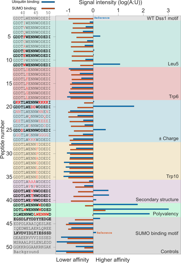
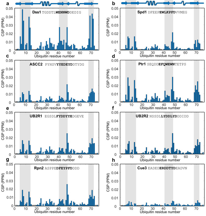
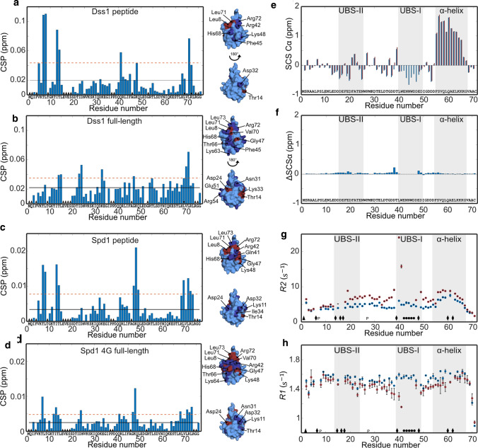
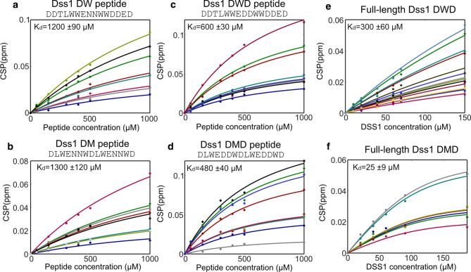
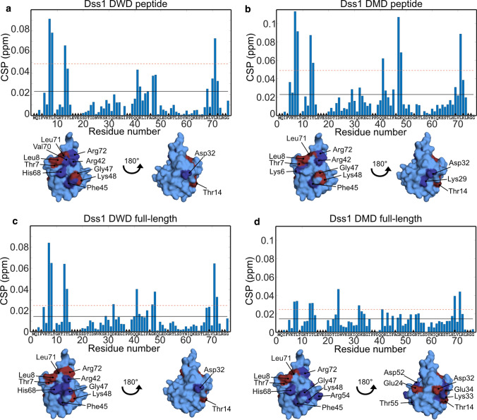
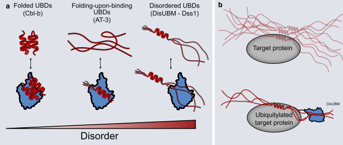
Ubiquitin is a small, globular protein that is conjugated to other proteins as a posttranslational event. A palette of small, folded domains recognizes and binds ubiquitin to translate and effectuate this posttranslational signal. Recent computational studies have suggested that protein regions can recognize ubiquitin via a process of folding upon binding. Using peptide binding arrays, bioinformatics, and NMR spectroscopy, we have uncovered a disordered ubiquitin-binding motif that likely remains disordered when bound and thus expands the palette of ubiquitin-binding proteins. We term this motif Disordered Ubiquitin-Binding Motif (DisUBM) and find it to be present in many proteins with known or predicted functions in degradation and transcription. We decompose the determinants of the motif showing it to rely on features of aromatic and negatively charged residues, and less so on distinct sequence positions in line with its disordered nature. We show that the affinity of the motif is low and moldable by the surrounding disordered chain, allowing for an enhanced interaction surface with ubiquitin, whereby the affinity increases ~ tenfold. Further affinity optimization using peptide arrays pushed the affinity into the low micromolar range, but compromised context dependence. Finally, we find that DisUBMs can emerge from unbiased screening of randomized peptide libraries, featuring in de novo cyclic peptides selected to bind ubiquitin chains. We suggest that naturally occurring DisUBMs can recognize ubiquitin as a posttranslational signal to act as affinity enhancers in IDPs that bind to folded and ubiquitylated binding partners.

Keywords: Context; Cyclic peptide; Deep mutational scanning; IDP; NMR; SLiM; UBM; Ubiquitin.


Conflict of interest statement

The authors declare that they have no conflicts of interests.

Figures

Fig. 1
Fig. 1
Properties of the disordered ubiquitin-binding motif (DisUBM). Several features of the motif were investigated by a membrane-bound peptide array. The features are indicated to the right (and in different colors), and sequences are shown to the left. Bold sequences indicate peptides with emission intensities larger than the Dss1 UBS1 peptide. The blue and orange bars show emission signals from the peptides incubated with ubiquitin and SUMO, respectively. The emission intensities are normalized to the known binding motifs for ubiquitin (Pep 1) and SUMO (Pep 47), respectively. Ubiquitin binding (blue bars), sumo binding (red bars)
Fig. 2
Fig. 2
DisUBMs in different IDPs bind to the same patch on ubiquitin. a-h: CSP per residue of 100 µM 15N-labeled ubiquitin upon addition of 1 mM 15-mer peptides analyzed by 1H-15N-HSQC NMR. The secondary structure of ubiquitin is shown in the top cartoon and names and sequences of peptides indicated in each plot. The gray bars indicate the interaction site on ubiquitin as defined by the Dss1 peptide. The bolded part of the sequences indicates the proposed binding motif. The black triangles in the bar plot designate unassigned residues
Fig. 3
Fig. 3
The context of the DisUBM affects binding. a CSP per residue of 100 µM 15N-labeled ubiquitin added 3000 µM Dss1 peptide. The black line indicates the average CSP and the dashed red line the average CSP + one standard deviation. Black triangles indicate unassigned residues, and black circles line broadening upon addition of Dss1. Surface representations of ubiquitin with affected residues in dark blue (above average CSP) and red (above average + one standard deviation) highlighted are shown to the right. b Same as a but with 440 µM full-length Dss1. c Same as a but with 1000 µM Spd1 peptide. d Same as a but with 290 µM 4G Spd1. e SCS of the Cα atoms of 1 mM 13C-15 N-labeled Dss1 alone (blue) or with 2 mM ubiquitin (red). Gray bars indicate the transient helix and ubiquitin-binding sites in Dss1 as identified by Paraskevopoulos et al. [36]. f Differences between the SCSs shown in e of Dss1 with and without ubiquitin. g R2 and h R1 values of 500 µM 15 N-labeled Dss1 without (blue) and with (red) 1 mM ubiquitin. The error bars are standard deviations from triplicate recordings on the same sample. Triangles are unassigned residues, diamonds residues with peak overlap, “p” indicates prolines and circles peaks that disappear upon addition of ubiquitin
Fig. 4
Fig. 4
Increased affinity for ubiquitin by aromatics, negative charge and chain context. Changes in CSP with the concentrations of peptide or protein. Each fit line and corresponding colored data points reflect the CSPs of individual NMR signals from 1H-15 N-HSQC spectra of 100 µM 15 N-labeled ubiquitin upon titration with either peptides (ad) or proteins (e and f). Peptide and protein names are shown at the top. All data points in one titration were fitted using a global fit to obtain the Kd values reported on each plot
Fig. 5
Fig. 5
Binding surfaces on ubiquitin of optimized Dss1 variants. CSP per residue of 100 µM 15 N-labeled ubiquitin with a 300 µM DWD peptide, b 500 µM DMD peptide, c 150 µM full-length DWD Dss1, d 90 µM full-length DMD Dss1. Residues with CSP values above average and above average plus one standard deviation are shown on the surface of ubiquitin in dark blue and red, respectively. Black lines indicate mean CSP value, the red dashed line the mean + one standard deviation. The black triangles indicate unassigned residues, black circles line broadening or peak coalescence
Fig. 6
Fig. 6
Cellular effects of DisUBM variants. a Yeast growth assays with wild-type and dss1Δ S. pombe cells. Serial fivefold dilutions of the indicated strains were spotted in droplets onto solid media and incubated at 29 °C or 37 °C. b Yeast cells expressing GFP and the indicated GFP-tagged Dss1 variants were lysed and used for immunoprecipitation (IP) assays using GFP-trap resin. Samples of the input and the precipitated material were analyzed by SDS-PAGE and western blotting using antibodies to the 19S proteasome subunit Mts4/Rpn1, the 20S proteasome α-subunits, and GFP
Fig. 7
Fig. 7
DisUBMs feature in de novo cyclic peptides from unbiased screens. a De novo cyclic peptides from trillion-scale random cyclic peptide library are enriched in DisUBMs. Shown are the 20 highest-ranked cyclic peptides after 4 rounds of RaPID selection against K48Ub2 or K48Ub4. Cyclic peptides containing DisUBMs have motif highlighted: XΩXXXΩX, where Ω is F/Y/W, X is any amino acid, and motif contains at least one E/D. Figure adapted from [45]. b Binding of natural DisUBM and de novo cyclic peptide induce similar chemical shift perturbations (CSP) in monomeric and proximal ubiquitin of K48Ub2, respectively. CSP one standard deviation above the mean for Dss1 and Spd1, and CSP > 0.3 for K48Ub2 [45] are shown in cyan (structures based on pdb 1aar). c Deep mutational scanning of cyclic peptides Ub2i and Ub2ii binding to K48Ub2 reveals that mutations which increase DisUBM character improve binding. In particular, mutation to D/E between the two aromatic amino acids of DisUBM ΩXXXΩ
Fig. 8
Fig. 8
Disorder in ubiquitin binding. a Canonical UBDs (red) are known to have stable secondary or tertiary structures both in isolation and when binding ubiquitin (blue) (left). Recently, it was shown that some UBDs only fold upon binding to ubiquitin (middle). This study has revealed that a ubiquitin-binding motif, DisUBM occurs frequently in IDPs and that this binds ubiquitin without adopting any fixed secondary structure (right). b The DisUBM motif is suggested to act as an affinity enhancer when IDPs bind to ubiquitylated folded binding partners. The folded binding partner could be ubiquitin itself

References

    1. Komander D, Rape M (2012) The ubiquitin code. Annu Rev Biochem 81:203–229. 10.1146/annurev-biochem-060310-170328 - PubMed
    1. Yau R, Rape M (2016) The increasing complexity of the ubiquitin code. Nat Cell Biol 18:579–586 - PubMed
    1. Schreiner P, Chen X, Husnjak K et al (2008) Ubiquitin docking at the proteasome through a novel pleckstrin-homology domain interaction. Nature 453:548–552. 10.1038/nature06924 - PMC - PubMed
    1. Wu X, Liu S, Sagum C et al (2019) Crosstalk between Lys63- and Lys11-polyubiquitin signaling at DNA damage sites is driven by Cezanne. Genes Dev 33:1702–1717. 10.1101/gad.332395.119 - PMC - PubMed
    1. Hofmann RM, Pickart CM (1999) Noncanonical MMS2-encoded ubiquitin-conjugating enzyme functions in assembly of novel polyubiquitin chains for DNA repair. Cell 96:645–653. 10.1016/S0092-8674(00)80575-9 - PubMed

LinkOut - more resources