

Life-threatening gastrointestinal bleeding caused by jejunal heterotopic gastric mucosa in an adult dog: a rare case report
- PMID: 35974373
- PMCID: PMC9380381
- DOI: 10.1186/s12917-022-03415-0
Life-threatening gastrointestinal bleeding caused by jejunal heterotopic gastric mucosa in an adult dog: a rare case report
Abstract
Background: Heterotopic gastric mucosa has been scarcely reported in the veterinary literature. Its presence can be asymptomatic or associated with various clinical signs ranging from apathy, vomiting, to abdominal pain. This report illustrates the presence of heterotopic gastric mucosa in the jejunum of an adult dog. It is the first to describe severe anemia, requiring acute blood transfusion, following intestinal hemorrhage caused by heterotopic gastric mucosa.
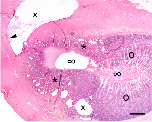
Case presentation: A twelve-year-old, intact male Maltese dog was presented with a history of apathy, vomiting and anemia. The dog was on a strict diet for recurrent diarrhea, food intolerance and skin allergy. Clinical examination revealed severe anemic mucous membranes and painful abdominal palpation. Blood examination confirmed severe regenerative anemia. Ultrasonography showed an intestinal neoplasm, gall bladder sludge and non-homogeneous liver parenchyma. Three-view thoracic radiographs failed to show any metastatic lesions or enlarged lymph nodes. After initial stabilization and blood transfusion, a midline exploratory laparotomy was performed. Three different masses were found in the jejunum. Resection and anastomosis of approximately 40 cm of jejunum was performed, followed by liver and lymph node biopsy and placement of an esophagostomy tube. Two days after surgery the dog started to clinically improve and was discharged from the hospital on the sixth day after surgery. Histopathology revealed the intestinal masses to be heterotopic gastric mucosa associated with intramural cystic distensions, multifocal ulceration and bleeding into the intestinal lumen. Two years after surgery, the dog did not have a recurrence of anemia or gastrointestinal signs.
Conclusions: This case demonstrates that heterotopic gastric mucosa can be considered one of the differential diagnoses in case of severe anemia due to gastrointestinal hemorrhage and suspected intestinal tumors. Although in most described cases in literature the finding seems to be incidental on necropsy, our report shows that heterotopic gastric mucosa can be the etiology of life-threatening signs. In addition, because no recurrent diarrhea episodes occurred after surgical resection of the ectopic tissue, it is likely that the heterotopic gastric mucosa was the cause of the food intolerance signs in this dog.
Keywords: Anemia; Case report; Differential diagnosis; Ectopic tissue; Heterotopic gastric mucosa; Intestinal bleeding; Intestinal mass.
© 2022. The Author(s).
Conflict of interest statement
The authors declare that they have no competing interests.
Figures


Similar articles
-
Heterotopic gastric mucosa associated with abdominal abscess formation, hypotension, and acute abdominal pain in a puppy.J Vet Emerg Crit Care (San Antonio). 2014 Nov-Dec;24(6):745-50. doi: 10.1111/vec.12249. Epub 2014 Nov 11. J Vet Emerg Crit Care (San Antonio). 2014. PMID: 25388790
-
Long-term intestinal bleeding in a child: a rare case of heterotopic gastric mucosa in the jejunum.BMJ Case Rep. 2016 Nov 25;2016:bcr2016216949. doi: 10.1136/bcr-2016-216949. BMJ Case Rep. 2016. PMID: 27888219 Free PMC article.
-
Epiploic foramen entrapment in a dog.Vet Surg. 2023 Nov;52(8):1237-1244. doi: 10.1111/vsu.13975. Epub 2023 Jun 9. Vet Surg. 2023. PMID: 37293954
-
[Clinical characteristics and literature review of ectopic gastric mucosa in the small intestine].Zhonghua Yi Xue Za Zhi. 2024 Jun 4;104(21):1998-2002. doi: 10.3760/cma.j.cn112137-20240312-00559. Zhonghua Yi Xue Za Zhi. 2024. PMID: 38825944 Review. Chinese.
-
Rectal red blood loss in a healthy toddler is not always a juvenile polyp.Acta Gastroenterol Belg. 2017 Jan-Mar;80(1):67-70. Acta Gastroenterol Belg. 2017. PMID: 29364101 Review.
Cited by
-
Assessment of the effect of prokinetic drugs on transit time and gastrointestinal cleanliness in capsule endoscopy.BMC Vet Res. 2025 Jul 2;21(1):417. doi: 10.1186/s12917-025-04862-1. BMC Vet Res. 2025. PMID: 40604869 Free PMC article.
-
Jejunal perforation and septic abdomen resulting from a choristoma in a dog.Can Vet J. 2024 Jan;65(1):29-32. Can Vet J. 2024. PMID: 38164377 Free PMC article.
References
-
- Wang H, Tan Y, Liu D. A rare heterotopic gastric mucosa appearing between the muscularis mucosae and submucosa. Rev Esp Enferm Dig. 2019;111:712–713. - PubMed
Publication types
MeSH terms
LinkOut - more resources
Full Text Sources
Medical
