Review
. 2022 Oct;118(4):295-308.
doi: 10.1111/mmi.14974. Epub 2022 Aug 24.

Nuclear entry and egress of parvoviruses

Affiliations
Review

Nuclear entry and egress of parvoviruses

Salla Mattola et al. Mol Microbiol. 2022 Oct.

Abstract

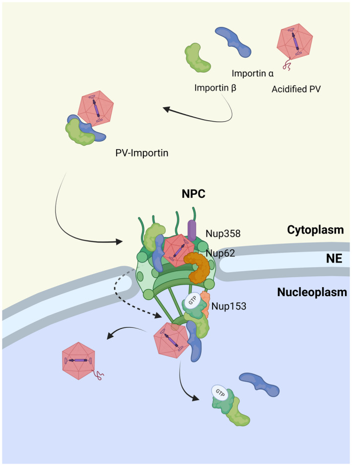
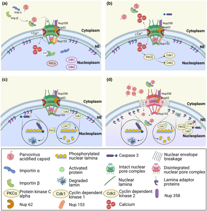
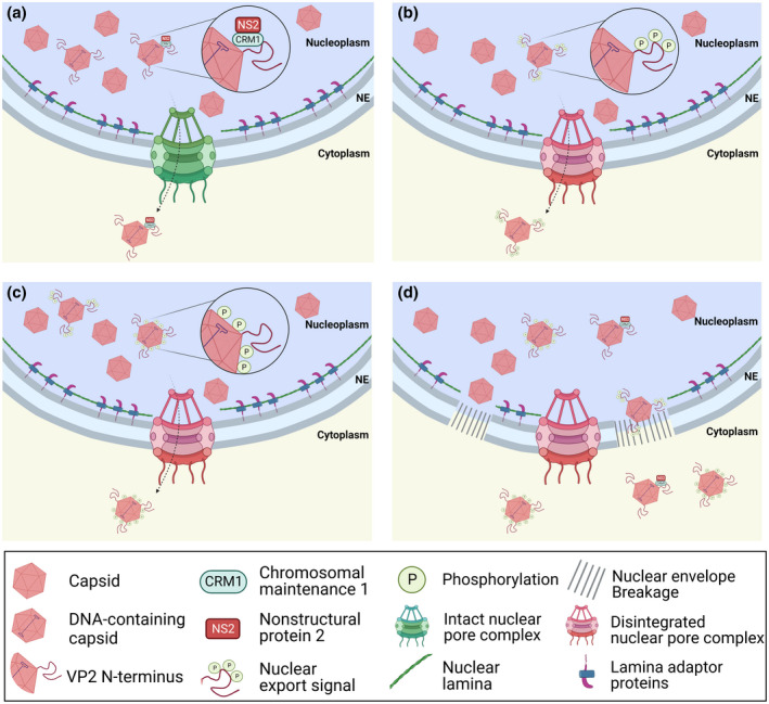
Parvoviruses are small non-enveloped single-stranded DNA viruses, which depend on host cell nuclear transcriptional and replication machinery. After endosomal exposure of nuclear localization sequence and a phospholipase A2 domain on the capsid surface, and escape into the cytosol, parvovirus capsids enter the nucleus. Due to the small capsid diameter of 18-26 nm, intact capsids can potentially pass into the nucleus through nuclear pore complexes (NPCs). This might be facilitated by active nuclear import, but capsids may also follow an alternative entry pathway that includes activation of mitotic factors and local transient disruption of the nuclear envelope. The nuclear entry is followed by currently undefined events of viral genome uncoating. After genome release, viral replication compartments are initiated and infection proceeds. Parvoviral genomes replicate during cellular S phase followed by nuclear capsid assembly during virus-induced S/G2 cell cycle arrest. Nuclear egress of capsids occurs upon nuclear envelope degradation during apoptosis and cell lysis. An alternative pathway for nuclear export has been described using active transport through the NPC mediated by the chromosome region maintenance 1 protein, CRM1, which is enhanced by phosphorylation of the N-terminal domain of VP2. However, other alternative but not yet uncharacterized nuclear export pathways cannot be excluded.

Keywords: import and export; nuclear envelope; nuclear pore complexes; nucleus; parvoviruses.


Figures

FIGURE 1
FIGURE 1
Nuclear entry of parvoviruses through the NPC. Cytoplasmic parvoviruses (PVs) that have undergone structural changes within the endosome bind to importin α (KPNA) /importin β (KPNB). This allows transport through cellular nuclear pore complexes (NPCs). Upon reaching the nuclear basket, the PV‐importin complex dissociates, releasing the capsid into the nucleoplasm. Figure created with BioRender.
FIGURE 2
FIGURE 2
Entry through the NE by increased nuclear envelope permeability. (a) Parvovirus capsids bound to importins (KPNA: Importin α/KPNB: Importin β) bind to Nups. The binding triggers exposure of PLA2 on VP1u inducing calcium efflux. (b) the release of calcium activates PKCα, which activates cdk2/cdk1. Caspase 3 is also activated. (c) Hyper‐phosphorylation of Lamin B by kinases as well as Lamin B‐cleavage by caspase 3 leads to its local degradation. (d) The formation of transient holes allows entry of NPC‐bound or cytosolic capsids or capsid‐importin complexes. Figures created with BioRender.
FIGURE 3
FIGURE 3
Nuclear egress of progeny capsids. Packaging of viral genomes inside capsids causes a conformational change exposing the VP2 N‐terminal on the capsid surface. (a) MVM capsids are actively exported out of the nucleus through NPCs mediated by the interaction between NS2 NES with CRM1 (Bodendorf et al., ; Eichwald et al., ; Engelsma et al., ; Fornerod et al., ; Maroto et al., ; Miller & Pintel, 2002). (b) The phosphorylation of the exposed VP2 N‐terminal end on the capsid surface acts as a nuclear export signal enhancing capsid export out of the nucleus (Maroto et al., 2004). (c) Phosphorylation of the capsid surface enhances capsid export (Wolfisberg et al., 2016). (d) Activation of apoptosis and necrosis affect the structure of the nuclear lamina, and capsids are released to the cytoplasm in late infection (Chen & Qiu, ; Nykky et al., , (Wolfisberg et al., 2016). Figures created with BioRender.

Similar articles

Cited by

References

    1. Abschuetz, A. , Kehl, T. , Geibig, R. , Leuchs, B. , Rommelaere, J. & Régnier‐Vigouroux, A. (2006) Oncolytic murine autonomous parvovirus, a candidate vector for glioma gene therapy, is innocuous to normal and immunocompetent mouse glial cells. Cell and Tissue Research, 325, 423–436. 10.1007/S00441-006-0199-Z/FIGURES/8 - DOI - PubMed
    1. Adeyemi, R.O. & Pintel, D.J. (2014) Parvovirus‐induced depletion of cyclin B1 prevents mitotic entry of infected cells. PLoS Pathogens, 10, e1003891. 10.1371/journal.ppat.1003891 - DOI - PMC - PubMed
    1. Adeyemi, R.O. , Landry, S. , Davis, M.E. , Weitzman, M.D. & Pintel, D.J. (2010) Parvovirus minute virus of mice induces a DNA damage response that facilitates viral replication. PLoS Pathogens, 6, 1–11. 10.1371/journal.ppat.1001141 - DOI - PMC - PubMed
    1. Agbandje‐McKenna, M. , Llamas‐Saiz, A.L. , Wang, F. , Tattersall, P. & Rossmann, M.G. (1998) Functional implications of the structure of the murine parvovirus, minute virus of mice. Structure, 6, 1369–1381. 10.1016/S0969-2126(98)00137-3 - DOI - PubMed
    1. Arora, R. , Malla, W.A. , Tyagi, A. , Mahajan, S. , Sajjanar, B. & Tiwari, A.K. (2021) Canine parvovirus and its non‐structural gene 1 as oncolytic agents: mechanism of action and induction of anti‐tumor immune response. Frontiers in Oncology, 11, 1290. 10.3389/FONC.2021.648873/BIBTEX - DOI - PMC - PubMed

Publication types

MeSH terms