Review
. 2022 Aug 14;35(4):328-337.
doi: 10.1055/s-0042-1744481. eCollection 2022 Jul.

Radiology of the Mesentery

Affiliations
Review

Radiology of the Mesentery

Patrick W O'Regan et al. Clin Colon Rectal Surg. .

Abstract

The recent description and re-classification of the mesentery as an organ prompted renewed interest in its role in physiological and pathological processes. With an improved understanding of its anatomy, accurately and reliably assessing the mesentery with non-invasive radiological investigation becomes more feasible. Multi-detector computed tomography is the main radiological modality employed to assess the mesentery due to its speed, widespread availability, and diagnostic accuracy. Pathologies affecting the mesentery can be classified as primary or secondary mesenteropathies. Primary mesenteropathies originate in the mesentery and subsequently progress to involve other organ systems (e.g., mesenteric ischemia or mesenteric volvulus). Secondary mesenteropathies describe disease processes that originate elsewhere and progress to involve the mesentery with varying degrees of severity (e.g., lymphoma). The implementation of standardized radiological imaging protocols, nomenclature, and reporting format with regard to the mesentery will be essential in improving the assessment of mesenteric anatomy and various mesenteropathies. In this article, we describe and illustrate the current state of art in respect of the radiological assessment of the mesentery.

Keywords: mesenteropathy; mesentery; organ; peritoneum; radiology.


Conflict of interest statement

Conflict of Interest None declared.

Figures

Fig. 1
Fig. 1
Image depicting ultrasonographic appearance in a patient with intra-abdominal free fluid (dashed arow). The presence of fluid allows the small bowel loops (double line arrow) and associated mesentery (solid arrow) to be clearly identified.
Fig. 2
Fig. 2
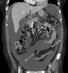
Photograph depicting the appearance of a coronal section of the abdomen obtained by computerised tomographic (CT) imaging of the abdomen. Intravenous contrast was administered and the CT was viewed in the portal phase. A large volume of intraperitoneal free fluid (double line arrow) is demonstrated. The free fluid delineates fatty and vascular structures of the small bowel region of the mesentery (dashed arrow) and sigmoid region of the mesentery (solid arrow).
Fig. 3
Fig. 3
Panel of photographs generated from computerized axial tomographic imaging of the abdomen and pelvis ( AC ) with intravenous contrast in the portal venous phase. The sagittal aspect of the abdomen is demonstrated showing a large volume of intraperitoneal free fluid. This fluid delineates the transverse mesocolon (solid arrow), small bowel region of the mesentery (double line arrow) and mesosigmoid (dashed arrow).
Fig. 4
Fig. 4
( A ) Photograph of T2 weighted coronal image of the of the abdomen generated from magnetic resonance imaging (MRI). The photograph demonstrates how MRI can be used to depict the anatomy of the small bowel region of the mesentery in normality (solid arrow). ( B ) Photograph of T1 fat-saturated coronal MRI with contrast demonstrating malrotation; the small bowel and adjoining mesentery are located on the right side of the abdomen (dashed arrow).
Fig. 5
Fig. 5
Panel of photographs generated from computerised axial tomographic imaging of the abdomen and pelvis ( AD ). Intravenous contrast was administered and axial images were taken in the arterial phase. The images progress cranio-caudally to demonstrate the superior mesenteric artery (solid arrow) in the mesentery adjoining the small bowel. The superior mesenteric artery is occluded by a mesenteric volvulus. These properties collectively result in the “vascular whirl” sign (dashed arrow). There is hypoattenuation of the small bowel wall (double line arrow) indicating small bowel ischemia.
Fig. 6
Fig. 6
Photograph depicting the axial appearance of the abdomen obtained by computerised tomographic (CT) imaging. Intravenous contrast was administered and the CT was viewed in the portal phase. The image includes a well-defined smooth homogenous soft tissue mass (solid arrow) in the mesentery adjoining the small bowel mesentery that encases but does not occlude the mesenteric vasculature (dashed arrow). These appearances are consistent with mesenteric lymphoma.

Similar articles

Cited by

References

    1. Coffey J C, O'Leary D P. The mesentery: structure, function, and role in disease. Lancet Gastroenterol Hepatol. 2016;1(03):238–247. doi: 10.1016/S2468-1253(16)30026-7. - DOI - PubMed
    1. Byrnes K G, Walsh D, Dockery P, McDermott K, Coffey J C. Anatomy of the mesentery: Current understanding and mechanisms of attachment. Semin Cell Dev Biol. 2019;92:12–17. doi: 10.1016/j.semcdb.2018.10.004. - DOI - PubMed
    1. Byrnes K G, Walsh D, Lewton-Brain P, McDermott K, Coffey J C. Anatomy of the mesentery: Historical development and recent advances. Semin Cell Dev Biol. 2019;92:4–11. doi: 10.1016/j.semcdb.2018.10.003. - DOI - PubMed
    1. Gray H. John W. Parker and Son; 1858. Anatomy, Descriptive and Surgical.
    1. Treves F.Lectures on the anatomy of the intestinal canal and peritoneum in man BMJ 18851(1264):580–583.10.1136/bmj.1.1264.580 - DOI - PMC - PubMed