. 2022 Jul 20;13(30):8781-8790.
doi: 10.1039/d2sc02198f. eCollection 2022 Aug 4.

All-in-one disulfide bridging enables the generation of antibody conjugates with modular cargo loading

Affiliations

All-in-one disulfide bridging enables the generation of antibody conjugates with modular cargo loading

Friederike M Dannheim et al. Chem Sci. .

Abstract

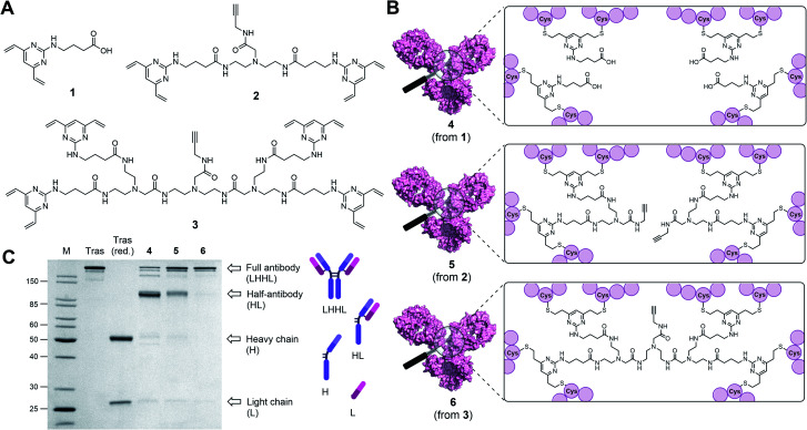
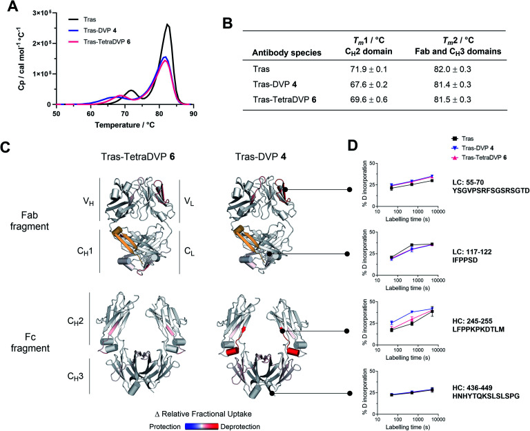
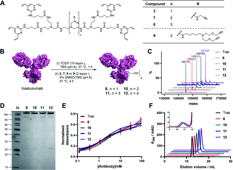
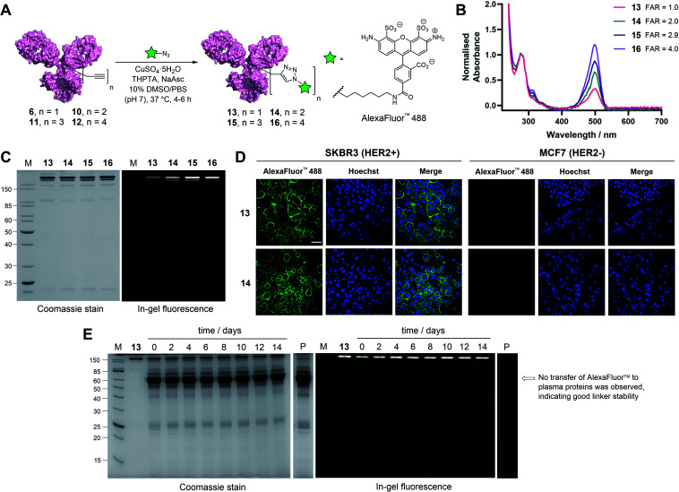
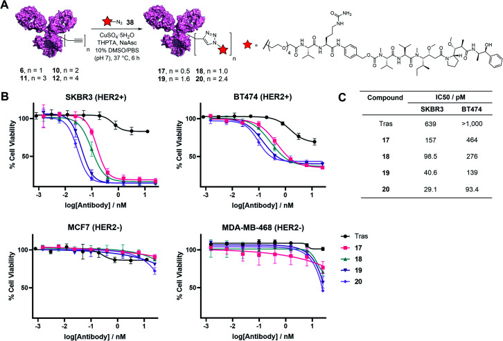
Antibody-drug conjugates (ADCs) are valuable therapeutic entities which leverage the specificity of antibodies to selectively deliver cytotoxins to antigen-expressing targets such as cancer cells. However, current methods for their construction still suffer from a number of shortcomings. For instance, using a single modification technology to modulate the drug-to-antibody ratio (DAR) in integer increments while maintaining homogeneity and stability remains exceptionally challenging. Herein, we report a novel method for the generation of antibody conjugates with modular cargo loading from native antibodies. Our approach relies on a new class of disulfide rebridging linkers, which can react with eight cysteine residues, thereby effecting all-in-one bridging of all four interchain disulfides in an IgG1 antibody with a single linker molecule. Modification of the antibody with the linker in a 1 : 1 ratio enabled the modulation of cargo loading in a quick and selective manner through derivatization of the linker with varying numbers of payload attachment handles to allow for attachment of either 1, 2, 3 or 4 payloads (fluorescent dyes or cytotoxins). Assessment of the biological activity of these conjugates demonstrated their exceptional stability in human plasma and utility for cell-selective cytotoxin delivery or imaging/diagnostic applications.


Conflict of interest statement

FMD, SJW and DRS are inventors on a patent application relating to the use of TetraDVP linkers for ADC synthesis.

Figures

Fig. 1
Fig. 1. Antibody modifications with DVP (1), BisDVP (2) or TetraDVP (3) bridging reagents lead to conjugates with varying homogeneity. (A) Structures of bridging reagents. (B) Structures of antibody–linker-conjugates. (C) Homogeneity analysis of antibody–linker conjugates by SDS-PAGE; M = molecular weight marker, Tras = trastuzumab, Tras (red.) = reduced trastuzumab. The band at ∼150 kDa indicates fully rebridged antibody (LHHL); the band at ∼75 kDa indicates half-antibody species (HL) caused by non-native rebridging.
Fig. 2
Fig. 2. Biophysical stability analysis of trastuzumab (Tras), trastuzumab–DVP conjugate 4 and trastuzumab–TetraDVP conjugate 6. (A) Differential scanning calorimetry results of unmodified, DVP– and TetraDVP–linked trastuzumab. (B) Average of the melting temperatures of the antibody variants for duplicate measurements for Tras–DVP and Tras–TetraDVP, triplicates for Tras. The errors are the standard deviations of the repeats. (C) Crystal structures (Fab: 1N8Z; Fc: 3AVE) coloured according to the relative change in fractional deuterium exchange for Tras–TetraDVP and Tras–DVP compared to Tras; the differences plotted are significant differences assessed by a t-test, with p-value < 0.01. The yellow region represents the residues that were missing in the peptide map. Reduced exchange is shown in blue, no change in white, and increased exchange in red. (D) Details of the uptake plots for selected regions; the error bars represent a Student's t distribution with 95% confidence interval based on triplicates.
Fig. 3
Fig. 3. Generation and analysis of TetraDVP–trastuzumab conjugates. (A) Structures of TetraDVP linkers 3, 7, 8 and 9 containing one, two, three or four alkyne moieties, respectively. (B) Reaction of trastuzumab with TetraDVPs 3, 7, 8 and 9. TBS = tris-buffered saline. (C) LC-MS analysis of trastuzumab and conjugates 6, 10, 11 and 12. Samples were deglycosylated with PNGase F prior to analysis. (D) Analysis of conjugates 6, 10, 11 and 12 by SDS-PAGE under reducing conditions; M = molecular weight marker. (E) Binding affinity comparison of trastuzumab, 6, 10, 11 and 12 by ELISA. Error bars represent the standard deviation of biological quadruplicates. (F) Size-exclusion chromatography (SEC) analysis of trastuzumab and TetraDVP conjugates.
Fig. 4
Fig. 4. Functional modification and stability analysis of TetraDVP-modified trastuzumab. (A) Reaction of TetraDVP conjugates 6, 10, 11 and 12 with AlexaFluor™ 488 azide to form fluorescent conjugates 13, 14, 15 and 16. PBS = phosphate-buffered saline. (B) UV/vis spectra of conjugates 13, 14, 15 and 16. Absorbance was normalised at 280 nm. Absorbance at 495 nm was used to calculate the fluorophore-to-antibody ratio (FAR). (C) Analysis of conjugates 13, 14, 15 and 16 by SDS-PAGE under reducing conditions; M = molecular weight marker. Left gel is after Coomassie staining, right gel is in-gel fluorescence measured before staining. (D) Live cell microscopy images of HER2-positive SKBR3 cells and HER2-negative MCF7 cells after treatment with 13 or 14 shows selective labelling of antigen-positive cells. Scale bar represents 50 μm. (E) Stability analysis of conjugate 13 in human plasma by SDS-PAGE; M = molecular weight marker, P = human plasma, days of incubation are depicted above the representative lane. Left gel is after Coomassie staining, right gel is in-gel fluorescence measured before staining. No transfer of AlexaFluor™ 488 to human serum albumin (66.5 kDa, indicated by the arrow) or any other plasma proteins is observed over the 14 day incubation period.
Fig. 5
Fig. 5. (A) Reaction of TetraDVP conjugates 6, 10, 11 and 12 with azide-functionalised MMAE 38 to form ADCs 17, 18, 19 and 20. PBS = phosphate-buffered saline. (B) Cytotoxicity of TetraDVP ADCs in HER2-positive (SKBR3 and BT474) and HER2-negative (MCF7 and MDA-MB-468) cell lines. Viability data shows the mean of three independent experiments and error bars represent the standard error of the mean (SEM). (C) Calculated IC50 values for TetraDVP ADCs in SKBR3 and BT474 cell lines.

Similar articles

Cited by

References

    1. Teicher B. A. Doroshow J. H. N. Engl. J. Med. 2012;367:1847–1848. doi: 10.1056/NEJMe1211736. - DOI - PubMed
    1. Beck A. Goetsch L. Dumontet C. Corvaïa N. Nat. Rev. Drug Discovery. 2017;16:315–337. doi: 10.1038/nrd.2016.268. - DOI - PubMed
    1. Cazzamalli S. Dal Corso A. Widmayer F. Neri D. J. Am. Chem. Soc. 2018;140:1617–1621. doi: 10.1021/jacs.7b13361. - DOI - PMC - PubMed
    1. Zammarchi F. Corbett S. Adams L. Tyrer P. C. Kiakos K. Janghra N. Marafioti T. Britten C. E. Havenith C. E. G. Chivers S. D'Hooge F. Williams D. G. Tiberghien A. Howard P. W. Hartley J. A. Van Berkel P. H. Blood. 2018;131:1094–1105. doi: 10.1182/blood-2017-10-813493. - DOI - PubMed
    1. Kaplon H. Chenoweth A. Crescioli S. Reichert J. M. mAbs. 2022;14:e2014296. doi: 10.1080/19420862.2021.2014296. - DOI - PMC - PubMed