. 2022 Aug;70(8):583-596.
doi: 10.1369/00221554221119543. Epub 2022 Aug 16.

Expression of Calbindin-D28K in the Developing and Adult Mouse Cochlea

Affiliations

Expression of Calbindin-D28K in the Developing and Adult Mouse Cochlea

Wenjing Liu et al. J Histochem Cytochem. 2022 Aug.

Abstract

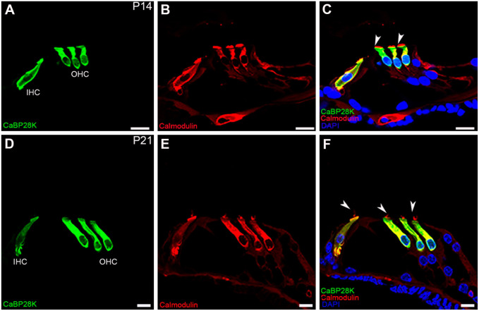
Herein, we aimed to use double-labeling immunofluorescence to describe the expression pattern of Calbindin-D28K (CaBP28K) in the mouse cochlea from late embryonic (E) stages to the adulthood. CaBP28K was expressed in the inner hair cells (IHCs) and the greater epithelial ridge (GER) at E17. In addition, its expression was observed in the interdental cells. On postnatal day 1 (P1), CaBP28K immunoreactivity was observed in the IHCs and outer hair cells (OHCs) and was also specifically expressed in the nucleus and the cytoplasm of spiral ganglion neurons (SGNs). At P8, CaBP28K labeling disappeared from the interdental cells, and the CaBP28K-positive domain within the GER shifted from the entire cytoplasm to only the apical and basal regions. At P14, CaBP28K immunoreactivity was lost from the GER; however, its expression in the IHCs and OHCs, as well as the SGNs, persisted into adulthood. The identification of CaBP28K in the hair cells (HCs) and cuticular plates, as well as SGNs, was confirmed by its colocalization with several markers for Sox2, Myosin VIIa, Phalloidin, and Tuj1. We also detected colocalization with calmodulin in the cytoplasm of both HCs and SGNs. Western blot revealed an increase in CaBP28K postnatal expression in the mouse cochlea.

Keywords: Ca2+-binding proteins; cochlear development; immunohistochemistry.


Conflict of interest statement

Competing Interests: The author(s) declared no potential conflicts of interest with respect to the research, authorship, and/or publication of this article.

Figures

Figure 1.
Figure 1.
CaBP28K immunolabeling in the mouse cochlea at E17 and E19. (A) A low-magnification view of the cross-sections of the mouse cochlea labeled with CaBP28K (green) at E17. CaBP28K labeling was mainly present in the GER and IHCs, no other structure was labeled in the mouse cochlea. Scale bar, 50 μm. (B) CaBP28K (green) and Sox2 (red) labeling in the apical turn of the mouse cochlea at E17. CaBP28K was localized in the cytosol of columnar epithelial cells of the GER, a portion of the GER that exhibited the nuclear staining pattern of Sox2 was not stained. Scale bar, 20 μm. (C) CaBP28K (green) and Sox2 (red) labeling in the middle turn of E17. The expression of CaBP28K emerged in the IHCs and the interdental cells in the spiral limbus, in addition to CaBP28K-labeled GER. Sox2-expressing cells localized in the most external part of the GER were immunonegative for CaBP28K. Scale bar, 20 μm. (D) Detail of CaBP28K labeling in the organ of Corti in the middle turn of E17. Sox2 labeled hair cell and supporting cell nuclei, Sox2-stained nucleus indicated the position of hair cells. CaBP28K was observed in the cytosol of IHC, the OHCs above three rows of the Deiters’ cell were not stained. Scale bar, 10 μm. (E) A low-magnification view of CaBP28K labeling in the medial turn of E17. No CaBP28K expression was observed in the SGNs and Sox2-labeled glial cells of Rosenthal’s canal. (F) CaBP28K (green) and Sox2 (red) labeling in the apical turn of the mouse cochlea at E19. IHCs remained unlabelled. Scale bar, 20 μm. (G) CaBP28K (green) and Sox2 (red) labeling in the middle turn of E19. CaBP28K immunostaining was observed in the GER, the IHCs, and the interdental cells. Scale bar, 20 μm. (H) Detail of E19 mouse organ of Corti double-labeled by CaBP28K (green) and Myosin VIIa (red). Colocalization of CaBP28K with Myosin VIIa in the IHC cytosol (yellow) could be detected in the merged image. The nucleus of IHCs (large arrow) showed labeling for CaBP28K. Non-specific staining was observed in the basilar membrane and the spiral vessel underneath the basilar membrane (arrowheads). Scale bar, 5 μm. (I) Detail of CaBP28K labeling in the organ of Corti in the basal turn at E19. The expression of CaBP28K in three rows of OHCs was marginally above the background level. Scale bar, 10 μm.Abbreviations: GER, greater epithelial ridge; IHC, inner hair cell; SGN, spiral ganglion neuron; SL, spiral limbus; IDC, the interdental cells; DC, Deiters cells; OHC, outer hair cell.
Figure 2.
Figure 2.
CaBP28K immunolabeling in the mouse cochlea at P1. (A) A low-magnification view of the cross-sections of the mouse cochlea labeled with CaBP28K (green) at P1. The expression of CaBP28K emerged in the IHCs and three rows of OHCs in the apical turn of the P1 mouse cochlea. Scale bar, 50 μm. (B) Detail of CaBP28K (green) labeling in the GER in the middle turn of P1. CaBP28K was not detectable in a few columnar inner supporting cells located medially to the IHCs. CaBP28K immunoreactivity was maintained in the interdental cells. Scale bar, 20 μm. (C) Detail of CaBP28K (green) labeling in the P1 organ of Corti. CaBP28K immunoreactivity was present in both the cytoplasm (yellow) and the nucleus of IHC and OHCs (large arrow), as demonstrated by co-labeling with Myosin VIIa (red). Scale bar, 10 μm. (D, E, F) Detail of CaBP28K (green) and phalloidin (red) expression in the P1 organ of Corti. Colocalization of CaBP28K with phalloidin was found in the cuticular plates (large arrow) of HCs, CaBP28K was not observed in the phalloidin-positive stereociliary bundle (red) of HCs (arrowheads). Scale bar, 10 μm. (G, H, I) Detail of CaBP28K (green) and Tuj1 (red) expression in the P1 SGNs and the merged image+DAPI. CaBP28K immunolabeling was present in a subpopulation of scattered SGNs. Both the cytoplasm and the nucleus of SGNs were stained. In CaBP28K-Tuj1 double-positive SGNs, CaBP28K labeling was colocalized with Tuj1 in the cytoplasm (arrowheads). Abbreviations: GER, greater epithelial ridge; IHC, inner hair cell; OHC, outer hair cell; SGN, spiral ganglion neuron; CP, cuticular plates; SL, spiral limbus; IDC, the interdental cells.
Figure 3.
Figure 3.
CaBP28K immunolabeling in the mouse cochlea at P5 and P8. (A) A low-magnification view of CaBP28K (green) labeling in the P5 cochlear section. Scale bar, 100 μm. (B) Detail of CaBP28K (green) labeling in the GER of P5. Sox2 (red) expression domain extended medially into the medial portion of the GER. Scale bar, 20 μm. (C) Detail of CaBP28K (green) and phalloidin (red) expression in the P5 organ of Corti. CaBP28K was co-labeled with phalloidin in the cuticular plates (yellow) of HCs. Phalloidin was clearly visible in the stereociliary bundles (arrowheads) of HCs. CaBP28K immunoreactivity was lost from hair cell nuclei. Scale bar, 10 μm. (D, E, F) Detail of CaBP28K (green) and Tuj1 (red) expression in the P5 SGNs and the merged image+DAPI. Most of the CaBP28K-stained SGN somata were colabeled with Tuj1, and the nucleus of SGNs expressed only CaBP28K (green). Scale bar, 10 μm. (G) An overview of CaBP28K (green) labeling in the cochlear section at P8. Labeling for CaBP28K was absent from the interdental cells. Scale bar, 100 μm. (H) Detail of CaBP28K (green) labeling in the P8 organ of Corti. Note that the thickened ridge of the GER has regressed, CaBP28K expression was restricted to the apical and basal parts of the GER, the inner sulcus started expressing CaBP28K. Scale bar, 10 μm. (I) Detail of CaBP28K (green) labeling in the P8 SGNs. CaBP28K expression remained in SGN somata and nuclei. Scale bar, 10 μm. Abbreviations: SGN, spiral ganglion neuron; GER, greater epithelial ridge; IHC, inner hair cell; OHC, outer hair cell; CP, cuticular plates.
Figure 4.
Figure 4.
Detail of CaBP28K (green) and phalloidin (red) expression in the P10 and P14 mouse organ of Corti. (A, B, C) Colocalization of CaBP28K with phalloidin in the cuticular plates (yellow) of P10 HCs, CaBP28K immunostaining was limited to the apical surface of the residual GER cells. CaBP28K and phalloidin were clearly colocalized in the cuticular plates (yellow) of P10 HCs. No immunoreactivity for CaBP28K was observed in the stereociliary bundles (arrowheads). (D, E, F) Colocalization of CaBP28K with phalloidin in the cuticular plates (yellow) of P14 HCs. CaBP28K completely disappeared from the GER, the stereociliary bundles (arrowheads) were still unstained. Scale bar, 10 μm. Abbreviations: GER, greater epithelial ridge; IHC, inner hair cell; OHC, outer hair cell; CP, cuticular plates
Figure 5.
Figure 5.
Double-immunofluorescence staining with CaBP28K (green) and calmodulin (red) in P14 and P21 mouse organ of Corti. (A, B, C) Co-expression of CaBP28K and calmodulin was detected in the cytoplasm (yellow) of P14 HCs. Note the stereociliary bundles (arrowheads) were labeled with calmodulin (red) but not with CaBP28K (green). (D, E, F) At P21, CaBP28K was colocalized with calmodulin in the cytoplasm (yellow) of HCs but not in the stereociliary bundles (arrowheads). Scale bar, 10 μm. Abbreviations: IHC, inner hair cell; OHC, outer hair cell
Figure 6.
Figure 6.
CaBP28K immunolabeling in the mouse cochlea at P28. (A, B, C) Colocalization of CaBP28K (green) with Myosin VIIa (red) in the cytoplasm (yellow) of P28 HCs. Scale bar, 10 μm. (D, E, F) Colocalization of CaBP28K (green) with phalloidin (red) in the cuticular plates (yellow) of P28 HCs. Scale bar, 10 μm. (G, H, I) Detail of CaBP28K (green) and Tuj1 (red) expression in the P28 SGNs and the merged image+DAPI. Many, but not all, CaBP28K-positive the cytoplasm of SGNs were double-labeled for Tuj1, and a few CaBP28K-positive SGNs somata (arrows) were not co-labeled with Tuj1.Abbreviations: IHC, inner hair cell; OHC, outer hair cell; SGN, spiral ganglion neuron.
Figure 7.
Figure 7.
Double-immunofluorescence staining with CaBP28K (green) and calmodulin (red) in adult (P28) mouse organ of Corti. (A, B) An overview of CaBP28K (green) and calmodulin (red) labeling in the middle turn of the adult cochlea. Scale bar, 50 μm. (C) The secondary antibodies showed no non-specific binding. Scale bar, 50 μm. (D, E, F) Co-expression of CaBP28K (green) and calmodulin (red) was detected in the cytoplasm of adult HCs. Scale bar, 10 μm. (G, H, I) Detail of calmodulin expression in the adult SGNs. Among the calmodulin-positive SGNs, only a subpopulation of calmodulin-positive SGN somata was dually stained (yellow), SGN nuclei were unstained. Scale bar, 20 μm.Abbreviations: SGN, spiral ganglion neuron; IHC, inner hair cell; OHC, outer hair cell;SL, spiral limbus.
Figure 8.
Figure 8.
Statistical analysis of CaBP28K protein levels in the mouse cochlea at P1, P7, P14 and P28. GAPDH, used as an internal control, was detected at a position corresponding to a molecular weight of 36 kDa. There were difference in expression of CaBP28K at the various developmental stages (P<0.05), and there were significant differences in the expression of CaBP28K between P1 compared with P7, P14 and P28 (P<0.05). ** P<0.05

Similar articles

Cited by

References

    1. Berggard T, Miron S, Onnerfjord P, Thulin E, Akerfeldt KS, Enghild JJ, Akke M, Linse S. Calbindin D28k exhibits properties characteristic of a Ca2+ sensor. J Biol Chem. 2002;277(19):16662–72. - PubMed
    1. Nejatbakhsh N, Feng ZP. Calcium binding protein-mediated regulation of voltage-gated calcium channels linked to human diseases. Acta Pharmacol Sin. 2011;32(6):741–8. - PMC - PubMed
    1. Dechesne CJ, Lavigne-Rebillard M, Brehier A, Thomasset M, Sans A. Appearance and distribution of neuron-specific enolase and calbindin (CaBP 28 kDa) in the developing human inner ear. Brain Res. 1988;469(1–2):221–30. - PubMed
    1. Imamura S, Adams JC. Changes in cytochemistry of sensory and nonsensory cells in gentamicin-treated cochleas. J Assoc Res Otolaryngol. 2003;4(2):196–218. - PMC - PubMed
    1. Fischer N, Johnson CL, Majerus A, Potrusil T, Riechelmann H, Schmutzhard J, Schrott-Fischer A, Glueckert R. Age-dependent calcium-binding protein expression in the spiral ganglion and hearing performance of C57BL/6J and 129/SvJ mice. ORL J Otorhinolaryngol Relat Spec. 2019;81(2–3):138–54. - PubMed