. 2022 Oct;42(10):1008-1027.
doi: 10.1002/cac2.12351. Epub 2022 Aug 16.

A non-metabolic function of hexokinase 2 in small cell lung cancer: promotes cancer cell stemness by increasing USP11-mediated CD133 stability

Affiliations

A non-metabolic function of hexokinase 2 in small cell lung cancer: promotes cancer cell stemness by increasing USP11-mediated CD133 stability

Juhong Wang et al. Cancer Commun (Lond). 2022 Oct.

Abstract

Background: Maintenance of cancer stem-like cell (CSC) stemness supported by aberrantly regulated cancer cell metabolism is critical for CSC self-renewal and tumor progression. As a key glycolytic enzyme, hexokinase 2 (HK2) plays an instrumental role in aerobic glycolysis and tumor progression. However, whether HK2 directly contribute to CSC stemness maintenance in small cell lung cancer (SCLC) is largely unclear. In this study, we aimed to investgate whether HK2 independent of its glycolytic activity is directly involved in stemness maintenance of CSC in SCLC.

Methods: Immunoblotting analyses were conducted to determine the expression of HK2 in SCLC CSCs and their differentiated counterparts. CSC-like properties and tumorigenesis of SCLC cells with or without HK2 depletion or overexpression were examined by sphere formation assay and xenograft mouse model. Immunoprecipitation and mass spectrometry analyses were performed to identify the binding proteins of CD133. The expression levels of CD133-associated and CSC-relevant proteins were evaluated by immunoblotting, immunoprecipitation, immunofluorescence, and immunohistochemistry assay. RNA expression levels of Nanog, POU5F1, Lin28, HK2, Prominin-1 were analyzed through quantitative reverse transcription PCR. Polyubiquitination of CD133 was examined by in vitro or in vivo ubiquitination assay. CD133+ cells were sorted by flow cytometry using an anti-CD133 antibody.

Results: We demonstrated that HK2 expression was much higher in CSCs of SCLC than in their differentiated counterparts. HK2 depletion inhibited CSC stemness and promoted CSC differentiation. Mechanistically, non-mitochondrial HK2 directly interacted with CD133 and enhanced CD133 expression without affecting CD133 mRNA levels. The interaction of HK2 and CD133 promoted the binding of the deubiquitinase ubiquitin-specific protease 11 (USP11) to CD133, thereby inhibiting CD133 polyubiquitylation and degradation. HK2-mediated upregulation of CD133 expression enhanced the expression of cell renewal regulators, SCLC cell stemness, and tumor growth in mice. In addition, HK2 expression was positively correlated with CD133 expression in human SCLC specimens, and their expression levels were associated with poor prognosis of SCLC patients.

Conclusions: These results revealed a critical non-metabolic function of HK2 in promotion of cancer cell stemness. Our findings provided new insights into the multifaceted roles of HK2 in tumor development.

Keywords: CD133; HK2; SCLC; USP11; cancer stem-like cell; metabolic enzyme; non-metabolic function; ubiquitylation.


Conflict of interest statement

The authors declare that they have no conflicts of interest.

Figures

FIGURE 1
FIGURE 1
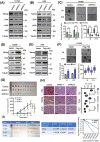
HK2 expression is elevated in SCLC CSCs and correlates with poor prognosis of SCLC patients. (A‐B) Glucose consumption (A) and lactate production (B) of H446 and H1048 cell monolayers or spheroids were determined. Data shown are the mean ± SD (n = 3). ***, P < 0.001. (C‐D) Immunoblotting analyses of the lysis of H446 (C) and H1048 (D) cell monolayers or spheroids were performed with the indicated antibodies. (E) Immunofluorescence analyses were performed with the indicated antibodies in monolayer H1048 cells or spheroid cells. DAPI was used for nuclear staining. Scale bars: 50 μm. (F‐G) Ninety human SCLC specimens with their adjacent tissues were analyzed by IHC with an anti‐HK2 antibody (F, top). Representative and magnified IHC images imagines of SCLC tissues and normal tissues were shown (F, bottom). IHC scores of HK2 expression were calculated (G). N, adjacent normal tissue; T, tumor tissue. Scale bars: 50 μm. (H) Kaplan‐Meier plots of the overall survival time of 90 SCLC patients with low (score ≤ 4) or high (score >4) expression levels of HK2. P values are calculated using a log‐rank test (two‐tailed). Abbreviations: HK2, hexokinase 2; SCLC, small cell lung cancer; CSC, cancer stem cell; SD, standard deviation; DAPI, 4',6‐diamidino‐2‐phenylindole; IHC, immunohistochemistry
FIGURE 2
FIGURE 2
HK2 promotes CSC stemness and tumor growth. (A‐B) Immunoblotting analyses of H1048 (A) and H69 (B) cells with or without HK2 depletion were performed with the indicated antibodies. (C) The sphere formation ability of H1048 cells or H1048 cells with or without HK2 depletion in their first and the second passage was examined. Representative images were displayed (top), and the diameters and number of spheres were determined (bottom). Data shown are the mean ± SD (n = 3). ***, P < 0.001. Scale bars: 100 μm. (D‐E) Immunoblotting analyses of H1048 (D) and H69 (E) cells with or without HK2 overexpression were performed with the indicated antibodies. (F) The sphere formation ability of H1048 cells with or without HK2 overexpression in their first and the second passage was examined. Representative images were displayed (top), and the diameters and number of spheres were determined (bottom). Data shown are the mean ± SD (n = 3). **, P < 0.01, ***, P < 0.001. Scale bars: 100 μm. (G) Immunodeficient mice (n = 10) were subcutaneously inoculated with an equal number (2 × 10⁶) of H1048 cells with or without HK2 shRNA expression. Tumor sizes (top) and volumes (bottom) were measured and calculated. Data represent the means ± SD of ten mice per group. ***, P < 0.001. (H) IHC staining of mouse tumor tissues derived from H1048 cells with or without HK2 shRNA expression was performed with the indicated antibodies. Representative images were displayed (left), and the IHC scores were calculated (right). Data shown are the mean ± SD (n = 5). ***, P < 0.001. Scale bars: 100 μm. (I) Different numbers of H1048 cells with or without HK2 overexpression were subcutaneously injected into NOD/SCID mice (n = 10). Images of the tumors are shown on the left. Scale bar (right): 1 cm. Stem cell frequencies were estimated as the ratio 1/x with the upper and lower 95% confidence intervals (middle). ELDA analysis was performed (right). Abbreviations: HK2, hexokinase 2; CSC, cancer stem cell; SD, standard deviation; IHC, immunohistochemistry; ELDA, extreme limiting dilution analysis
FIGURE 3
FIGURE 3
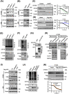
HK2 interacts with CD133 and inhibits polyubiquitylation‐dependent CD133 degradation. (A‐B) HK2 was depleted (A) or overexpressed (B) in H1048 or H69 cells. Immunoblotting analyses were performed with an anti‐CD133 antibody. (C‐D) H1048 cells with or without HK2 depletion (C) or overexpression (D) were treated with CHX (100 μg/mL) for the indicated periods of time. Immunoblotting analyses were performed with the indicated antibodies (left). Quantification of CD133 expression levels relative to tubulin expression levels is shown (right). (E‐F) H1048 cells with or without HK2 depletion (E) or overexpression (F) were treated with MG132 (50 μmol/L) for 8 hours. Immunoprecipitation with an anti‐CD133 antibody and immunoblotting analyses were performed with the indicated antibodies. (G) Purified His‐HK2 was incubated with purified GST or GST‐CD133 together with glutathione‐sepharose 4B beads. GST pulldown assay and immunoblotting analyses were performed with the indicated antibodies. (H) Mitochondrial and non‐mitochondrial fractions of H1048 monolayers or spheroids were prepared and subjected to immunoprecipitation with an anti‐HK2 antibody. Immunoblotting analyses were performed with the indicated antibodies. (I) RNAi‐resistant WT HK2, inactive HK2 D209/657A, or HK2 △N16 was expressed in H1048 and H69 cells with depletion of endogenous HK2. Immunoblotting analyses were performed with the indicated antibodies. (J) H1048 cells with or without WT HK2 or inactive HK2 D209/657A overexpression were treated with MG132 (50 μmol/L) for 8 hours. Immunoprecipitation and immunoblotting analyses were performed with the indicated antibodies. (K) H1048 cells with or without expression of inactive HK2 D209/657A were treated with CHX (100 μg/mL) for the indicated periods of time. Immunoblotting analyses were performed with the indicated antibodies (upper). Quantification of CD133 expression levels relative to tubulin expression levels is shown (lower). Abbreviations: HK2, hexokinase 2; CHX, cycloheximide; IP, immunoprecipitation; WCL, whole cell lysate; WT, wide type
FIGURE 4
FIGURE 4
The interaction between HK2 and CD133 induces binding of USP11 to CD133 and inhibits CD133 polyubiquitylation and degradation. (A) SCLC monolayer or spheroid cells were lysed. Immunoprecipitation and immunoblotting analyses were performed with the indicated antibodies. (B) 293T cells expressing HA‐Ub were transfected with or without a Flag‐CD133 plasmid, followed by treatment with MG132 (50 μmol/L) for 8 hours. CD133 purified by immunoprecipitation was incubated with or without bacterially purified GST‐USP11. Immunoblotting analyses were performed with the indicated antibodies (upper). The purification efficiency was determined using SDS‐PAGE and Coomassie brilliant blue staining (lower). (C) shRNA resistant WT Flag‐USP11 or inactive Flag‐USP11(C318S) mutant was expressed in H1048 USP11 depletion cells. Immunoblotting analyses were performed with the indicated antibodies. (D) H1048 cells expressing two different USP11 shRNAs or a control shRNA were treated with MG132 (50 μmol/L) for 8 hours. Immunoprecipitation and immunoblotting analyses were performed with the indicated antibodies. (E) Two different USP11 shRNAs or a control shRNA were expressed in H1048 cells. Immunoblotting analyses were performed with the indicated antibodies. (F) H1048 cells expressing two different USP11 shRNAs or a control shRNA were examined using a first and the second passage sphere formation assay. Representative images were displayed (upper). The diameters and number of spheres were determined (lower). Data shown are the mean ± SD (n = 3). ***, P < 0.001. Scale bars: 100 μm. (G) 293T cells were transfected with or without Flag‐CD133. Purified CD133 was incubated with or without His‐HK2, followed by incubation with purified GST‐USP11. GST pulldown assay and immunoblotting analyses were performed (upper). The purification efficiency was determined using SDS‐PAGE and coomassie brilliant blue staining (lower). (H) H1048 CSCs with endogenous HK2 depletion and with or without reconstituted expression of RNAi‐resistant WT Flag‐rHK2 or an inactive Flag‐rHK2 D209/657A mutant were analyzed by immunoprecipitation and immunoblotting with the indicated antibodies. (I) RNAi‐resistant WT HK2, inactive HK2 D209/657A, or HK2 △N16 was expressed in H1048 cells with depletion of endogenous HK2. Immunoprecipitation and immunoblotting analyses were performed with the indicated antibodies. Abbreviations: HK2, hexokinase 2; USP11, ubiquitin‐specific protease 11; SCLC, small cell lung cancer; SDS‐PAGE; sodium dodecyl sulfate polyacrylamide gel electrophoresis; shRNA, short hairpin RNA; SD, standard deviation; WT, wide type; IP, immunoprecipitation; WCL, whole cell lysate
FIGURE 5
FIGURE 5
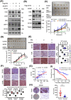
HK2 promotes cancer stem‐like properties via CD133. (A) H1048 cells with or without the indicated expression of Flag‐HK2, USP11 shRNA, and CD133 were analyzed via immunoblotting with the indicated antibodies. (B) H1048 cells with or without expression of HK2 shRNA and Flag‐CD133 were analyzed via immunoblotting with the indicated antibodies. (C) H1048 cells with or without the indicated combined expression of Flag‐HK2, USP11 shRNA, and CD133 were subcutaneously injected into the flank regions of nude mice. Tumor sizes (upper) and volumes (lower) were measured and calculated. Data represent the means ± SD of five mice per group. **, P < 0.01, ***; P < 0.001. (D) H1048 cells with or without HK2 shRNA expression or combined expression of HK2 shRNA and Flag‐CD133 were subcutaneously injected into the flank regions of nude mice. Tumor sizes (top) and volumes (bottom) were measured and calculated. Data represent the means ± SD of eight mice per group. ***, P < 0.001. (E‐F) IHC staining of tumor tissues was performed with the indicated antibodies. Representative images were shown (left or upper). IHC scores (right or lower) were calculated. Data represent the means ± SD (n = 5). *, P < 0.05; ***, P < 0.001. Scale bars: 100 μm. (G) Correlation between HK2 and CD133 expression in an SCLC tissue microarray was analyzed using a two‐tailed Pearson correlation coefficient. (H) Representative and magnified IHC images of CD133 protein expression (left) in paired SCLC tumor and adjacent normal tissues from a tissue array were shown. Analysis of the IHC scores of CD133 expression was performed (right). N, adjacent normal tissue; T, tumor tissue. Scale bars: 50 μm. (I) Kaplan–Meier survival curves were compared using a log‐rank test in 90 patients. The expression of CD133 was classified as low (score ≤ 4) or high (score >4). Abbreviations: HK2, hexokinase 2; USP11, ubiquitin‐specific protease 11; shRNA, short hairpin RNA; SCLC, small cell lung cancer; SD, standard deviation; IHC, immunohistochemistry
FIGURE 6
FIGURE 6
A schematic displays the mechanism underlying HK2‐regulated cancer cell stemness. In cancer stem cells, HK2 expression is increased. Non‐mitochondrial HK2 binds to CD133 and subsequently promotes the interaction between USP11 and CD133, leading to inhibition of CD133 polyubiquitlytion and increase of CD133 stability and CD133 maintenance (left). In differentiated cancer cells, HK2 binds to outer membrane of mitochondria, promoting ubiquitin‐proteasome system‐mediated CD133 degradation. The light green parts represent the cytoplasm. The red solid arrows represent positive regulation or increased expression of the indicated factors. Black solid arrows represent decrease of gene expression. The black dotted line indicates that the regulation will not occur

Similar articles

Cited by

References

    1. Yang S, Zhang Z, Wang Q. Emerging therapies for small cell lung cancer. J Hematol Oncol. 2019;12(1):47. - PMC - PubMed
    1. van Meerbeeck JP, Fennell DA, De Ruysscher DK. Small‐cell lung cancer. Lancet. 2011;378(9804):1741–55. - PubMed
    1. Bunn PA, Jr. , Minna JD, Augustyn A, Gazdar AF, Ouadah Y, Krasnow MA, et al. Small Cell Lung Cancer: Can Recent Advances in Biology and Molecular Biology Be Translated into Improved Outcomes? J Thorac Oncol. 2016;11(4):453–74. - PMC - PubMed
    1. Semenova EA, Nagel R, Berns A. Origins, genetic landscape, and emerging therapies of small cell lung cancer. Genes Dev. 2015;29(14):1447–62. - PMC - PubMed
    1. Farago AF, Keane FK. Current standards for clinical management of small cell lung cancer. Transl Lung Cancer Res. 2018;7(1):69–79. - PMC - PubMed