. 2022 Nov;9(32):e2201358.
doi: 10.1002/advs.202201358. Epub 2022 Aug 17.

Implantable Electroceutical Approach Improves Myelination by Restoring Membrane Integrity in a Mouse Model of Peripheral Demyelinating Neuropathy

Affiliations

Implantable Electroceutical Approach Improves Myelination by Restoring Membrane Integrity in a Mouse Model of Peripheral Demyelinating Neuropathy

Aseer Intisar et al. Adv Sci (Weinh). 2022 Nov.

Abstract

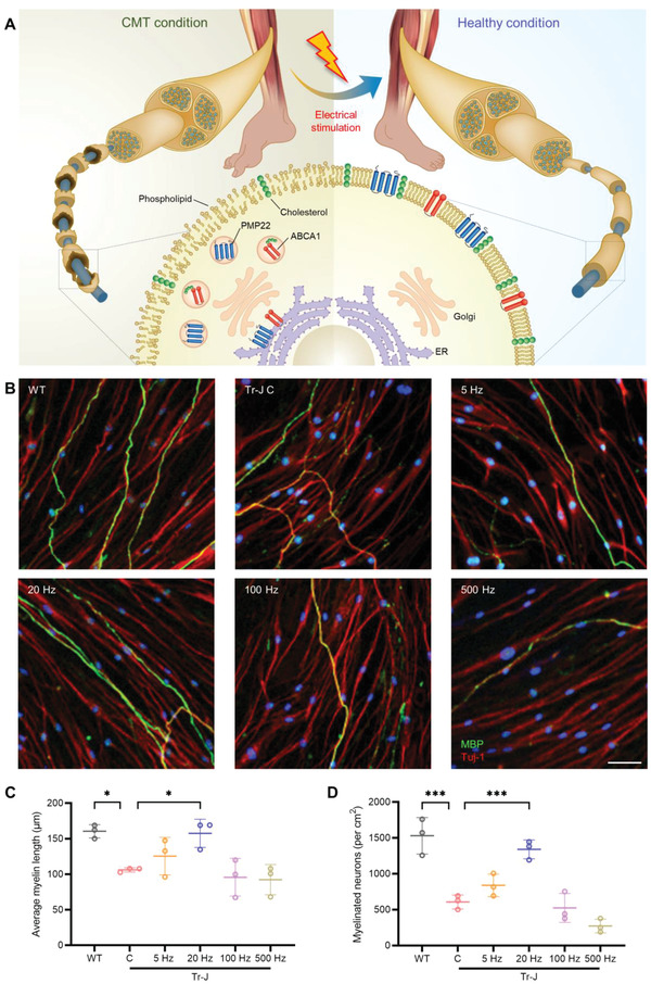
Although many efforts are undertaken to treat peripheral demyelinating neuropathies based on biochemical interventions, unfortunately, there is no approved treatment yet. Furthermore, previous studies have not shown improvement of the myelin membrane at the biomolecular level. Here, an electroceutical treatment is introduced as a biophysical intervention to treat Charcot-Marie-Tooth (CMT) disease-the most prevalent peripheral demyelinating neuropathy worldwide-using a mouse model. The specific electrical stimulation (ES) condition (50 mV mm-1 , 20 Hz, 1 h) for optimal myelination is found via an in vitro ES screening system, and its promyelinating effect is validated with ex vivo dorsal root ganglion model. Biomolecular investigation via time-of-flight secondary ion mass spectrometry shows that ES ameliorates distribution abnormalities of peripheral myelin protein 22 and cholesterol in the myelin membrane, revealing the restoration of myelin membrane integrity. ES intervention in vivo via flexible implantable electrodes shows not only gradual rehabilitation of mouse behavioral phenotypes (balance and endurance), but also restored myelin thickness, compactness, and membrane integrity. This study demonstrates, for the first time, that an electroceutical approach with the optimal ES condition has the potential to treat CMT disease and restore impaired myelin membrane integrity, shifting the paradigm toward practical interventions for peripheral demyelinating neuropathies.

Keywords: Charcot-Marie-Tooth disease; PMP22; cholesterol; electroceuticals; myelin membrane integrity; myelination; peripheral demyelinating neuropathies.


Conflict of interest statement

The authors declare no conflict of interest.

Figures

Figure 1
Figure 1
Schematic diagram of the treatment approach and the results of in vitro screening. A) Schematic showing the novel approach to improve myelination in CMT disease via ES and the biomolecular changes that precede the improvement in myelination after ES. ES ameliorates the abnormalities associated with aggregated peripheral myelin protein 22 (PMP22) and ATP binding cassette subfamily A member 1 (ABCA1) near the ER, as well as dispersed cholesterol distribution in the CMT myelin membrane. B) Representative immunostained images of WT and Tr‐J neuron‐SC cocultures at 35 DIV, with the latter treated with ES at 5, 20, 100, and 500 Hz. Neurons were stained with beta III tubulin (Tuj‐1) and SCs were stained with MBP. Scale bar, 50 µm. C) Average myelin length (n = 3) in the control WT and Tr‐J cocultures, and Tr‐J cocultures treated with ES at different frequencies. D) Density of myelinated neurons (n = 3) in the control WT and Tr‐J cocultures, and Tr‐J cocultures treated with ES at different frequencies. The average myelin length was measured from the MBP (green) channel from 10× images using the NeuronJ plugin in ImageJ. Images from three biologically distinct samples per condition were used, and the average of ten myelin segments per image was taken for each sample. Myelinated neurons per cm2 were measured using the colocalization of Tuj‐1 (red) and MBP (green) channels from 10× images using ImageJ, and images from three biologically distinct samples per condition were used. The data are expressed as the mean ± s.d. *p < 0.05, **p < 0.005, ***p < 0.0005.
Figure 2
Figure 2
ES ameliorates demyelination in Tr‐J ex vivo. A) Schematic illustration of DRG explant extraction from the spinal column of an adult mouse and the subsequent culture. B) Representative immunostained images of WT, Tr‐J control, and Tr‐J ES DRGs. The image in the inset shows the enlarged region marked in the respective images. Scale bars, 500 µm (main) and 100 µm (inset). C) Average myelin length, D) percentage of myelinated neurons, and E) percentage of fragmented myelin segments in WT, Tr‐J control, and Tr‐J ES DRGs (n = 3). F) Percentage of myelin segments in the DRG in each 200 µm radial slice, starting from the edge of the explant (n = 3). G) Percentage of myelin segments in a slice 200 µm from the edge of the explant (n = 3). H) Percentage of myelin segments in a slice 1000 µm from the edge of the explant (n = 3). The average myelin length was measured from the MBP (green) channel from 4× images using the NeuronJ plugin in ImageJ. Images from three biologically distinct samples per condition were used, and the average of ten myelin segments per image was taken for each sample. Myelinated neurons per cm2 were measured using the colocalization of Tuj‐1 (red) and MBP (green) channels from 4× images using ImageJ, and images from three biologically distinct samples per condition were used. The percentage of fragmented myelin and percentage of myelin in 200 µm slices were measured using the MBP (green) channel from the 4× images of three biologically distinct samples per condition. The data are expressed as the mean ± s.d. *p < 0.05, **p < 0.005, ***p < 0.0005.
Figure 3
Figure 3
Amelioration of abnormalities in PMP22/ABCA1 distribution, ER stress, and cholesterol biosynthesis in Tr‐J after ES. A) Representative immunostained images of the colocalization of PMP22 with ER (calnexin used as marker) in the myelin of WT, Tr‐J control, and Tr‐J ES cocultures. The arrowhead shows the PMP22 retention around the ER, and the arrows show the intermittent PMP22 distribution along the myelin. Scale bar, 25 µm. B) Representative immunostained images of the colocalization of PMP22 with ABCA1 in the myelin of WT, Tr‐J control, and Tr‐J ES cocultures. The arrowheads show the PMP22 and ABCA1 colocalization and retention around the ER. Scale bar, 25 µm. C) Density of myelin segments with PMP22 restricted around the ER (n = 3). D) Density of myelin segments with uniform PMP22 distribution (n = 3). E) Density of myelin segments with ABCA1 restricted around the ER (n = 3). F) Density of myelin segments with uniform ABCA1 distribution (n = 3). Quantitative reverse transcription polymerase chain reaction (RT‐qPCR) analysis for gene expression related to G) ER stress and H) cholesterol biosynthesis (n = 3). Quantification of PMP22 and ABCA1 distribution was performed using individual ABCA1 (red) and PMP22 (pink) channels from 10× images using ImageJ and using 4′,6‐diamidino‐2‐phenylindole (blue) as a reference to identify the perinuclear region. In each quantification, images from three biologically distinct samples per condition were used. The data are expressed as the mean ± s.d. *p < 0.05, **p < 0.005, ***p < 0.0005.
Figure 4
Figure 4
Imaging myelin lipids via time‐of‐flight secondary ion mass spectrometry (ToF‐SIMS) and size distribution of cholesterol‐rich fragments. A) A schematic summarizing the process of ToF‐SIMS imaging. Representative SIMS images of total lipids, cholesterol, and sphingomyelin in the WT, Tr‐J control, and Tr‐J ES samples from B) in vitro coculture and C) ex vivo DRG explant. Scale bars, 100 µm. D) Frequency and F) percentage of cholesterol‐rich fragments of different size groups in in vitro coculture (n = 3). E) Frequency and G) percentage of cholesterol‐rich fragments of different size groups in ex vivo DRG explants (n = 3). Statistical comparison (two‐way ANOVA) was performed between the following groups: WT versus Tr‐J C and Tr‐J C versus Tr‐J ES. For each condition, images from three biologically distinct samples were used. The data are expressed as the mean ± s.d. *p < 0.05, **p < 0.005, ***p < 0.0005, #p < 0.0001.
Figure 5
Figure 5
ES improves the behavioral phenotypes of Tr‐J mice. A) Implantation of the cuff electrode onto the sciatic nerve of a Tr‐J mouse. The connecting wires are routed via the subcutaneous layer, and a head port is secured onto the scalp. B) ES is provided on the first day of the week, 30 min per day for 3 consecutive weeks, via a head port connected to a stimulator. C) At the end of each week, rotarod and treadmill tests are conducted to evaluate the physical balance and running endurance, respectively. The maximum D) time and E) RPM achieved in the rotarod test at the end of each week during a 3 week ES treatment period (n = 3). The maximum F) time and G) distance achieved in the treadmill test at the end of each week during a 3 week ES treatment period (n = 3). The week 1 to week 3 change in the H) rotarod test time, I) rotarod test RPM, J) treadmill test time and K) treadmill test distance for the three groups (n = 3). Behavioral tests were conducted on three biologically distinct mice of the same age, and the average of two tests per mouse was recorded. The data are expressed as the mean ± s.d. *p < 0.05, **p < 0.005, ***p < 0.0005.
Figure 6
Figure 6
ES ameliorates demyelination and PMP22/ABCA1 distribution abnormality in vivo. A) Representative transmission electron microscopy (TEM) images of cross‐sections of sciatic nerves. Scale bar, 500 nm. B) Myelin thickness measured from TEM images (n = 3). C) The g‐ratio measured from TEM images (n = 3). D) Relationship between g‐ratio and axon diameter (n = 3). E) Representative TEM images at higher magnification showing the interperiodic distance. Scale bar, 20 nm. F) Measurement of the interperiodic distance from TEM images (n = 3). G) Representative 2D and 3D confocal images of teased longitudinal sections of sciatic nerves immunostained against MBP and Tuj‐1. Scale bar, 10 µm. H) Representative images of longitudinal sections of sciatic nerves immunostained against PMP22 and ABCA1. The white arrow points to PMP22/ABCA1 aggregation in the perinuclear region. I) Percentage of myelin segments with PMP22/ABCA1 aggregates in the perinuclear region (n = 3). For myelin thickness, g‐ratio, and interperiodic distance, the average of five measurements was taken from the TEM images of three biologically distinct samples. The percentage of myelin segments with aggregates was counted from 20× images of three biologically distinct samples per condition. The data are expressed as the mean ± s.d. *p < 0.05, **p < 0.005, ***p < 0.0005.

Similar articles

Cited by

References

    1. Barreto L. C., Oliveira F. S., Nunes P. S., de Franca Costa I. M., Garcez C. A., Goes G. M., Neves E. L., de Souza Siqueira Quintans J., de Souza Araujo A. A., Neuroepidemiology 2016, 46, 157. - PubMed
    1. a) Brennan K. M., Bai Y., Shy M. E., Neurosci. Lett. 2015, 596, 14; - PubMed
    2. b) Murphy S. M., Laura M., Fawcett K., Pandraud A., Liu Y. T., Davidson G. L., Rossor A. M., Polke J. M., Castleman V., Manji H., Lunn M. P., Bull K., Ramdharry G., Davis M., Blake J. C., Houlden H., Reilly M. M., J. Neurol., Neurosurg. Psychiatry 2012, 83, 706. - PMC - PubMed
    1. Morena J., Gupta A., Hoyle J. C., Int. J. Mol. Sci. 2019, 20, 3419. - PubMed
    1. a) Passage E., Norreel J. C., Noack‐Fraissignes P., Sanguedolce V., Pizant J., Thirion X., Robaglia‐Schlupp A., Pellissier J. F., Fontes M., Nat. Med. 2004, 10, 396; - PubMed
    2. b) Pareyson D., Reilly M. M., Schenone A., Fabrizi G. M., Cavallaro T., Santoro L., Vita G., Quattrone A., Padua L., Gemignani F., Visioli F., Laurà M., Radice D., Calabrese D., Hughes R. A. C., Solari A., Lancet Neurol. 2011, 10, 320. - PubMed
    1. a) Fledrich R., Abdelaal T., Rasch L., Bansal V., Schutza V., Brugger B., Luchtenborg C., Prukop T., Stenzel J., Rahman R. U., Hermes D., Ewers D., Mobius W., Ruhwedel T., Katona I., Weis J., Klein D., Martini R., Bruck W., Muller W. C., Bonn S., Bechmann I., Nave K. A., Stassart R. M., Sereda M. W., Nat. Commun. 2018, 9, 3025; - PMC - PubMed
    2. b) Zhou Y., Bazick H., Miles J. R., Fethiere A. I., Salihi M. O. A., Fazio S., Tavori H., Notterpek L., Exp. Neurol. 2019, 321, 113031. - PubMed

Publication types

MeSH terms

LinkOut - more resources