. 2022 Oct;9(28):e2200750.
doi: 10.1002/advs.202200750. Epub 2022 Aug 17.

Aberrant Cholesterol Metabolism and Wnt/β-Catenin Signaling Coalesce via Frizzled5 in Supporting Cancer Growth

Affiliations

Aberrant Cholesterol Metabolism and Wnt/β-Catenin Signaling Coalesce via Frizzled5 in Supporting Cancer Growth

Shaoqin Zheng et al. Adv Sci (Weinh). 2022 Oct.

Abstract

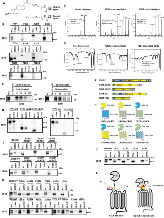
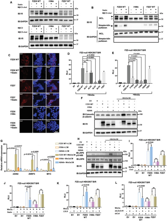
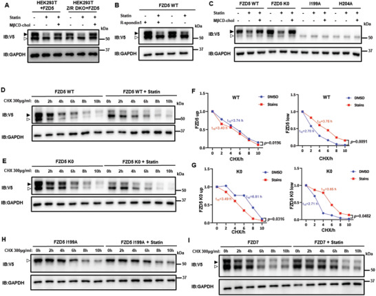
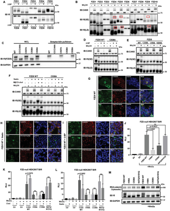
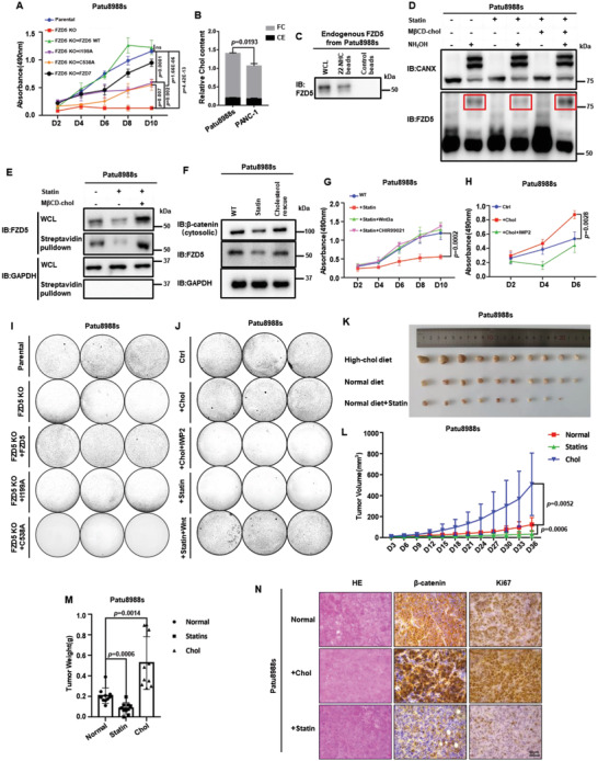
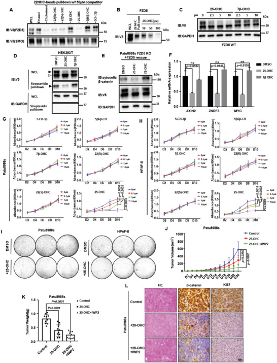
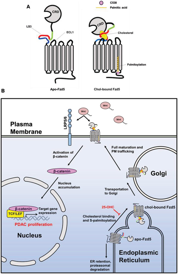
Frizzled (Fzd) proteins are Wnt receptors and play essential roles in development, homeostasis, and oncogenesis. How Wnt/Fzd signaling is coupled to physiological regulation remains unknown. Cholesterol is reported as a signaling molecule regulating morphogen such as Hedgehog signaling. Despite the elusiveness of the in-depth mechanism, it is well-established that pancreatic cancer specially requires abnormal cholesterol metabolism levels for growth. In this study, it is unexpectedly found that among ten Fzds, Fzd5 has a unique capacity to bind cholesterol specifically through its conserved extracellular linker region. Cholesterol-binding enables Fzd5 palmitoylation, which is indispensable for receptor maturation and trafficking to the plasma membrane. In Wnt-addicted pancreatic ductal adenocarcinoma (PDAC), cholesterol stimulates tumor growth via Fzd5-mediated Wnt/β-catenin signaling. A natural oxysterol, 25-hydroxylsterol competes with cholesterol and inhibits Fzd5 maturation and Wnt signaling, thereby alleviating PDAC growth. This cholesterol-receptor interaction and ensuing receptor lipidation uncover a novel mechanism by which Fzd5 acts as a cholesterol sensor and pivotal connection coupling lipid metabolism to morphogen signaling. These findings further suggest that cholesterol-targeting may provide new therapeutic opportunities for treating Wnt-dependent cancers.

Keywords: Wnt/β-catenin signaling; Frizzled receptor; cholesterol; pancreatic cancer.


Conflict of interest statement

The authors declare no conflict of interest.

Figures

Figure 1
Figure 1
Cholesterol specifically and reversibly binds to Fzd5, depending primarily on conserved residues at the extracellular linker and loop. A) Demonstration of synthesized 22‐NHC beads and control beads. B) In vitro pulldown assay and WB of Fzd1‐10 by 22‐NHC or control beads. C) Mass‐spectrometry detection of Fzd5‐bound cholesterol (m/z range: 200–600). The cholesterol signature peak at m/z 437.1657 (combined particle of cholesterol, formic acid, and photon) was clearly seen in Fzd5‐associated lipids. Pure cholesterol and Fzd7‐associated lipids are used as positive and negative controls, respectively. D) FT‐IR showing the stretching vibration of ‐OH and ‐CH3/CH2 and the bending vibration of ‐CH3/CH2 (cholesterol signature IR absorption) were clearly seen in Fzd5‐associated lipids. Pure cholesterol and Fzd7‐associated lipids are used as positive and negative controls, respectively. E) 22‐NHC beads pulldown and competition assay of Fzd5 and Smo by cholesterol as a competitor. F) Schematic of various Fzd5 truncation constructs. FL: full‐length; ΔCRD: deletion of CRD; ΔECD: deletion of extracellular domain; ΔC: deletion of the carboxyl terminus; 7TM: transmembrane domain only. G) 22‐NHC beads pulldown assay of Fzd5 truncation constructs depicted in (F). H) Schematic of CRD‐swapped chimeric Fzds. (i.e.: CRD5+ΔCRD7 represents the chimeric protein consisting of Fzd5 CRD and Fzd7 ΔCRD.) I) 22‐NHC beads pulldown assay of the chimeric Fzds depicted in (H). J) 22‐NHC beads pulldown assay of Fzd5 full‐length or truncation of linker segment 1–4 (ΔLS1‐4). K) 22‐NHC beads pulldown assay of Fzd5 WT and point‐mutations of all conserved residues on LS3. L) Working model of cholesterol binding to Fzd5: owing to the steric hindrance, cholesterol could not insert into the hydrophobic grove in the CRD as PA does; the exposed hydrophobic part of cholesterol appears to be covered up by the hydrophobic and aromatic residues in the extracellular linker and loop regions, which form a “cove” with CRD possibly acting as “cap” to wrap around cholesterol. WCL: whole cell lysate.
Figure 2
Figure 2
Cholesterol regulates Fzd5 protein levels at the PM and Fzd5‐mediated Wnt/β‐catenin signaling. A) Upper panel shows the mature (upper) band and the immature (lower) band distribution of V5‐tagged Fzd5 WT, I199A, and Fzd7 under normal culture, cholesterol starvation by statin, and rescue conditions in HEK293T cells. Lower panel shows acute cholesterol depletion by MβCD and rescue in HEK293T cells. B) Surface biotin labeling assay of Fzd5 WT, I199A, and Fzd7 under normal, cholesterol starvation (by statin treatment) and rescue conditions (by MβCD‐cholesterol complex treatment). Solid arrow head: mature Fzd; hollowed arrow head: immature Fzd. C) Confocal fluorescent imaging of SNAP‐tagged Fzd5, Fzd7, and Fzd5 I199A on the plasma membrane under normal and statin treatment conditions in HEK293T cells. SNAP‐tagged proteins are labelled with cell‐impermeable SNAP‐Surface549. Hoechst labels the nucleus. All images in the panel are in the same scale. D) TOPFlash assay in Fzd‐null HEK293T while transfecting Fzd5 WT, ΔLS1‐4 mutants, and Fzd7. Error bars mean ± SD, n = 3 replicates, by one‐way ANOVA analysis. E) TOPFlash assay in Fzd‐null HEK293T while transfecting Fzd5 WT, Fzd5 cholesterol binding loss‐of‐function point mutations, and Fzd7. Error bars mean ± SD, n = 3 replicates, by one‐way ANOVA analysis. F) Cytosolic β‐catenin assay of Fzd5 WT, I199A, and H204A under LCM or Wnt3a CM treatment in Fzd‐null HEK293T cells. G) RT‐qPCR assay of Wnt target gene AXIN2, ZNRF3, and MYC in Fzd‐null HEK293T cells. Error bars mean ± SD, n = 3 replicates, by one‐way ANOVA analysis. H) LRP6 phosphorylation assay of Fzd5 WT, I199A, and H204A under LCM or Wnt3a CM treatment in Fzd‐null HEK293T cells. I,J) TOPFlash assay in Fzd‐null HEK293T while transfecting Fzd5 WT, I199A, and Fzd7 under cholesterol addition (I) or cholesterol starvation treatment (J). Error bars mean ± SD, n = 3 replicates, by two‐tailed unpaired student's t‐test analysis. K,L) TOPFlash assay in Fzd‐null HEK293T while transfecting Fzd5 WT, I199A, and Fzd7 under LDLR overexpression (K) or knockdown (L) conditions. Error bars mean ± SD, n = 3 replicates, by two‐tailed unpaired student's t‐test analysis. RLU: relative luciferase unit. ns: not significant.
Figure 3
Figure 3
Cholesterol regulates Fzd5 maturation and stability independent of ubiquitination‐lysosomal degradation pathway. A) The mature/immature band distribution of Fzd5 in HEK293T or HEK293T Z/R DKO under normal, statin treatment and cholesterol rescue conditions. B) The mature/immature band distribution of Fzd5 under the treatment of statin or R‐spondin1 in HEK293T cells. C) The mature/immature band distributions of Fzd5 WT, K0, I199A, and H204A under normal, statin treatment and cholesterol rescue conditions in HEK293T cells. D,E) Pulse‐chase assay of Fzd5 WT (D) and K0 (E) under DMSO or statin treatment in HEK293T cells. F,G) The quantification by ImageJ of the upper/lower protein and the half‐lives in (D) and (E), respectively. By two‐tailed unpaired student's t‐test analysis. H,I) Pulse‐chase assay of Fzd5 I199A (H) and Fzd7 (I) under DMSO or statin treatment. Solid arrow head: mature Fzd; hollowed arrow head: immature Fzd. ns: not significant.
Figure 4
Figure 4
Cholesterol binding enables Fzd5 S‐palmitoylation, which is required for Fzd5 maturation. A) The band patterns of Fzd1‐10 under 2‐bromopalmitate (2‐BP) treatment in HEK293T cells. B) APE‐assay of Fzd1‐10 for palmitoylation detection in HEK293T cells. Among 10 subtypes, Fzd4, Fzd5, and Fzd9 showed clear band migration due to palmitoylation (highlighted by red boxes). C) Surface biotin labeling of the Cys mutations at Fzd5 cytoplasmic tail. CCC/AAA: C537A/C538A/C539A. D) APE‐assay of Fzd5 WT and C538A under the treatment of 2‐BP in HEK293T cells. E) APE‐assay of Fzd5 WT, C538A, I199A, and H204A in HEK293T cells. F) APE‐assay of Fzd5 WT and C538A under normal, statin treatment and cholesterol rescue conditions in HEK293T cells. G–I). Fluorescent microscopic images showing Fzd5 WT subcellular localizations under normal (G), statin treatment (H), and 2‐BP treatment (I) conditions in HEK293T cells. SNAP‐Fzd5 is labelled by SNAP Cell Oregon Green. CANX (ER), GOLGA2 (cis‐Golgi) and STX6 (trans‐Golgi) are immunofluorescent. All images are in the same scale. J) TOPFlash assay in Fzd‐null HEK293T while transfecting Fzd5 WT, I199A, C538A, and N47Q/N151Q. Error bars mean ± SD, n = 3 replicates, by one‐way ANOVA analysis. K,L) TOPFlash assay in Fzd‐null HEK293T while transfecting Fzd5 WT, I199A, C538A, and N47Q/N151Q under additional cholesterol treatment (K) or statin treatment (L). Error bars mean ± SD, n = 3 replicates, by two‐tailed unpaired student's t‐test analysis. M) Cytosolic β‐catenin assay of Fzd5 WT, I199A, C538A, and N47Q/N151Q under LCM or Wnt3a CM treatment in Fzd‐null HEK293T cells. SE: short exposure. LE: long exposure. RLU: relative luciferase unit. ns: not significant.
Figure 5
Figure 5
Cholesterol modulates RNF43‐mutant PDAC growth via Fzd5‐mediated Wnt/β‐catenin activity. A) MTT cell growth assay for Patu8988s Fzd5‐KO cell line and rescues. Error bars mean ± SD, n = 3 replicates, by two‐way ANOVA analysis. B) Quantities of cholesterol contents in PDAC cells. FC: free cholesterol; CE: cholesterol ester. Error bars mean ± SD, n = 3 replicates, by two‐tailed unpaired student's t‐test analysis. C) 22‐NHC pulldown assay of endogenous Fzd5 extracted from Patu8988s cells. D) APE‐assay of endogenous Fzd5 in Patu8988s cells upon cholesterol starvation and repletion treatments. Stripes in the red boxes present palmitoylated Fzd5. CANX is used as control. E) Surface biotin labeling assay of endogenous Fzd5 upon cholesterol starvation and repletion treatments. F) Cytosolic β‐catenin assay of Patu8988s cell under normal, statin treatment and cholesterol rescue conditions. G) MTT cell growth assay for Patu8988s cells under normal, statin, statin+Wnt3a, and statin+CHIR99021 treatment conditions. Error bars mean ± SD, n = 3 replicates, by two‐way ANOVA analysis. H) MTT cell growth assay for Patu8988s cell under normal, cholesterol addition, cholesterol addition+IWP2 (Porcupine inhibitor) conditions. Error bars mean ± SD, n = 3 replicates, by two‐way ANOVA analysis. I) Colony formation assay of parental, Fzd5‐KO and rescued Patu8988s cells. Each set was triplicated. J) Colony formation assay of Patu8988s cell under normal, additional cholesterol, additional cholesterol+IWP2, statin, statin+Wnt treatment conditions. Each set was triplicated. K) The PDAC tumors formed by subcutaneous implantations of Patu8988s cells. From top to bottom, the rows represent high‐cholesterol diet fed group, normal diet fed group, and normal diet plus pravastatin fed group, respectively. n = 10 for each group. L) Subcutaneously implanted tumor size measurement by days. Error bars mean ± SD, n = 10 for each group, by two‐way ANOVA analysis. M) Subcutaneously implanted tumor weight measurements after section at Day36. Error bars mean ± SD, n = 10 for each group, by one‐way ANOVA analysis. N) Immunohistochemistry of subcutaneously implanted tumors and hematoxylin‐eosin staining. For all MTT experiments, D: day. 6‐well plates are used for all colony formation assays. Each circle represents a whole well of a 6‐well plate. ns: not significant.
Figure 6
Figure 6
25‐hydroxysterol alleviates PDAC tumor burden by inhibiting cholesterol‐Fzd5‐Wnt/β‐catenin axis. A) 22‐NHC beads pulldown and competition assay. Ectopically expressed V5‐tagged Fzd5 or Smo was pulled down by 22‐NHC and competed by various oxysterols. B) 22‐NHC beads pulldown and competition assay of V5‐tagged Fzd5 by different doses of 25‐OHC. C) WB assay showing 25‐OHC inhibits Fzd5 maturation at different doses. D) Surface biotin labeling assay of Fzd5 under oxysterol treatments. E) WB assay showing 25‐OHC reduces cytosolic β‐catenin level in Patu8988s cells. F) RT‐qPCR assay showing treatment of 25‐OHC inhibited Wnt target genes including AXIN2, ZNRF3, and MYC in Patu8988s cells. Error bars mean ± SD, n = 3 replicates, by one‐way ANOVA analysis. G,H). MTT cell growth assay of Patu8988s (G) and HPAF‐II (H) under various oxysterol treatments. Error bars mean ± SD, n = 3 replicates, by two‐way ANOVA analysis (If the p‐value is not specified, it is greater than 0.05). I) Colony formation assay of Patu8988s and HPAF‐II cells upon 25‐OHC treatment. Each set was triplicated. J) Subcutaneously implanted tumor size measurements by days of control group, 25‐OHC treatment group, and 25‐OHC+IWP2 treatment group. Error bars mean ± SD, n = 10 for each group, by two‐way ANOVA analysis. K) Subcutaneously implanted tumor weight measurements after section on Day24. Error bars mean ± SD, n = 10 for each group, by one‐way ANOVA analysis. L) Immunohistochemistry of subcutaneously implanted tumors and hematoxylin‐eosin staining. For all MTT experiments, D represents day. 6‐well plates are used for colony formation assay. Each circle represents a whole well of a 6‐well plate. ns: not significant.
Figure 7
Figure 7
Working model of cholesterol affecting RNF43‐mutant PDAC growth through Fzd5‐mediated Wnt/β‐catenin signaling. A) Cholesterol binding to Fzd5 enables the conserved C538 to be palmitoylated. B) Cholesterol‐coupled palmitoylation allows Fzd5 for ER exit and maturation in Golgi, thus potentiates RNF43‐mutant PDAC growth through Fzd5‐mediated Wnt/β‐catenin signaling. 25‐OHC competes with cholesterol and inhibits Fzd5 maturation.

Similar articles

Cited by

References

    1. MacDonald B. T., Tamai K., He X., Dev. Cell 2009, 17, 9. - PMC - PubMed
    1. MacDonald B. T., He X., Cold Spring Harbor Perspect. Biol. 2012, 4, a007880. - PMC - PubMed
    1. a) Yang S., Wu Y., Xu T. H., de Waal P. W., He Y., Pu M., Chen Y., DeBruine Z. J., Zhang B., Zaidi S. A., Popov P., Guo Y., Han G. W., Lu Y., Suino‐Powell K., Dong S., Harikumar K. G., Miller L. J., Katritch V., Xu H. E., Shui W., Stevens R. C., Melcher K., Zhao S., Xu F., Nature 2018, 560, 666; - PubMed
    2. b) Tsutsumi N., Mukherjee S., Waghray D., Janda C. Y., Jude K. M., Miao Y., Burg J. S., Aduri N. G., Kossiakoff A. A., Gati C., Garcia K. C., Elife 2020, 9, e58464. - PMC - PubMed
    1. Xu Q., Wang Y., Dabdoub A., Smallwood P. M., Williams J., Woods C., Kelley M. W., Jiang L., Tasman W., Zhang K., Nathans J., Cell 2004, 116, 883. - PubMed
    1. a) Flanagan D. J., Phesse T. J., Barker N., Schwab R. H., Amin N., Malaterre J., Stange D. E., Nowell C. J., Currie S. A., Saw J. T., Beuchert E., Ramsay R. G., Sansom O. J., Ernst M., Clevers H., Vincan E., Stem Cell Rep. 2015, 4, 759; - PMC - PubMed
    2. b) Steinhart Z., Pavlovic Z., Chandrashekhar M., Hart T., Wang X., Zhang X., Robitaille M., Brown K. R., Jaksani S., Overmeer R., Boj S. F., Adams J., Pan J., Clevers H., Sidhu S., Moffat J., Angers S., Nat. Med. 2017, 23, 60; - PubMed
    3. c) Hao H. X., Jiang X., Cong F., Cancers 2016, 8, 54. - PMC - PubMed

Publication types

LinkOut - more resources