. 2022 Sep;26(18):4847-4858.
doi: 10.1111/jcmm.17515. Epub 2022 Aug 17.

Adiponectin gene therapy prevents islet loss after transplantation

Affiliations

Adiponectin gene therapy prevents islet loss after transplantation

Chengshi Wang et al. J Cell Mol Med. 2022 Sep.

Abstract

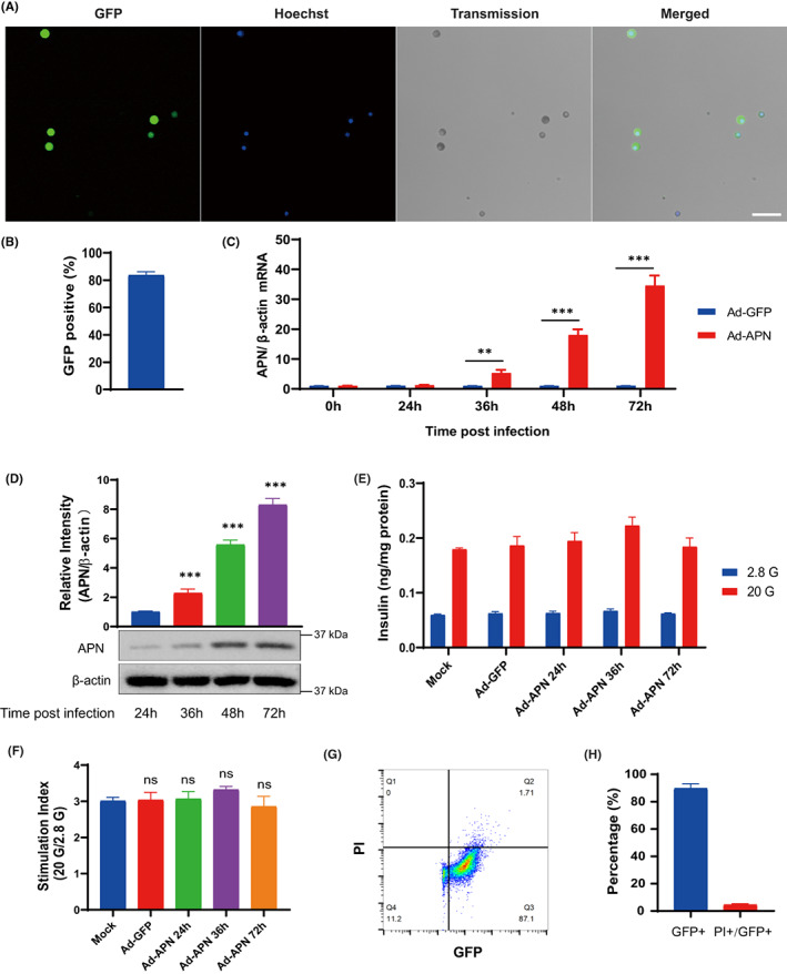
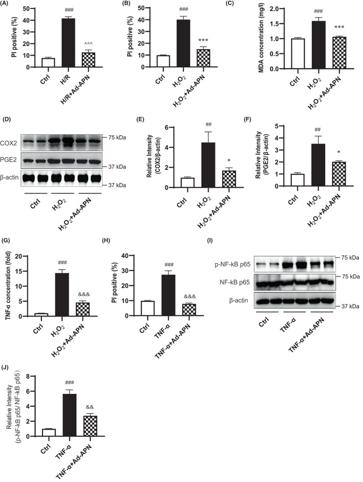
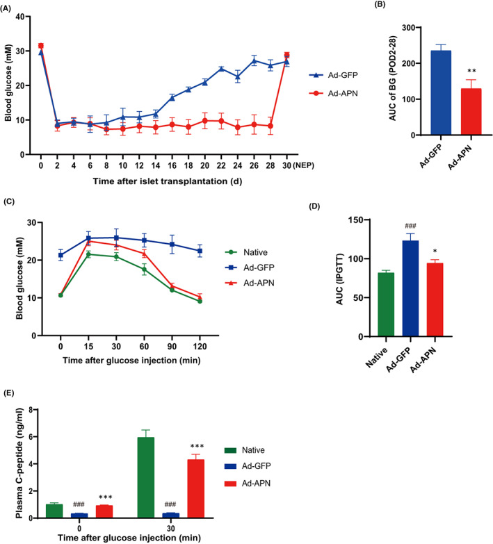
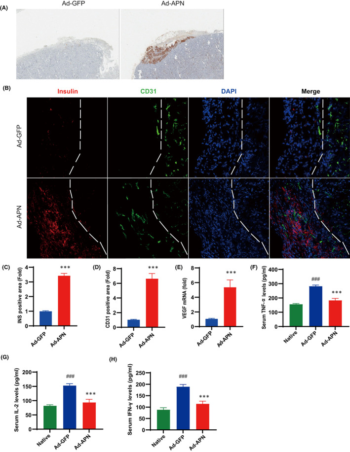
Significant pancreatic islet dysfunction and loss shortly after transplantation to the liver limit the widespread implementation of this procedure in the clinic. Nonimmune factors such as reactive oxygen species and inflammation have been considered as the primary driving force for graft failure. The adipokine adiponectin plays potent roles against inflammation and oxidative stress. Previous studies have demonstrated that systemic administration of adiponectin significantly prevented islet loss and enhanced islet function at post-transplantation period. In vitro studies indicate that adiponectin protects islets from hypoxia/reoxygenation injury, oxidative stress as well as TNF-α-induced injury. By applying adenovirus mediated transfection, we now engineered islet cells to express exogenous adiponectin gene prior to islet transplantation. Adenovirus-mediated adiponectin transfer to a syngeneic suboptimal islet graft transplanted under kidney capsule markedly prevented inflammation, preserved islet graft mass and improved islet transplant outcomes. These results suggest that adenovirus-mediated adiponectin gene therapy would be a beneficial clinical engineering approach for islet preservation in islet transplantation.

Keywords: Adiponectin; gene therapy; hypoxia/reoxygenation injury; inflammation; islet transplantation; oxidative stress.


Conflict of interest statement

P‐OB is founder and CEO of the biotech company Biocrine AB.

Figures

FIGURE 1
FIGURE 1
Adenovirus‐mediated adiponectin transduction is not toxic to islet cells. (A) Typical fluorescence microphotographs of primary islet cells infected with Ad‐Adiponectin‐GFP (Ad‐APN, MOI = 100) at 72 h post‐transfection. Scale bar = 50 μm. (B) Quantification of the percentage of GFP positive cells among islet cells infected with Ad‐APN for 72 h. n > 200 cells from three independent pancreatic islet isolations were analysed. (C) qPCR analysis of adiponectin mRNA levels in islet cells infected with Ad‐GFP or Ad‐APN at indicated time post‐transfection (n = 5). (D) Western blot analysis of the expression levels of adiponectin in islet cells infected with Ad‐APN at indicated time points post‐transfection (n = 4). (E) GSIS of islet cells under indicated treatments (n = 4). (F) Stimulation index calculated by the ratio of insulin secretion at 20 mmol/L glucose (20 G) to insulin secretion at 2.8 mmol/L glucose (2.8 G) in islet cells with indicated treatments (n = 4). (G and H) Flow cytometry analysis and quantification of adiponectin gene transduction efficiency (percentage of GFP+ cells) and the apoptotic rate (percentage of PI+ cells among GFP+ cells) of islet cells infected with Ad‐APN at 72 h post‐transfection (n = 4). Values are mean ± SEM; ns, not significant; **p < 0.01; ***p < 0.001.
FIGURE 2
FIGURE 2
Islet cells expressing exogenous adiponectin gene are resistant to hypoxia/reoxygenation injury. (A and B) Flow cytometry analysis of the apoptotic (PI positive cells) rate of islet cells with indicated treatments (n = 5). (C) Measurement of MDA concentrations in islet cells with indicated treatments (n = 5). (D–F) Western blot analysis and quantification of the expression levels of COX2 and PGE2 in islet cells with indicated treatments (n = 5). (G) Measurement of TNF‐α levels in the culture medium of islet cells with indicated treatments (n = 5). (H) Flow cytometry analysis of the apoptotic (PI positive cells) rate of islet cells with indicated treatments (n = 5). (I and J) Western blot analysis and quantification of the expression levels of p‐NF‐kB p65 and NF‐kB p65 in islet cells with indicated treatments (n = 4). Values are mean ± SEM; ## p < 0.01, ### p < 0.001 versus Ctrl group; ^^^ p < 0.001 versus H/R group; *p < 0.05, ***p < 0.001 versus H2O2 group; && p < 0.01, &&& p < 0.001 versus TNF‐α group.
FIGURE 3
FIGURE 3
Adiponectin transduction improves islet transplant outcomes. (A) Monitoring of blood glucose levels after islet transplantation until nephrectomy (NEP) was performed at post‐transplant day (POD) 30 (n = 6). (B) AUC of blood glucose levels from POD2 to POD28 (n = 6). (C and D) IPGTT on native Balb/c mice (Ctrl), diabetic Balb/c mice transplanted with Ad‐GFP islets (Ad‐GFP) or Ad‐APN islets (Ad‐APN) and AUC of blood glucose levels (n = 6). (E) Plasma C‐peptide levels of indicated animals at 0 and 30 min after glucose injection (n = 6). Values are mean ± SEM; ### p < 0.001 versus Ctrl group; *p < 0.001, ***p < 0.001 versus Ad‐GFP group.
FIGURE 4
FIGURE 4
Adiponectin transduction reduces inflammatory responses and prevents islet graft loss. (A) Immunohistochemical analysis of insulin at implantation sites of the mouse at POD 30. (B) Fluorescent staining of insulin (red) and CD31 (green) in implantation sites of the mouse at POD 30. DAPI was used to stain the cell nucleus (blue). (C and D) Quantification of insulin and CD31 positive areas. (E) qPCR analysis of VEGF mRNA levels in islet grafts (n = 5). (F–H) Measurement of the levels of serum TNF‐α, IL‐2 and IFN‐γ (n = 5). Values are mean ± SEM; ### p < 0.001 versus Native group; ***p < 0.001 versus Ad‐GFP group.

Similar articles

Cited by

References

    1. Ryan EA, Paty BW, Senior PA, et al. Five‐year follow‐up after clinical islet transplantation. Diabetes. 2005;54(7):2060‐2069. doi:10.2337/diabetes.54.7.206054/7/2060 [pii]. - DOI - PubMed
    1. Biarnés M, Montolio M, Nacher V, Raurell M, Soler J, Montanya E. Beta‐cell death and mass in syngeneically transplanted islets exposed to short‐ and long‐term hyperglycemia. Diabetes. 2002;51(1):66‐72. doi:10.2337/diabetes.51.1.66 - DOI - PubMed
    1. Davalli AM, Ogawa Y, Ricordi C, Scharp DW, Bonner‐Weir S, Weir GC. A selective decrease in the beta cell mass of human islets transplanted into diabetic nude mice. Transplantation. 1995;59(6):817‐820. - PubMed
    1. Sklavos MM, Bertera S, Tse HM, et al. Redox modulation protects islets from transplant‐related injury. Diabetes. 2010;59(7):1731‐1738. doi:10.2337/db09-0588 - DOI - PMC - PubMed
    1. Zhu H, Wang J, Jiang H, et al. Bilirubin protects grafts against nonspecific inflammation‐induced injury in syngeneic intraportal islet transplantation. Exp Mol Med. 2010;42(11):739‐748. doi:10.3858/emm.2010.42.11.075 - DOI - PMC - PubMed

Publication types