

In the footsteps of sea stars: deciphering the catalogue of proteins involved in underwater temporary adhesion
- PMID: 35975651
- PMCID: PMC9382459
- DOI: 10.1098/rsob.220103
In the footsteps of sea stars: deciphering the catalogue of proteins involved in underwater temporary adhesion
Abstract
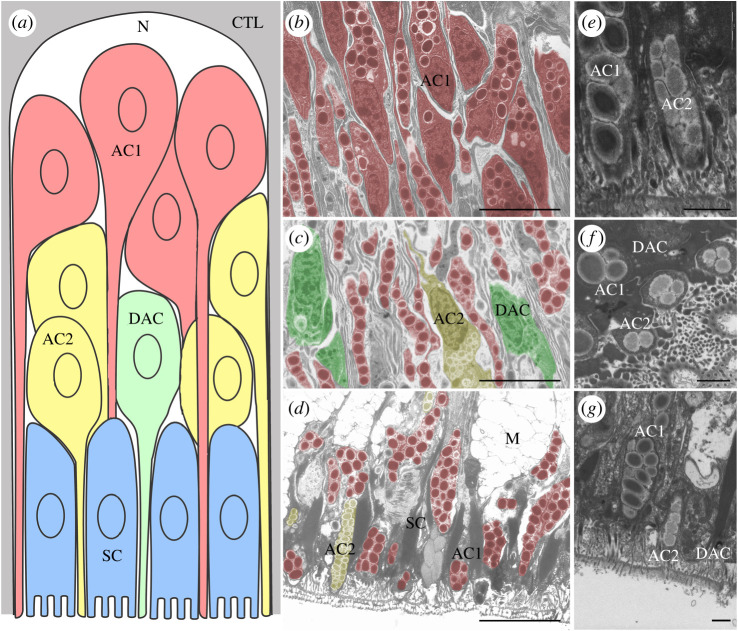
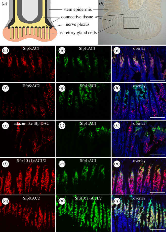
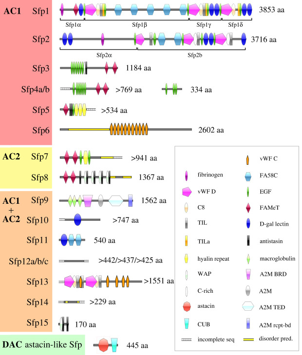
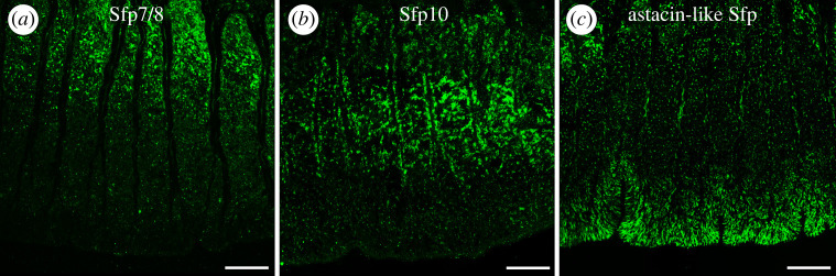
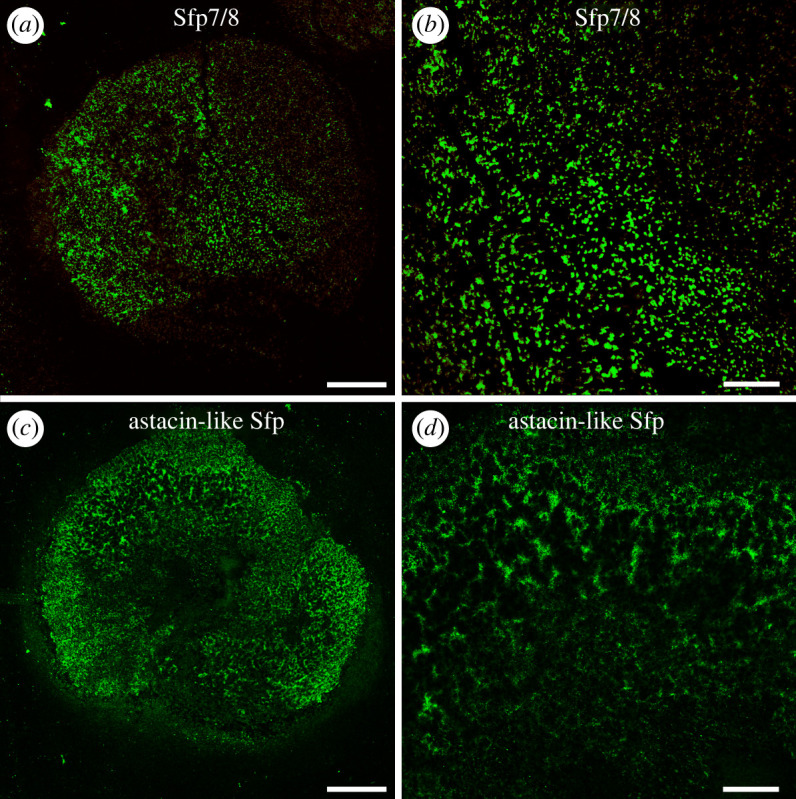
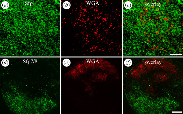
Sea stars adhere strongly but temporarily to underwater substrata via the secretion of a blend of proteins, forming an adhesive footprint that they leave on the surface after detachment. Their tube feet enclose a duo-gland adhesive system comprising two types of adhesive cells, contributing different layers of the footprint and de-adhesive cells. In this study, we characterized the catalogue of sea star footprint proteins (Sfps) in the species Asterias rubens to gain insights in their potential function. We identified 16 Sfps and mapped their expression to type 1 and/or type 2 adhesive cells or to de-adhesive cells by double fluorescent in situ hybridization. Based on their cellular expression pattern and their conserved functional domains, we propose that the identified Sfps serve different functions during attachment, with two Sfps coupling to the surface, six providing cohesive strength and the rest forming a binding matrix. Immunolabelling of footprints with antibodies directed against one protein of each category confirmed these roles. A de-adhesive gland cell-specific astacin-like proteinase presumably weakens the bond between the adhesive material and the tube foot surface during detachment. Overall, we provide a model for temporary adhesion in sea stars, including a comprehensive list of the proteins involved.
Keywords: Asteroidea; Echinodermata; duo-gland adhesive system; proteomics and transcriptomics; sea star footprint protein; tube feet.
Conflict of interest statement
The authors declare that they have no competing financial interests or personal relationships that could have appeared to influence the work reported in this paper.
Figures
Similar articles
-
Interspecies comparison of sea star adhesive proteins.Philos Trans R Soc Lond B Biol Sci. 2019 Oct 28;374(1784):20190195. doi: 10.1098/rstb.2019.0195. Epub 2019 Sep 9. Philos Trans R Soc Lond B Biol Sci. 2019. PMID: 31495313 Free PMC article.
-
Characterisation of the carbohydrate fraction of the temporary adhesive secreted by the tube feet of the sea star Asterias rubens.Mar Biotechnol (NY). 2011 Jun;13(3):484-95. doi: 10.1007/s10126-010-9319-6. Epub 2010 Oct 1. Mar Biotechnol (NY). 2011. PMID: 20886254
-
Micro- and nanostructure of the adhesive material secreted by the tube feet of the sea star Asterias rubens.J Struct Biol. 2008 Oct;164(1):108-18. doi: 10.1016/j.jsb.2008.06.007. Epub 2008 Jun 24. J Struct Biol. 2008. PMID: 18625322
-
Properties of temporary adhesion systems of marine and freshwater organisms.J Exp Biol. 2018 Aug 30;221(Pt 16):jeb182717. doi: 10.1242/jeb.182717. J Exp Biol. 2018. PMID: 30166319 Review.
-
Adhesive secretions in the Platyhelminthes.Adv Parasitol. 2001;48:101-224. doi: 10.1016/s0065-308x(01)48006-7. Adv Parasitol. 2001. PMID: 11013756 Review.
Cited by
-
Research Progress on Starfish Outbreaks and Their Prevention and Utilization: Lessons from Northern China.Biology (Basel). 2024 Jul 17;13(7):537. doi: 10.3390/biology13070537. Biology (Basel). 2024. PMID: 39056729 Free PMC article. Review.
-
Structural and evolutionary insights into astacin metallopeptidases.Front Mol Biosci. 2023 Jan 4;9:1080836. doi: 10.3389/fmolb.2022.1080836. eCollection 2022. Front Mol Biosci. 2023. PMID: 36685277 Free PMC article.
-
The Involvement of Cell-Type-Specific Glycans in Hydra Temporary Adhesion Revealed by a Lectin Screen.Biomimetics (Basel). 2022 Oct 15;7(4):166. doi: 10.3390/biomimetics7040166. Biomimetics (Basel). 2022. PMID: 36278723 Free PMC article.
-
Recurrent evolution of adhesive defence systems in amphibians by parallel shifts in gene expression.Nat Commun. 2024 Jul 10;15(1):5612. doi: 10.1038/s41467-024-49917-3. Nat Commun. 2024. PMID: 38987280 Free PMC article.
-
Placozoan secretory cell types implicated in feeding, innate immunity and regulation of behavior.bioRxiv [Preprint]. 2025 Jan 16:2024.09.18.613768. doi: 10.1101/2024.09.18.613768. bioRxiv. 2025. Update in: PLoS One. 2025 Apr 22;20(4):e0311271. doi: 10.1371/journal.pone.0311271. PMID: 39372748 Free PMC article. Updated. Preprint.
References
-
- Hermans CO. 1983. The duo-gland adhesive system. Oceanogr. Mar. Biol. 21, 283-339.
-
- Flammang P. 1996. Adhesion in echinoderms. In Echinoderm studies 5 (eds M Jangoux, JM Lawrence), pp. 1-60. Boca Raton, FL: CRC Press.
Publication types
MeSH terms
Substances
Associated data
Grants and funding
LinkOut - more resources
Full Text Sources
Miscellaneous
