. 2022 Dec 16;66(7):959-975.
doi: 10.1042/EBC20220046.

Controlling amyloid formation of intrinsically disordered proteins and peptides: slowing down or speeding up?

Affiliations

Controlling amyloid formation of intrinsically disordered proteins and peptides: slowing down or speeding up?

Yong Xu et al. Essays Biochem. .

Abstract

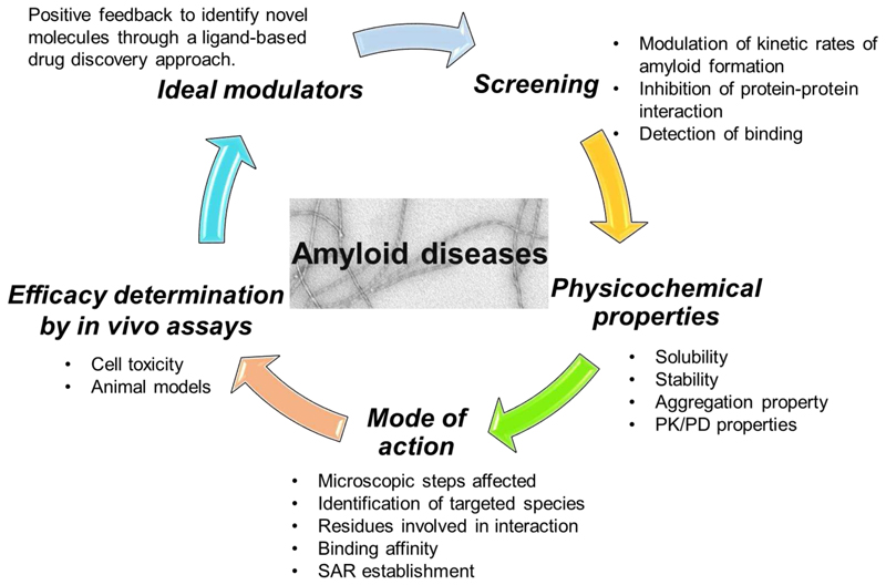
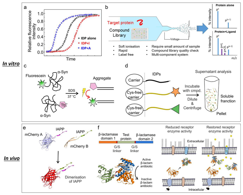
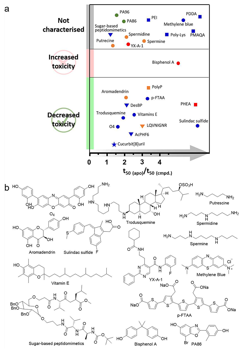
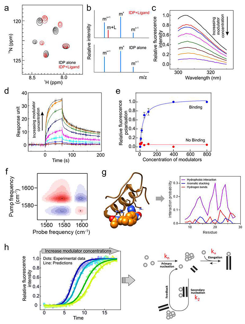
The pathological assembly of intrinsically disordered proteins/peptides (IDPs) into amyloid fibrils is associated with a range of human pathologies, including neurodegeneration, metabolic diseases and systemic amyloidosis. These debilitating disorders affect hundreds of millions of people worldwide, and the number of people affected is increasing sharply. However, the discovery of therapeutic agents has been immensely challenging largely because of (i) the diverse number of aggregation pathways and the multi-conformational and transient nature of the related proteins or peptides and (ii) the under-development of experimental pipelines for the identification of disease-modifying molecules and their mode-of-action. Here, we describe current approaches used in the search for small-molecule modulators able to control or arrest amyloid formation commencing from IDPs and review recently reported accelerators and inhibitors of amyloid formation for this class of proteins. We compare their targets, mode-of-action and effects on amyloid-associated cytotoxicity. Recent successes in the control of IDP-associated amyloid formation using small molecules highlight exciting possibilities for future intervention in protein-misfolding diseases, despite the challenges of targeting these highly dynamic precursors of amyloid assembly.

Keywords: accelerator; amyloid; energy landscape; inhibitor; protein aggregation; small-molecule modulator.


Conflict of interest statement

Conflicts of interest

All authors declare they have no competing interests.

Figures

Fig. 1
Fig. 1. Linked steps in a campaign of modulator discovery towards IDPs involved in protein aggregation into amyloid.
The order of the steps for modulator discovery does not matter, as all are needed for successful modulator discovery. PK (Pharmacokinetics), PD (Pharmacodynamics) and SAR (Structure-Activity Relationship).
Fig. 2
Fig. 2. Different approaches have been applied for the discovery of small-molecule modulators against IDPs.
(a) Fluorescence-based ThT assays. These assays are usually practiced in miniaturised formats with low cost. However, they require extremely pure proteins/peptides to achieve kinetic data with high reproducibility. Also the assay conditions (such as buffer, temperature, shaking or non-shaking, etc.) need to be carefully controlled. (b) Screening by nESI-MS. Under soft ionisation conditions, the non-covalent interactions between protein and ligand can be maintained. nESI-MS is label-free, can be used in multi-component systems, and confirms the integrity of the compound library at the same time. However caution should be exercised due to the inherent drawbacks of nESI-MS, such as non-specific binding during the electrospray process. (c) FRET-based assays have been developed for HTS of large library to identify small-molecule modulators in vitro. This figure was adapted from ref [46]. (d) Synergistic Aggregation Modulator Assay (SynAggreg) is an in vitro HT platform for the study of protein aggregation and the effect of modulators on protein aggregation [47]. This figure was adapted from ref [47]. (e) A bimolecular fluorescence complementary assay (BiFC) in a constructed E. coli system was developed to monitor the initial transient dimerization stage [35]. Specifically, mCherry protein was split and fused into amyloidogenic peptides/proteins, and a strong fluorescent signal can be detected if the fused biomolecules self-assemble into dimers. This figure was adapted from ref [35]. (f) β-Lactamase tripartite fusion system in E. coli has been introduced to screen inhibitors that prevent protein aggregation [36]. The bioassay can be configured in a 48-well format and has a simple phenotypic antibiotic resistance readout which directly links to the aggregation events of the test proteins/peptides. This figure was adapted from ref [36]. In vivo screening using different organisms have also been reported, such as C. elegans [48, 49]. One of the most important features of these systems is that they are able to investigate the amyloid-forming protein/peptide induced toxicity including those transiently populated low molecular weight oligomers.
Fig. 3
Fig. 3. Accelerators of IDPs.
(a) Relative t50 of the accelerators and their effect on IDP-induced cytotoxicity. Blue, red, green and orange represent Aβ, hIAPP, α-Syn and multiple targets, respectively. Circle, triangle, square and star represent small molecules, peptides, polymers and macrocycle, respectively. Of note, there are still a large number of accelerators whose effect on IDP-mediated cytotoxicity are not available. It would be interesting to determine the effect of these molecules, which might help to build a greater in-depth view of this possible therapeutic strategy. (b) Chemical structures of small-molecule accelerators discussed in this review.
Fig. 4
Fig. 4. Biochemical and biophysical approaches used for the characterisation of the interaction between IDPs and small molecules.
(a) Representative 2D NMR spectrum of an IDP with (red) and without (black) a small molecule. (b) nESI mass spectra show the mass addition of a ligand to the target, suggesting the interaction between the target and the ligand. (c) Fluorescence quenching caused by ligand binding. Stern-Volmer analysis of the fluorescence titration data enables extraction of the Kd. These data were taken from ref [32]. (d) SPR measurement showing ligand binding (Kd) as well as the association (kon) and dissociation (koff) rate constants. These data were taken from ref [32]. (e) Fluorescence polarisation (FP) can provide indications of binding/no binding of ligands. Fitting the FP titration can yield the binding affinity of the ligand. (f) 2D FTIR can provide binding site information of the inhibitor at residue-level resolution. (g) MD simulations can provide detailed descriptions of the conformational ensembles of IDPs and specific information about the IDP-ligand interactions at an atomic-level. MD simulation can predict the favoured interactions formed between IDP and ligand, such as hydrogen bonding, aromatic interaction, charge-charge interaction, etc. (see [29]). (h) Measurement of amyloid formation using ThT fluorescence can be used to determine the mechanism of amyloid formation and the species that bind ligands that affect the rate of assembly into amyloid. These data were taken from ref [32].

Similar articles

Cited by

References

    1. Iadanza MG, Jackson MP, Hewitt EW, Ranson NA, Radford SE. A new era for understanding amyloid structures and disease. Nat Rev Mol Cell Biol. 2018;19:755–73. - PubMed
    1. Knowles TPJ, Vendruscolo M, Dobson CM. The amyloid state and its association with protein misfolding diseases. Nat Rev Mol Cell Biol. 2014;15:384–96. - PubMed
    1. Gallardo R, Ranson NA, Radford SE. Amyloid structures: much more than just a cross-β fold. Curr Opin Struct Biol. 2020;60:7–16. - PubMed
    1. Tanzi RE, Bertram L. Twenty years of the Alzheimer’s disease amyloid hypothesis: a genetic perspective. Cell. 2005;120:545–55. - PubMed
    1. Milardi D, Gazit E, Radford SE, Xu Y, Gallardo RU, Caflisch A, et al. Proteostasis of islet amyloid polypeptide: A molecular perspective of risk factors and protective strategies for type II diabetes. Chem Rev. 2021;121:1845–93. doi: 10.1021/acs.chemrev.0c00981. - DOI - PMC - PubMed

Publication types