. 2022 Oct;61(10):958-971.
doi: 10.1002/mc.23453. Epub 2022 Aug 17.

TGFβ1 regulates HRas-mediated activation of IRE1α through the PERK-RPAP2 axis in keratinocytes

Affiliations

TGFβ1 regulates HRas-mediated activation of IRE1α through the PERK-RPAP2 axis in keratinocytes

Saie Mogre et al. Mol Carcinog. 2022 Oct.

Abstract

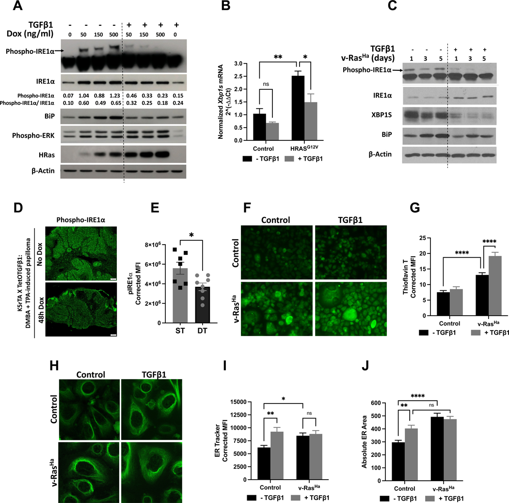
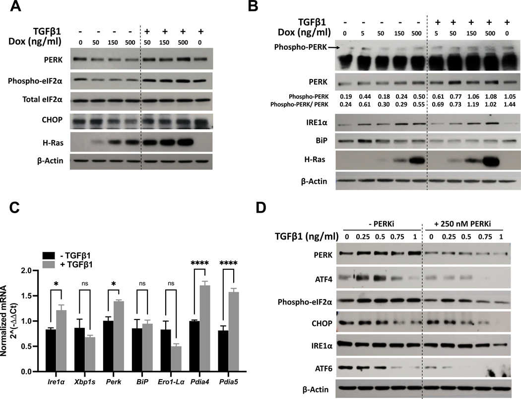
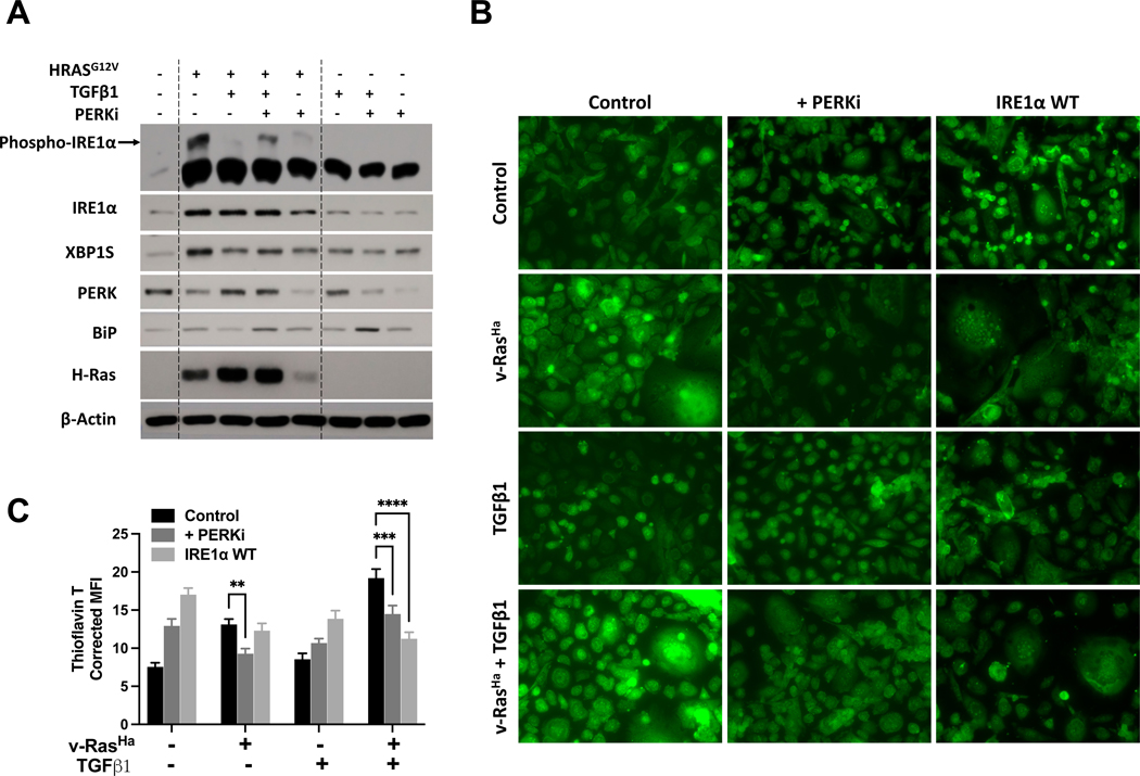
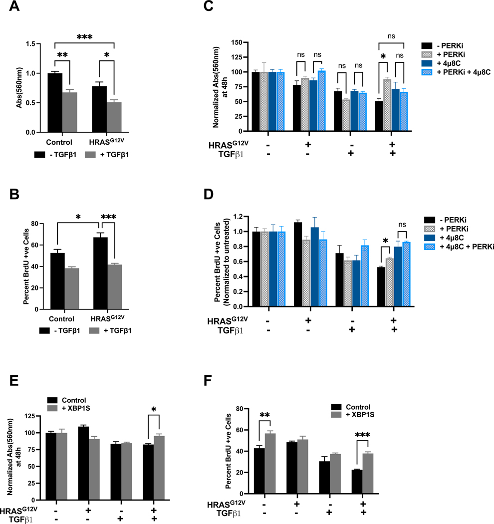
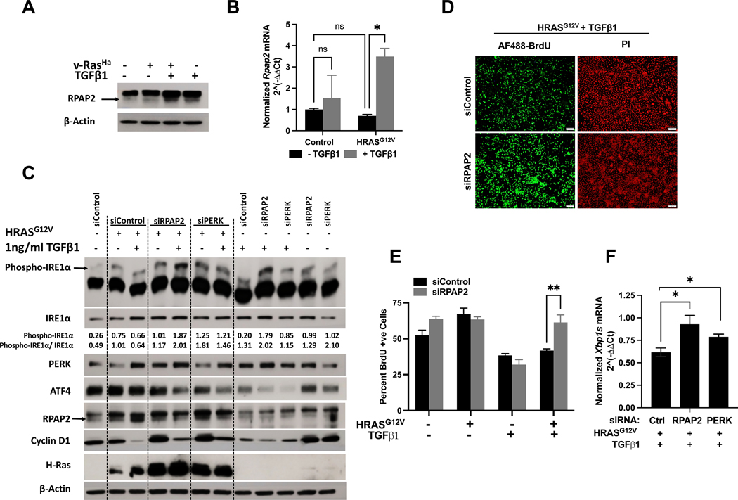
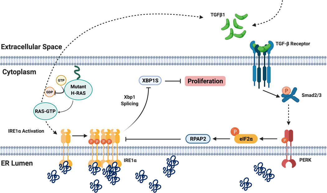
Transforming Growth Factor β1 (TGFβ1) is a critical regulator of tumor progression in response to HRas. Recently, TGFβ1 has been shown to trigger ER stress in many disease models; however, its role in oncogene-induced ER stress is unclear. Oncogenic HRas induces the unfolded protein response (UPR) predominantly via the Inositol-requiring enzyme 1α (IRE1α) pathway to initiate the adaptative responses to ER stress, with importance for both proliferation and senescence. Here, we show a role of the UPR sensor proteins IRE1α and (PKR)-like endoplasmic reticulum kinase (PERK) to mediate the tumor-suppressive roles of TGFβ1 in mouse keratinocytes expressing mutant forms of HRas. TGFβ1 suppressed IRE1α phosphorylation and activation by HRas both in in vitro and in vivo models while simultaneously activating the PERK pathway. However, the increase in ER stress indicated an uncoupling of ER stress and IRE1α activation by TGFβ1. Pharmacological and genetic approaches demonstrated that TGFβ1-dependent dephosphorylation of IRE1α was mediated by PERK through RNA Polymerase II Associated Protein 2 (RPAP2), a PERK-dependent IRE1α phosphatase. In addition, TGFβ1-mediated growth arrest in oncogenic HRas keratinocytes was partially dependent on PERK-induced IRE1α dephosphorylation and inactivation. Together, these results demonstrate a critical cross-talk between UPR proteins that is important for TGFβ1-mediated tumor suppressive responses.

Keywords: ER stress; HRas; IRE1α; PERK; TGFβ1; proliferation; unfolded protein response.


Figures

Figure 1:
Figure 1:. TGFβ1 blocks HRas-induced IRE1α phosphorylation without a corresponding reduction in ER stress.
(A) Phos-tag Western blot for phospho-IRE1α and Western blot analysis of total IRE1α, BiP, Phospho-ERK, and HRas in K5RasG12V primary keratinocytes treated simultaneously with increasing doses (0 – 500 ng/ml) of doxycycline and 1 ng/ml TGFβ1 for 48h. Numbers represent phospho- and total IRE1α densitometry normalized to β-actin. (B) qPCR analysis showing spliced Xbp1 mRNA expression 48h after addition of 1 ng/ml TGFβ1 to K5RasG12V primary keratinocytes treated with 500 ng/ml doxycycline. (C) Phos-tag Western blot for phospho-IRE1α and Western blot analysis of total IRE1α, XBP1S, and BiP in C57BL/6 primary keratinocytes expressing v-RasHa and treated with 1 ng/ml TGFβ1 for 1, 3, and 5 days. (D) Representative immunofluorescence images and (E) area-corrected mean fluorescence intensity (MFI) for phospho-ser724-IRE1α in DMBA-TPA-induced benign papilloma with and without overexpression of TGFβ1 for 48h. Scale bar is 100 μm. Statistical significance was determined by t-test using Welch’s correction. (F) Representative fluorescence confocal images and (G) area-corrected mean fluorescence intensity (MFI) of thioflavin-T-stained C57-T keratinocytes expressing v-RasHa and treated with 1 ng/ml TGFβ1 for 48h. Scale bar is 25 μm. (H) Representative fluorescence confocal images, (I) area-corrected mean fluorescence intensity (MFI), and (J) ER area of primary FVB/n keratinocytes expressing v-RasHa and treated with 1 ng/ml TGFβ1 for 48h and stained with ER Tracker™ Green. Scale bar is 20 μm. Data represent mean ± SEM from 3 biological replicates, or at least 50 cells per condition. Statistical significance was determined by two-way ANOVA and Tukey post-hoc test for multiple comparisons at p < 0.05.
Figure 2:
Figure 2:. TGFβ1 activates PERK signaling.
(A) Western blot analysis of PERK, phospho- and total eIF2α, CHOP, and HRas in K5RasG12V primary keratinocytes treated simultaneously with increasing doses (0 – 500 ng/ml) of doxycycline and 1 ng/ml TGFβ1 for 48h. (B) Phos-tag Western blot for phospho-PERK and Western blot analysis of total PERK, IRE1α, BiP, and HRas in K5Ras-T keratinocytes treated with 0 – 500 ng/ml doxycycline with and without 1 ng/ml TGFβ1 for 48h. Numbers represent phospho- and total PERK densitometry normalized to β-actin. (C) qPCR analysis of ER stress markers in primary K5RasG12V keratinocytes treated with 1 ng/ml TGFβ1 for 48h. Statistical significance was determined by Student’s t-test at p < 0.05. (D) Western blot of PERK, ATF4, phospho-eIF2α, CHOP, IRE1α, and ATF6 in K5Ras-T cells pre-treated with 250 nM PERK inhibitor (PERKi) and increasing doses (0 – 1 ng/ml) of TGFβ1 for 48h.
Figure 3:
Figure 3:. Inhibition of PERK signaling partially rescued TGFβ1-dependent IRE1α dephosphorylation and accumulation of unfolded proteins in HRas keratinocytes.
(A) Phos- tag Western blot for phospho-IRE1α and Western blot analysis of total IRE1α, XBP1S, PERK, BiP, and HRas in K5RasG12V primary keratinocytes treated simultaneously with 500 ng/ml doxycycline and 1 ng/ml TGFβ1 for 48h. Keratinocytes were pre-treated with 250 nM PERK inhibitor (PERKi) for 1h where indicated before the addition of doxycycline and TGFβ1. (B) Representative fluorescence confocal images and (C) area-corrected mean fluorescence intensity (MFI) of thioflavin-T-stained C57-T keratinocytes expressing v-RasHa and treated with 1 ng/ml TGFβ1. Keratinocytes were pre-treated with 250 nM PERKi for 1h where indicated. Overexpression of wildtype human IRE1α (IRE1α WT) was induced with 500 ng/ml doxycycline for 24h before treatment with 1 ng/ml of TGFβ1 for additional 48h. Data represent mean ± SEM for at least 30 cells. Scale bar is 25 μm. Statistical significance was determined by two-way ANOVA and Tukey post-hoc test for multiple comparisons at p < 0.05.
Figure 4:
Figure 4:. TGFβ1-PERK-dependent dephosphorylation of IRE1α in HRas keratinocytes regulates cell proliferation.
(A) MTT absorbance at 560 nm and (B) percent BrdU positive K5Ras-T keratinocytes treated simultaneously with 500 ng/ml doxycycline and 1 ng/ml TGFβ1 for 48h. (C) Normalized MTT absorbance at 560 nm and (D) percent BrdU positive K5Ras-T keratinocytes pre-treated with 250 nM PERKi for 1h or 20 μM 4μ8C for 24h, followed by 500 ng/ml doxycycline and 1 ng/ml TGFβ1 for 48h. Data were normalized to controls within each treatment group. Data represent mean ± SEM from 3 biological replicates. Statistical significance was determined by two-way ANOVA and Tukey post-hoc test for multiple comparisons at p < 0.05. (E) Normalized MTT absorbance at 560 nm and (F) Percent BrdU positive K5Ras-T keratinocytes overexpressing spliced Xbp1 mRNA treated simultaneously with 500 ng/ml doxycycline and 1 ng/ml TGFβ1 for 48h. Statistical significance was determined by two-way ANOVA and Šidák post-hoc test for multiple comparisons at p < 0.05.
Figure 5:
Figure 5:. TGFβ1-PERK-RPAP2 axis governs IRE1α activity in HRas keratinocytes.
(A) Western blot of RPAP2 expression in FVB/n primary keratinocytes expressing v-RasHa and treated with 1 ng/ml TGFβ1 for 48h. (B) qPCR analysis of K5RasG12V primary keratinocytes treated simultaneously with 500 ng/ml doxycycline and 1 ng/ml TGFβ1 for 48h. Data represent mean ± SEM from 3 biological replicates. Statistical significance was determined by two-way ANOVA and Tukey post-hoc test for multiple comparisons at p < 0.05. (C) Phos-tag Western blot for phospho-IRE1α and Western blot of total IRE1α, PERK, ATF4, RPAP2, Cyclin D1, and HRas in RPAP2 and PERK-depleted K5Ras-T keratinocytes. Keratinocytes were treated with 500 ng/ml doxycycline and 1 ng/ml TGFβ1 4 days after transfection of siRNAs against RPAP2 and PERK. Numbers represent phospho- and total IRE1α densitometry normalized to β-actin. (D) Representative fluorescence images of AF488-BrdU (green) and propidium iodide (PI) (red) and (E) percent BrdU positive control and RPAP2 knockdown K5Ras-T keratinocytes. Scale bar is 100 μm. Data represent mean ± SEM from 3 biological replicates. Statistical significance was determined by two-way ANOVA and Šidák post-hoc test for multiple comparisons at p < 0.05. (F) qPCR analysis showing spliced Xbp1 mRNA expression in K5Ras-T RPAP2 and PERK knockdown keratinocytes treated with 500 ng/ml doxycycline and 1 ng/ml TGFβ1 for 48h. Data represent mean ± SEM from 3 biological replicates. Statistical significance was determined by Student’s t-test at p < 0.05.
Figure 6:
Figure 6:. Proposed model of TGFβ1-regulation of IRE1α phosphorylation in oncogenic HRas keratinocytes.
TGFβ1 suppresses HRas-induced IRE1α phosphorylation and activation in mouse keratinocytes resulting in reduced Xbp1 splicing and XBP1S protein notwithstanding the increasing accumulation of unfolded protein in the ER lumen. This uncoupling of ER stress and IRE1α activation is mediated by TGFβ1-PERK signaling. PERK activation by TGFβ1 specifically upregulates an IRE1α phosphatase, RPAP2. The decrease in Xbp1 splicing by TGFβ1-PERK-RPAP2 contributes to the significantly lower proliferation of HRas keratinocytes. Created with BioRender.com.

Similar articles

Cited by

References

    1. Chalmers F, Mogre S, Son J, Blazanin N, Glick AB. The multiple roles of the unfolded protein response regulator IRE1α in cancer. Molecular carcinogenesis. 2019;58(9):1623–1630. doi:10.1002/mc.23031 - DOI - PMC - PubMed
    1. Hetz C, Zhang K, Kaufman RJ. Mechanisms, regulation and functions of the unfolded protein response. Nature Reviews Molecular Cell Biology. 2020/08/01 2020;21(8):421–438. doi:10.1038/s41580-020-0250-z - DOI - PMC - PubMed
    1. Bertolotti A, Zhang Y, Hendershot LM, Harding HP, Ron D. Dynamic interaction of BiP and ER stress transducers in the unfolded-protein response. Nature Cell Biology. 2000;2(6):326–332. doi:10.1038/35014014 - DOI - PubMed
    1. Adams CJ, Kopp MC, Larburu N, Nowak PR, Ali MMU. Structure and Molecular Mechanism of ER Stress Signaling by the Unfolded Protein Response Signal Activator IRE1. Frontiers in Molecular Biosciences. 2019;6:11. - PMC - PubMed
    1. Ali MMU, Bagratuni T, Davenport EL, et al. Structure of the Ire1 autophosphorylation complex and implications for the unfolded protein response. The EMBO journal. 2011;30(5):894–905. doi:10.1038/emboj.2011.18 - DOI - PMC - PubMed

Publication types

MeSH terms