. 2022 Sep 1;128(3):671-680.
doi: 10.1152/jn.00111.2022. Epub 2022 Aug 17.

Reduced activity of vertically acting motoneurons during convergence

Affiliations

Reduced activity of vertically acting motoneurons during convergence

Mark M G Walton. J Neurophysiol. .

Abstract

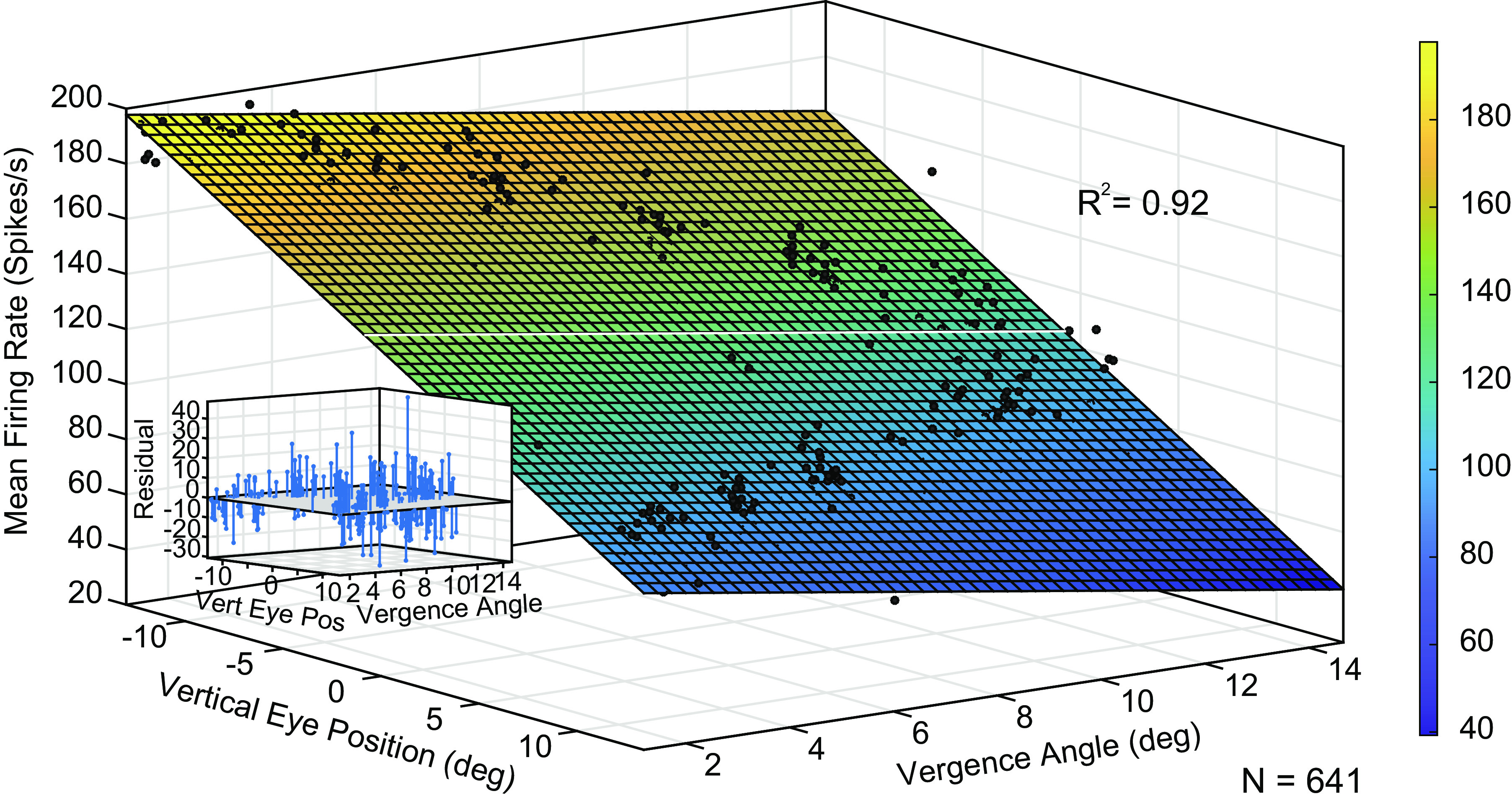
Previous studies have revealed unexpected relationships between the firing rates of horizontally acting motoneurons and vergence. During a vergence task, for example, antidromically identified abducens internuclear neurons show a negative correlation between vergence angle and firing rate, which is the opposite of the modulation displayed by the medial rectus motoneurons to which they project. For a given horizontal eye position, medial rectus motoneurons discharge at a higher rate if the eyes are converged than if the same eye position is reached during a task that requires version; paradoxically, however, the horizontal rectus eye muscles show corelaxation during vergence. These complex and unexpected relationships inspired the present author to investigate whether the tonic firing rates of vertically acting motoneurons in oculomotor nucleus are correlated with vergence angle. Monkeys were trained to fixate a single, randomly selected, visual target among an array of 60 red plus-shaped LEDs, arranged at 12 different distances in three-dimensional space. The targets were arranged to permit dissociation of vertical eye position and vergence angle. Here I report, for the first time, that most vertically acting motoneurons in oculomotor nucleus show a significant negative correlation between tonic firing rate and vergence angle. This suggests the possibility that there may be a general corelaxation of extraocular muscles during vergence.NEW & NOTEWORTHY An array of 60 plus-shaped LEDs, positioned at various locations in three-dimensional space, was used to elicit conjugate and disjunctive saccades while single neurons in oculomotor nucleus were recorded from rhesus monkeys. This study demonstrates that most vertically acting motoneurons in oculomotor nucleus discharge at a lower rate when the eyes are converged.

Keywords: monkey; motoneurons; neurophysiology; oculomotor nucleus; vergence.


Conflict of interest statement

No conflicts of interest, financial or otherwise, are declared by the author.

Figures

None
Graphical abstract
Figure 1.
Figure 1.
Schematic representation of the target array used for the present study. A: top view of the five triangle-shaped LED boards. The actual size of each LED was related to the distance, such that all targets subtended 1° of visual angle, as seen by the monkey. B: side view of one of the LED boards. Note that this arrangement of LEDs ensures that vertical eye position could be dissociated from vergence angle, because those two variables are not linearly related. The longer scale bar shows the distance from the farthest targets to the monkey’s eye (48.5 cm).
Figure 2.
Figure 2.
Vertical rate-position curves for six vertically acting motoneurons in oculomotor nucleus, including three with downward preferred directions (A, C, and E) and three with upward preferred directions (B, D, and F). To emphasize the nonlinearities in this figure, the regression lines were based on eye positions on the preferred side of 0° (<0° for inferior rectus motoneurons, >0° for inferior oblique and superior rectus motoneurons). The actual regression lines are shown as solid gray lines. All six neurons show a deflection point near 0°; as eye position moves further into the off direction, the firing rate does not decrease as much as one would expect, based on a linear fit to the eye positions in the on-direction. To understand why this occurs, refer to the arrangement of targets shown in Fig. 1B; for each triangle-shaped LED board, vergence demand increases as the vertical position tends toward zero. Thus, any sensitivity to vergence angle will result in a deflection point, near 0°, in the vertical rate position curves. To help the reader to more easily appreciate the nonlinearities, the regression lines have been extended to show the expected firing rates for various off-direction eye positions (dashed gray lines). Note the small number of data points that are outside the range of target positions. These were times when the animal looks away from the target array, either to a point above the array or below it. The vergence angle was always small in these cases, and including them resulted in slightly better fits.
Figure 3.
Figure 3.
Planar fit for one example inferior rectus motoneuron, recorded from monkey N1 (female). Note the obvious tilt along the vergence angle axis; higher vergence angles are associated with lower firing rates. Inset shows the residuals for this fit. This is the same neuron shown in Fig. 2E. Note that, despite the nonlinearity for this neuron when only vertical eye position is used to predict the firing rate, the data points fall on (or very close to) a plane when vergence angle is added to the model.
Figure 4.
Figure 4.
Relationship between vergence angle and predicted firing rates when the vertical eye position is near 0 (see Eq. 2, results). AC: relationship for three example neurons, including one from each monkey. Filled circles indicate predicted firing rates for various vergence angles at a vertical eye position of 0, computed using Eq. 2. Open circles show the mean observed firing rates obtained when the monkey was fixating the nearest target. D: mean observed firing rates for the nearest target location, plotted as a function of the predicted firing rate for the smallest vergence angle used in this analysis (<6°). Each data point represents results from a single neuron. Note that most data points fall below the unity line, indicating that, for the same vertical eye position, the firing rates were lower when the eyes were converged than the predicted firing rate when the eyes were diverged.
Figure 5.
Figure 5.
Example plane fit for one neuron for which data were collected both during performance of the saccade-vergence task (data points outside of the red rectangle) and while the monkey sat in complete darkness (data points within the red rectangle). Vertical eye positions in darkness ranged from ∼10° down to ∼25° up, but the vergence angle was consistently small. The firing rates were consistently higher when the eyes were diverged than when the same vertical eye positions occurred while the eyes were converged.
Figure 6.
Figure 6.
Comparison of mean firing rates during fixations when the eyes were converged by least 10° and during fixations in darkness when the vergence angle was less than 5°. This analysis was based on 20 neurons, including eleven from monkey N1 (circles), five from monkey N2 (squares), and four from monkey N3 (triangles). The asterisk shows the neuron depicted in Fig. 5. Vertical eye positions were similar in both conditions (−3° to 3°). Red filled circles: cells for which the mean firing rate was significantly higher when the eyes were diverged. Blue filled circles: cells for which the mean firing rate was significantly lower when the eyes were diverged. Red open circles: cells for which there was a significant negative correlation between vergence angle and firing rate during performance of the saccade-vergence task but no significant difference when firing rates were compared for converged fixations versus diverged eye positions in darkness. Black open circle: a cell for which there was no significant effect of vergence angle for either of these analyses.

Similar articles

Cited by

References

    1. Mays LE, Porter JD. Neural control of vergence eye movements: activity of abducens and oculomotor neurons. J Neurophysiol 52: 743–761, 1984. doi:10.1152/jn.1984.52.4.743. - DOI - PubMed
    1. Miller JM, Davison RC, Gamlin PD. Motor nucleus activity fails to predict extraocular muscle forces in ocular convergence. J Neurophysiol 105: 2863–2873, 2011. doi:10.1152/jn.00935.2010. - DOI - PMC - PubMed
    1. Gamlin PD, Gnadt JW, Mays LE. Abducens internuclear neurons carry an inappropriate signal for ocular convergence. J Neurophysiol 62: 70–81, 1989. doi:10.1152/jn.1989.62.1.70. - DOI - PubMed
    1. Miller JM, Bockisch CJ, Pavlovski DS. Missing lateral rectus force and absence of medial rectus co-contraction in ocular convergence. J Neurophysiol 87: 2421–2433, 2002. doi:10.1152/jn.00566.2001. - DOI - PubMed
    1. Demer JL, Clark RA. Magnetic resonance imaging of differential compartmental function of horizontal rectus extraocular muscles during conjugate and converged ocular adduction. J Neurophysiol 112: 845–855, 2014. doi:10.1152/jn.00649.2013. - DOI - PMC - PubMed

Publication types

LinkOut - more resources