. 2022 Sep 20;66(9):e0059222.
doi: 10.1128/aac.00592-22. Epub 2022 Aug 17.

Moxifloxacin-Mediated Killing of Mycobacterium tuberculosis Involves Respiratory Downshift, Reductive Stress, and Accumulation of Reactive Oxygen Species

Affiliations

Moxifloxacin-Mediated Killing of Mycobacterium tuberculosis Involves Respiratory Downshift, Reductive Stress, and Accumulation of Reactive Oxygen Species

Somnath Shee et al. Antimicrob Agents Chemother. .

Abstract

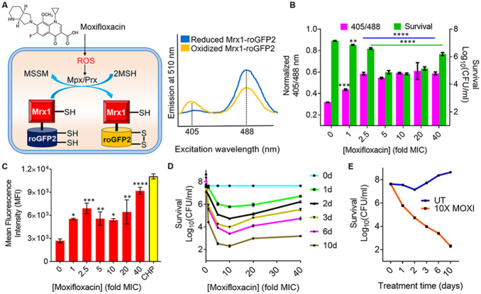
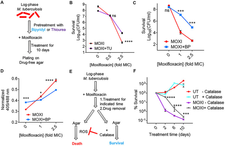
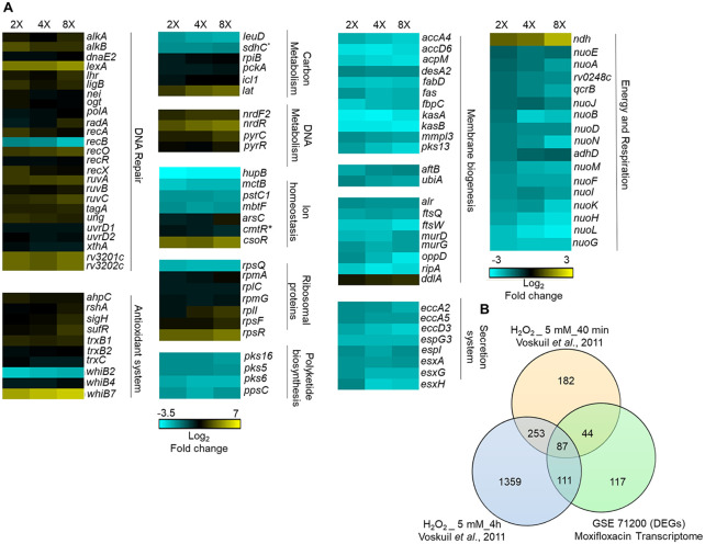
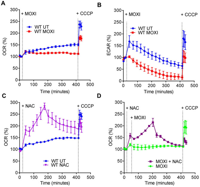
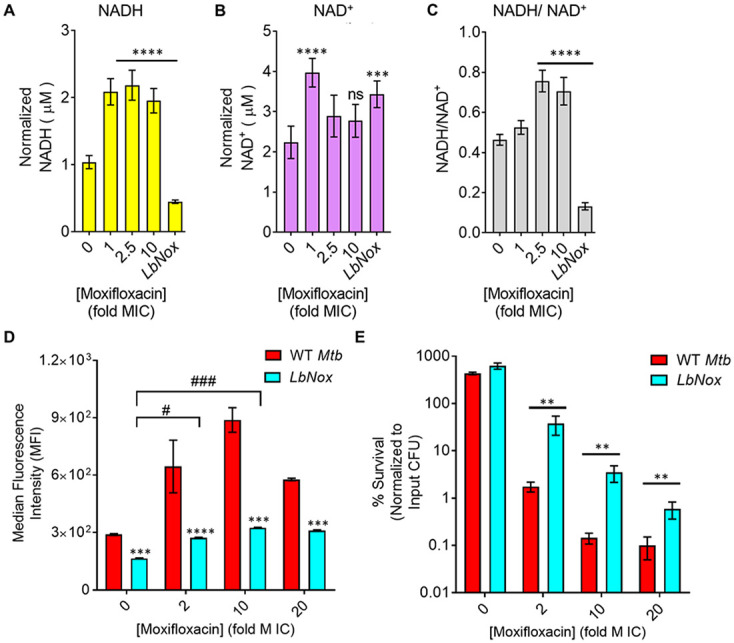
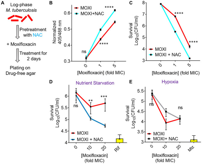
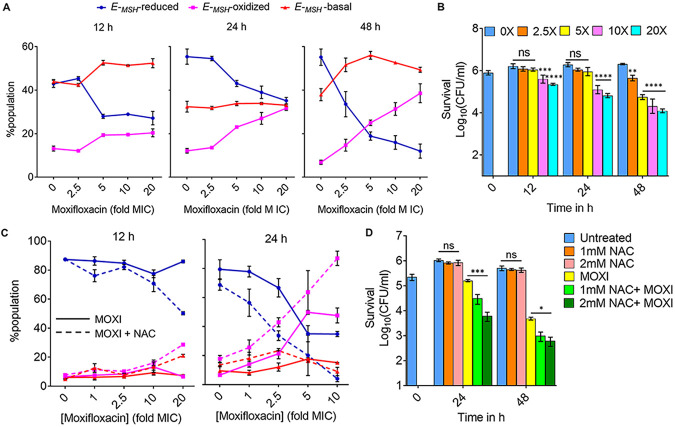
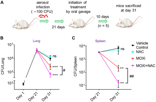
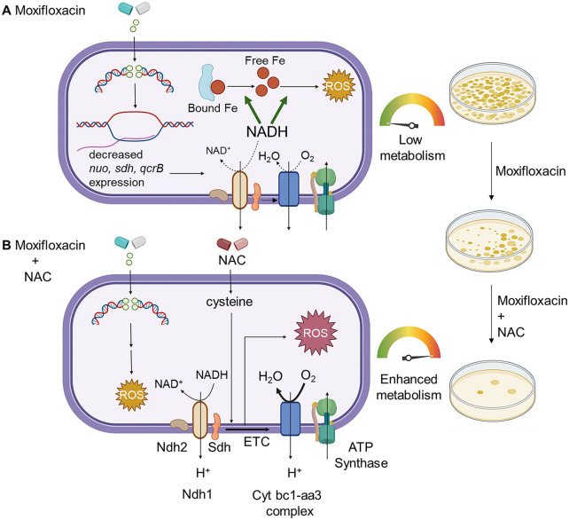
Moxifloxacin is central to treatment of multidrug-resistant tuberculosis. Effects of moxifloxacin on the Mycobacterium tuberculosis redox state were explored to identify strategies for increasing lethality and reducing the prevalence of extensively resistant tuberculosis. A noninvasive redox biosensor and a reactive oxygen species (ROS)-sensitive dye revealed that moxifloxacin induces oxidative stress correlated with M. tuberculosis death. Moxifloxacin lethality was mitigated by supplementing bacterial cultures with an ROS scavenger (thiourea), an iron chelator (bipyridyl), and, after drug removal, an antioxidant enzyme (catalase). Lethality was also reduced by hypoxia and nutrient starvation. Moxifloxacin increased the expression of genes involved in the oxidative stress response, iron-sulfur cluster biogenesis, and DNA repair. Surprisingly, and in contrast with Escherichia coli studies, moxifloxacin decreased expression of genes involved in respiration, suppressed oxygen consumption, increased the NADH/NAD+ ratio, and increased the labile iron pool in M. tuberculosis. Lowering the NADH/NAD+ ratio in M. tuberculosis revealed that NADH-reductive stress facilitates an iron-mediated ROS surge and moxifloxacin lethality. Treatment with N-acetyl cysteine (NAC) accelerated respiration and ROS production, increased moxifloxacin lethality, and lowered the mutant prevention concentration. Moxifloxacin induced redox stress in M. tuberculosis inside macrophages, and cotreatment with NAC potentiated the antimycobacterial efficacy of moxifloxacin during nutrient starvation, inside macrophages, and in mice, where NAC restricted the emergence of resistance. Thus, NADH-reductive stress contributes to moxifloxacin-mediated killing of M. tuberculosis, and the respiration stimulator (NAC) enhances lethality and suppresses the emergence of drug resistance.

Keywords: N-acetyl cysteine; NADH; ROS; antimycobacterial; fluoroquinolone; moxifloxacin; oxidative stress; redox biosensor; reductive stress; resistance; respiration.


Conflict of interest statement

The authors declare no conflict of interest.

Figures

FIG 1
FIG 1
Effect of moxifloxacin on biosensor oxidation, ROS level, and bacterial survival. (A) Moxifloxacin increases oxidative stress, which increases the ratio of oxidized (MSSM) to reduced (MSH) mycothiol via mycothiol-dependent peroxiredoxin (Prx) or peroxidase (Mpx). Oxidation of Mrx1-roGFP2 increases fluorescence intensity for excitation at ~400 nm and decreases it for excitation at ~490 nm. (B) Strain M. tuberculosis roGFP2 was exposed to the indicated concentrations of moxifloxacin (1× MIC = 0.5 μM) for 48 h, and the ratiometric response of the biosensor and survival following treatment were determined. (C) Cells were treated as for panel B, and ROS were quantified by flow cytometry using CellROX deep red dye. Cumene hydroperoxide (CHP; 10 mM) served as a positive control. Data represent mean fluorescence intensity of the dye. (D) Exponentially growing M. tuberculosis H37Rv was treated with moxifloxacin at the indicated concentrations for the indicated times; survival was assessed by determining CFU. (E) Time-kill curves for M. tuberculosis treated with 10× MIC of moxifloxacin. Error bars represent standard deviations from the mean. Data represent at least two independent experiments performed in at least duplicate. Statistical significance was calculated against the no-treatment control (****, P < 0.0001; ***, P < 0.001; **, P < 0.01; *, P < 0.05).
FIG 2
FIG 2
ROS mitigation reduces moxifloxacin-mediated killing of M. tuberculosis. (A) Plan for detecting thiourea (TU) and bipyridyl (BP) effects on moxifloxacin lethality. (B) Exponentially growing M. tuberculosis H37Rv cultures were either left untreated or treated with 10 mM TU for 1 h before addition of the indicated concentrations of moxifloxacin (MOXI; 1× MIC = 0.5 μM) for 10 days followed by determination of CFU. (C) Effect of bipyridyl. M. tuberculosis as for panel B was untreated or treated with 250 μM BP for 15 min prior to addition moxifloxacin as for panel B. (D) The Mrx1-roGFP2 biosensor ratiometric response was determined after 48 h treatment of M. tuberculosis cultures with the indicated concentrations of MOXI alone or with 250 μM BP. (E) M. tuberculosis cultures were treated with moxifloxacin, the drug was removed by washing, and cells were plated on drug-free 7H11 agar with or without catalase followed by CFU determination. (F) M. tuberculosis cultures were treated with 1× MIC of moxifloxacin for the indicated times, washed, and plated with or without catalase (17.5 U/mL of agar). Percentage survival was calculated relative to CFU of cultures at 0 h. Statistical significance was calculated between the drug-alone group and the drug + catalase group. Statistical considerations were as in Fig. 1.
FIG 3
FIG 3
Whole-genome transcriptome profiling of M. tuberculosis treated with moxifloxacin. (A) Heat map showing gene expression changes due to 16-h treatment of M. tuberculosis with moxifloxacin. DEGs exhibited a 2-fold change across all three treatment conditions (2×, 4×, and 8× MIC of moxifloxacin; 1× MIC = 0.4 μM); the color code for the fold change is at the bottom of the second column (yellow, upregulated genes; turquoise, downregulated genes). Genes are grouped according to function. For genes belonging to bioenergetics processes, the color code for the fold change is at the bottom of the fourth column. *, sdhC and cmtR are deregulated in two treatment conditions (2× and 4×). (B) Venn diagram showing transcriptome overlap between moxifloxacin-mediated (green circle) and H2O2-mediated stress for M. tuberculosis (44); DEGs obtained with treatment with 5 mM H2O2 for 40 min and 5 mM H2O2 for 4 h are shown in beige and blue circles, respectively.
FIG 4
FIG 4
Moxifloxacin-mediated respiration arrest reversed by NAC. (A) OCR (pmol/min). Exponentially growing M. tuberculosis cultures were either left untreated (UT) or treated with 10× MIC of moxifloxacin (MOXI; 5 μM) for the indicated times; black dotted lines indicate when MOXI, NAC (1 mM), or CCCP (10 μM) was added. Determination was done with a Seahorse XFp analyzer. Data are percentages of third baseline values. (B) ECAR (mpH/min) (H+ production or extracellular acidification due to glycolytic and TCA flux). Determination was as for OCR, with data representing percentages of third baseline values. (C and D) NAC (1 mM) addition enhanced OCR of (C) untreated and (D) MOXI-treated cells. Data are representative of two independent experiments performed in triplicate.
FIG 5
FIG 5
Dissipation of NADH reductive stress diminishes moxifloxacin-induced ROS increase and lethality with M. tuberculosis. (A and B) Detection of (A) NADH and (B) NAD+ levels. M. tuberculosis was treated with moxifloxacin (1× MIC = 0.5 μM) for 2 days, and NADH and NAD+ levels were determined by an alcohol dehydrogenase-based redox cycling assay. (C) NADH/NAD+ ratio. Untreated M. tuberculosis expressing LbNox served as a control. P was determined by unpaired two-tailed Student's t test analyzed relative to the untreated control. (D) ROS response to moxifloxacin. Wild-type M. tuberculosis H37Rv (WT Mtb) and cells expressing LbNox were exposed to the indicated concentrations of moxifloxacin for 48 h, and ROS were quantified by flow cytometry using CellROX deep red. (E) Cultures of exponentially growing wild-type M. tuberculosis (WT Mtb) and LbNox were treated with the indicated concentrations of moxifloxacin for 48 h, and survival was assessed by determining CFU. Statistical considerations were as for Fig. 1 (****, P < 0.0001; *** and ###, P < 0.001; **, P < 0.01; #, P < 0.05; ns, not significant).
FIG 6
FIG 6
N-acetyl cysteine increases oxidative stress and moxifloxacin-mediated killing of M. tuberculosis. NAC (1 mM) was administered 1 h before addition of moxifloxacin (MOXI; 1× MIC = 0.5 μM) at the indicated concentrations followed by 48 h of incubation. (A) Experimental plan. (B) Oxidative stress, measured by the ratiometric response of the Mrx1-roGFP2 biosensor. (C) Bacterial survival, measured by plating on 7H11 agar. (D) Effect of NAC and moxifloxacin combination with dormant bacilli. M. tuberculosis cultures were starved of nutrients for 14 days and then treated with moxifloxacin for 5 days in the presence or absence of NAC (1 mM) before determination of survival. Rifampicin (Rif; 25 μM) served as a positive control. (E) NAC (1 mM) was added when cultures were placed in Vacutainer tubes followed by the treatment conditions indicated in Fig. S2A and D. Metronidazole (Mtz; 10 mM) served as a positive control. Statistical considerations were as for Fig. 1.
FIG 7
FIG 7
Moxifloxacin-induced oxidative shift in EMSH and killing of M. tuberculosis inside macrophages. THP-1 macrophages, infected with Mtb-roGFP2 (MOI = 1:10), were treated with moxifloxacin (MOXI; 1× MIC = 0.5 μM) immediately after infection and incubated for the indicated times. (A) Approximately 10,000 infected macrophages were analyzed by flow cytometry to quantify changes in the EMSH of M. tuberculosis subpopulations. (B) Bacterial survival kinetics after MOXI treatment of THP-1 macrophages infected with Mtb-roGFP2 (CFU determination). (C) Mtb-roGFP2-infected THP-1 macrophages were treated with MOXI at the indicated concentrations in the presence or absence of NAC (1 mM) immediately after infection and incubated for the indicated times; analysis was as for panel A. (D) THP1 macrophages, infected by Mtb-roGFP2, were treated with NAC (1 mM or 2 mM), MOXI (10 μM), or the combination of NAC plus MOXI at those concentrations. After the indicated incubation times, the bacterial load in the macrophages was determined by plating on drug-free agar. P was determined by two-tailed Student's t test compared to MOXI-alone treatment at each time point. Statistical considerations were as for Fig. 1.
FIG 8
FIG 8
NAC decreases MDR M. tuberculosis survival in mice when combined with moxifloxacin. (A) Experimental protocol. (B and C) Bacterial CFU were enumerated from lungs and spleen at the indicated times. P was determined by unpaired two-tailed Student's t test relative to vehicle control treatment (**, P ≤ 0.01; ***, P ≤ 0.001; ns, not significant). Statistical significance between moxifloxacin (MOXI) alone and MOXI + NAC treatment is also shown (#, P < 0.05; ##, P ≤ 0.01). Error bars represent standard deviations from the mean of bacterial burden in 5 mice per group.
FIG 9
FIG 9
Moxifloxacin-mediated killing of M. tuberculosis involves accumulation of NADH-dependent ROS, which is further enhanced by NAC. (A) Moxifloxacin enters M. tuberculosis and traps gyrase on DNA as reversible, bacteriostatic drug-enzyme-DNA complexes in which the DNA is broken. The bacterium responds by downregulating the expression of genes involved in respiration. The transcriptional changes result in reduced rate of respiration. NADH levels and the ratio of NADH to NAD+ increase. NADH increases the free Fe2+ pool by releasing Fe from ferritin-bound forms and keeps it in a reduced state. ROS damage macromolecules in a self-amplifying process, as indicated by exogenous catalase blocking killing when added after removal of moxifloxacin. (B) Addition of N-acetyl cysteine to cells stimulates respiration and provides more ROS from moxifloxacin-mediated lesions. NAC alone does not induce ROS or trigger death. The additional ROS increases killing by moxifloxacin. Repair of moxifloxacin-mediated lesions, NADH dissipation, Fe sequestration, and ROS detoxification mechanisms contribute to survival. The image was created using BioRender.com.

Similar articles

Cited by

References

    1. Lange C, Chesov D, Heyckendorf J, Leung CC, Udwadia Z, Dheda K. 2018. Drug-resistant tuberculosis: an update on disease burden, diagnosis and treatment. Respirology 23:656–673. 10.1111/resp.13304. - DOI - PubMed
    1. WHO. 2019. Global tuberculosis report 2019. World Health Organization, Geneva, Switzerland.
    1. WHO. 2013. Global tuberculosis report 2013. World Health Organization, Geneva, Switzerland.
    1. Shandil RK, Jayaram R, Kaur P, Gaonkar S, Suresh BL, Mahesh BN, Jayashree R, Nandi V, Bharath S, Balasubramanian V. 2007. Moxifloxacin, ofloxacin, sparfloxacin, and ciprofloxacin against Mycobacterium tuberculosis: evaluation of in vitro and pharmacodynamic indices that best predict in vivo efficacy. Antimicrob Agents Chemother 51:576–582. 10.1128/AAC.00414-06. - DOI - PMC - PubMed
    1. Dong Y, Xu C, Zhao X, Domagala J, Drlica K. 1998. Fluoroquinolone action against mycobacteria: effects of C-8 substituents on growth, survival, and resistance. Antimicrob Agents Chemother 42:2978–2984. 10.1128/AAC.42.11.2978. - DOI - PMC - PubMed

Publication types