. 2022 Oct 27;190(3):1763-1776.
doi: 10.1093/plphys/kiac379.

The small PPR protein SPR2 interacts with PPR-SMR1 to facilitate the splicing of introns in maize mitochondria

Affiliations

The small PPR protein SPR2 interacts with PPR-SMR1 to facilitate the splicing of introns in maize mitochondria

Shi-Kai Cao et al. Plant Physiol. .

Abstract

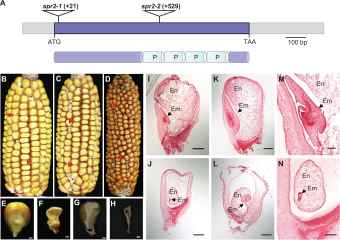
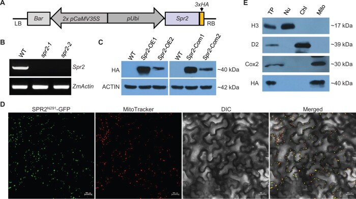
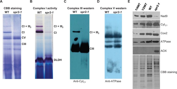
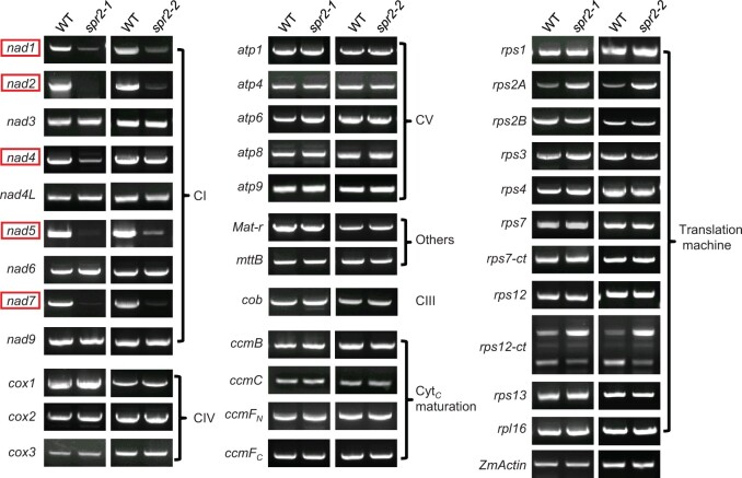
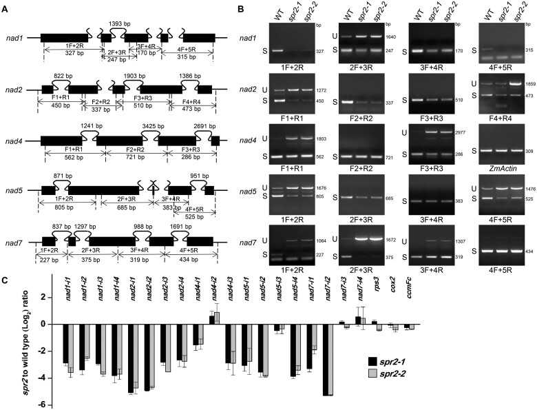
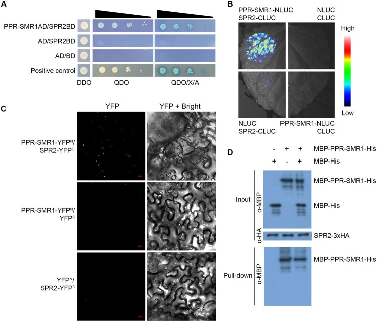
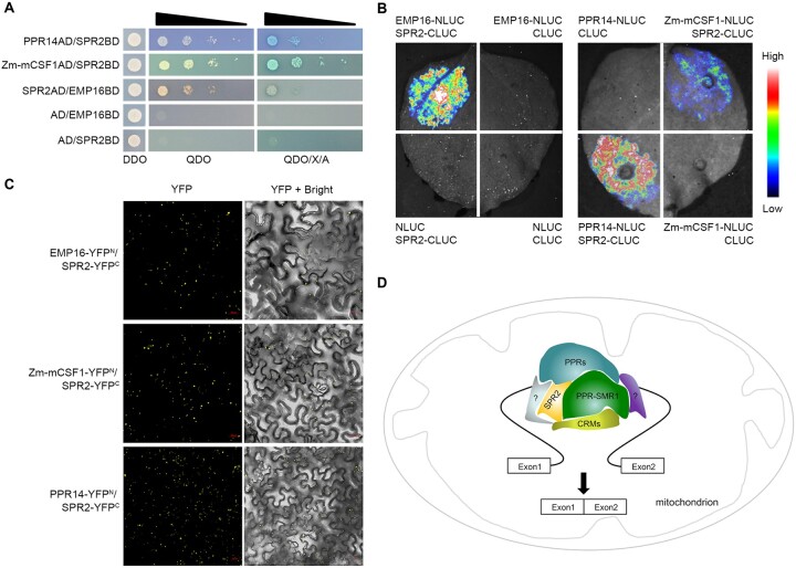
Splicing of plant mitochondrial introns is facilitated by numerous nucleus-encoded protein factors. Although some splicing factors have been identified in plants, the mechanism underlying mitochondrial intron splicing remains largely unclear. In this study, we identified a small P-type pentatricopeptide repeat (PPR) protein containing merely four PPR repeats, small PPR protein 2 (SPR2), which is required for the splicing of more than half of the introns in maize (Zea mays) mitochondria. Null mutations of Spr2 severely impair the splicing of 15 out of the 22 mitochondrial Group II introns, resulting in substantially decreased mature transcripts, which abolished the assembly and activity of mitochondrial complex I. Consequently, embryogenesis and endosperm development were arrested in the spr2 mutants. Yeast two-hybrid, luciferase complementation imaging, bimolecular fluorescence complementation, and semi-in vivo pull-down analyses indicated that SPR2 interacts with small MutS-related domain protein PPR-SMR1, both of which are required for the splicing of 13 introns. In addition, SPR2 and/or PPR-SMR1 interact with other splicing factors, including PPR proteins EMPTY PERICARP16, PPR14, and chloroplast RNA splicing and ribosome maturation (CRM) protein Zm-mCSF1, which participate in the splicing of specific intron(s) of the 13 introns. These results prompt us to propose that SPR2/PPR-SMR1 serves as the core component of a splicing complex and possibly exerts the splicing function through a dynamic interaction with specific substrate recognizing PPR proteins in mitochondria.


Figures

Figure 1
Figure 1
Mutation of Spr2 blocks embryogenesis and endosperm development. A, The top part represents the gene structure of Spr2, and the lower represents the protein structure of SPR2. The predicted SPR2 protein contains four P-type PPR motifs (P). Mu insertions in two alleles were indicated by triangles. B, A self-pollinated spr2-1/+ ear. Arrows point to spr2-1 mutant kernels. C, A self-pollinated spr2-2/+ ear. Arrows point to spr2-2 mutant kernels. D, Allelic test of spr2-1 and spr2-2 alleles. Arrows point to spr2 mutant kernels. E and F, Mature WT (E) and spr2-1 (F) kernels. G and H, Dissection of mature WT (G) and spr2-1 (H) kernels. I–N, Comparison of WT and spr2-1 kernel development at 10 DAP and 15 DAP. WT kernels at 10 DAP (I and M) and 15 DAP (K); spr2-1 kernels at 10 DAP (J and N) and 15 DAP (L). En, endosperm; Em, embryo. Scale bars, 1 mm in (E–H); 0.5 mm in (I–N).
Figure 2
Figure 2
Spr2 encodes a mitochondrion-localized PPR protein. A, Transgenic construct for overexpressing Spr2. LB: left border; RB: right border; 3 × HA: triple HA tag. B, Analysis of transcript levels of Spr2 in spr2 mutants by RT–PCR analysis using primer Spr2-F3 and Spr2-R3. RNAs were normalized against maize Actin gene (GRMZM2G126010). C, Analysis of protein abundance of SPR2 in overexpression lines (Spr2-OE) and complemented lines (Spr2-Com) by western blotting analysis. Maize ACTIN protein was used as a loading control. D, Subcellular localization of SPR2. Red fluorescence signals are from mitochondrial marker MitoTracker. Scale bar, 20 μm. E, Immunoblotting localization of SPR2 in total protein (TP), nuclear protein (Nu), mitochondrial protein (Mito), and chloroplast protein (Chl) extracts obtained from overexpression lines Spr2-OE1 probed with antibodies against H3 (Histone H3, nuclei marker), D2 (chloroplastic marker), Cox2 (mitochondrial marker), and HA.
Figure 3
Figure 3
The mutation of Spr2 abolished complex I assembly and activity. A, The BN-PAGE gels were stained with CBB. Crude mitochondrial extracts from endosperm and embryo of spr2-1 and WT immature kernels at 11 DAP. The positions of respiratory complexes I, III, V, and supercomplex I + III2 are indicated. CI: Complex I, CIII: Complex III, CV: Complex V. B, Detection of NADH dehydrogenase activity of complex I. Dihydrolipoamide dehydrogenase was used as a loading control. C and D, Accumulation of mitochondrial complex III and V in Spr2-1 mutant and WT by western blotting using specific antibodies against CytC1 (C) and ATPase (D). E, Immunodetection analysis with antibodies against Nad9, CytC1, Cox2, ATPase, and AOX.
Figure 4
Figure 4
Spr2 mutants affect expression of nad1, nad2, nad4, nad5, and nad7 in the mitochondria. RT–PCR analysis of transcript levels of 35 mitochondrion-encoded genes in WT and spr2 mutants. The substantial decrease of nad1, nad2, nad4, nad5, and nad7 transcripts occurs in spr2 alleles. Normalization was performed against ZmActin (GRMZM2G126010). The RNA was isolated from the same ear segregating for WT and spr2 mutants. CI: Complex I, CIII: Complex III, CIV: Complex IV, CV: Complex V.
Figure 5
Figure 5
Spr2 is involved in the splicing of multiple mitochondrial introns. A, Gene structure diagram of the maize nad1, nad2, nad4, nad5, and nad7 gene. Exons are shown as filled black boxes. The primers and expected amplification products are indicated. F: Forward primer, R: Reverse primer. B, RT–PCR analysis of inefficient splicing of mitochondrial introns of nad1, nad2, nad4, nad5, and nad7 in WT and spr2 mutants using primers as indicated in (A). Normalization was performed against ZmActin. S, intron spliced; U, intron unspliced; bp: base pair. C, RT-qPCR analysis of splicing efficiency of all 22 mitochondrial introns in spr2 alleles. The ratio of spliced to unspliced fragments was used to measure splicing efficiency. Data are means (±se) of three biological replicates.
Figure 6
Figure 6
SPR2 interacts with PPR-SMR1. A, Interaction analysis of SPR2 and PPR-SMR1 in Y2H assays. The combination of PPR-SMR1AD/SPR2BD was co-transfected into yeast strain Y2H Gold and spotted onto SD/–Leu/–Trp (DDO) medium and growth of diploid yeast colonies on SD/–Ade/–His/–Leu/–Trp (QDO) medium and QDO with added the X-α-gal and AbA (QDO/X/A) medium to reveal protein–protein interactions. Positive interaction was verified by growth on QDO and QDO/X/A plates. AD: GAL4 activation domain; BD: GAL4 DNA binding domain. B, LCI assays to determine interactions between SPR2 and PPR-SMR1. The combinations of PPR-SMR1–NLUC/SPR2–CLUC were co-infiltrated into 4-week-old N. benthamiana leaves. The luciferase signals were visualized using the Lumazone FA Pylon2048B system. The intensity of the fluorescent signals represents their interaction activities. C, BiFC analysis for interactions between SPR2 and PPR-SMR1. YFP is split into N-terminus (YFPN) and C-terminus (YFPC). The combination of PPR-SMR1–YFPN/SPR2–YFPC was co-infiltrated into 4-week-old N. benthamiana leaves using Agrobacterium. YFP signals were detected by a confocal laser microscope. Scale bars, 20 μm. D, Semi-in vivo pull-down assays for interactions between SPR2 and PPR-SMR1. Equal amounts of MBP-PPR-SMR1-His and MBP-His were combined with anti-HA magnetic beads preincubated with SPR2-HA. Pulled-down samples were analyzed by immunoblot with anti-MBP and anti-HA antibody. “+” and “−” indicate the presence and absence of corresponding proteins in the reactions, respectively. MBP: maltose binding protein; HA: human influenza hemagglutinin.
Figure 7
Figure 7
SPR2 interacts with multiple splicing factors. A, Interaction analysis of between SPR2 and EMP16, PPR14, and Zm-mCSF1 in Y2H assays. DDO medium: SD/–Leu/–Trp medium; QDO medium: SD/–Ade/–His/–Leu/–Trp medium; QDO/X/A: QDO medium with added the X-α-gal and AbA. AD: GAL4 activation domain; BD: GAL4 DNA binding domain. B, LCI assays to determine interactions between SPR2 and EMP16, PPR14, and Zm-mCSF1. The intensity of the fluorescent signals represents their interaction activities. C, BiFC analysis for interactions between SPR2 and EMP16, PPR14, and Zm-mCSF1 in N. benthamiana leaves. YFP signals were detected by a confocal laser microscope. Scale bars, 20 μm. D, A proposed working model of SPR2. Different colors represent various splicing factor. PPRs indicates other P-type PPR proteins, CRMs indicates CRM family proteins, and question mark indicates the other kind of mitochondrial splicing factors.

Similar articles

Cited by

References

    1. Barkan A, Small I (2014) Pentatricopeptide repeat proteins in plants. Annu Rev Plant Biol 65: 415–442 - PubMed
    1. Barkan A, Rojas M, Fujii S, Yap A, Chong YS, Bond CS, Small I (2012) A combinatorial amino acid code for RNA recognition by pentatricopeptide repeat proteins. PLoS Genet 8: e1002910. - PMC - PubMed
    1. Bonen L, Vogel J (2001) The ins and outs of group II introns. Trends Genet 17: 322–331 - PubMed
    1. Brown GG, Colas des Francs-Small C, Ostersetzer-Biran O (2014) Group II intron splicing factors in plant mitochondria. Front Plant Sci 5: 35. - PMC - PubMed
    1. Cai M, Li S, Sun F, Sun Q, Zhao H, Ren X, Zhao Y, Tan BC, Zhang Z, Qiu F (2017) Emp10 encodes a mitochondrial PPR protein that affects the cis-splicing of nad2 intron 1 and seed development in maize. Plant J 91: 132–144 - PubMed

Publication types

MeSH terms

LinkOut - more resources