. 2023 Mar;478(3):581-595.
doi: 10.1007/s11010-022-04536-8. Epub 2022 Aug 17.

Circadian disruption alters gut barrier integrity via a ß-catenin-MMP-related pathway

Affiliations

Circadian disruption alters gut barrier integrity via a ß-catenin-MMP-related pathway

Sung Yong Eum et al. Mol Cell Biochem. 2023 Mar.

Abstract

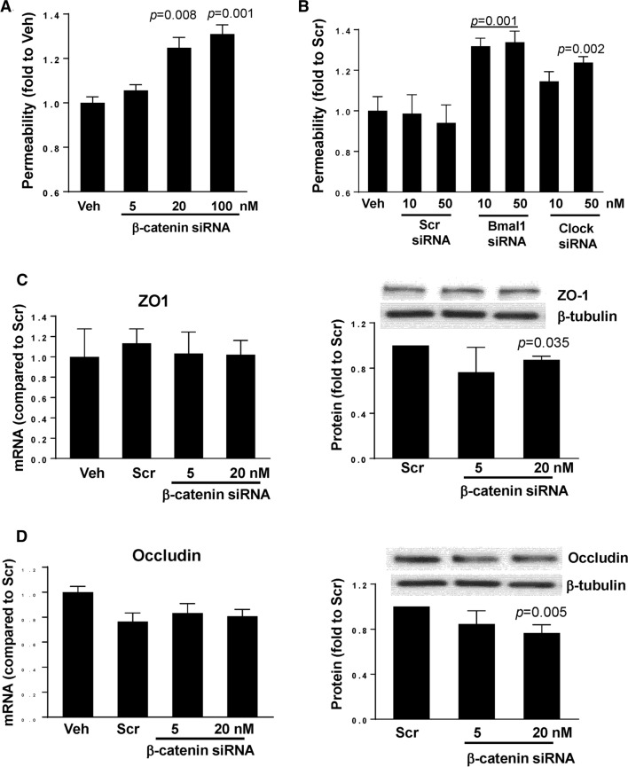
We evaluated the mechanistic link between circadian rhythms and gut barrier permeability. Mice were subjected to either constant 24-h light (LL) or 12-h light/dark cycles (LD). Mice housed in LL experienced a significant increase in gut barrier permeability that was associated with dysregulated ß-catenin expression and altered expression of tight junction (TJ) proteins. Silencing of ß-catenin resulted in disruption of barrier function in SW480 cells, with ß-catenin appearing to be an upstream regulator of the core circadian components, such as Bmal1, Clock, and Per1/2. In addition, ß-catenin silencing downregulated ZO-1 and occludin TJ proteins with only limited or no changes at their mRNA levels, suggesting post transcriptional regulation. Indeed, silencing of ß-catenin significantly upregulated expression of matrix metallopeptidase (MMP)-2 and MMP-9, and blocking MMP-2/9 activity attenuated epithelial disruption induced by ß-catenin silencing. These results indicate the regulatory role of circadian disruption on gut barrier integrity and the associations between TJ proteins and circadian rhythms, while demonstrating the regulatory role of ß-catenin in this process.

Keywords: Circadian clock genes; Circadian rhythm disruption; Circadian rhythm molecules; Intestinal barrier integrity; Tight junction proteins; ß-Catenin.


Conflict of interest statement

The authors report no competing interests.

Figures

Fig. 1
Fig. 1
Four weeks of constant light disrupts circadian rhythms in mice. A Summary actograms at baseline (before light manipulation) and two weeks after 12-h light/dark (LD) or circadian disruption (constant light, LL). Shaded areas, 18:00 (ZT12) to 6:00 (ZT0), indicate time of lights-off, or former lights-off for LL 2nd week. Numbers along the x-axis indicate hours of the day. Black areas are wheel revolutions binned each minute and reported here as the average for n = 19 mice per group. B Arrhythmicity of exercised mice maintained in LL as demonstrated by phase angle assessment. Phase angle is the difference in hours between the time of lights-off (or former lights-off for LL 2nd week), which was 18:00 (ZT12), and the time of activity onset; corresponding to uninterrupted activity on the wheel. In the LL group at the 2nd week, activity was significantly shifted ~ 30 min before lights-off. C Body temperature rhythm measured every 4 h over 24 h in LD and LL mice. D An increase in body mass in mice maintained in LL as compared to LD. Values in the LL group that are statistically different from those in the LD group at the corresponding time points at *p < 0.05, **p < 0.01, and **p < 0.001. Values are mean ± SEM
Fig. 2
Fig. 2
Expression of circadian clock molecules in the gut of the circadian rhythm disrupted mice. The mRNA and protein expression of circadian clock molecules, β-catenin, Bmal1, and Clock were measured in the intestinal epithelial cell-enriched fractions of the colon of mice exposed to 12-h light/dark cycling (LD) or constant 24-h light (LL) for 4 weeks. A mRNA expression of β-catenin, Bmal1 and Clock was assessed using RT-PCR. B, C Protein expression levels from the same mice as (A). Protein expression level of β-catenin, Bmal1 and Clock was measured by immunoblotting (B) and band intensity was quantified by densitometric analysis using Image J program (C). p indicates the level of statistical significance in the LL group compared to the LD group for individual genes and proteins. Values are mean ± SEM; n = 4–5
Fig. 3
Fig. 3
Circadian rhythm disruption alters ileum and colon permeability. Mice were treated as in Figs. 1 and 2. A Left panel, representative images of ex vivo permeability to FITC-dextran 4 kDa through the ileum segment in mice maintained under 12-h light/dark (LD) or constant light (LL) for 4 weeks. B Ex vivo permeability in the colon in the same mice as in A. Left panels, gut sections stained for nuclei (blue) and FITC (red). Right panels, quantitative data from ex vivo permeability measurements. p indicates the level of statistical significance in the LL group compared to the LD group. Values are mean ± SEM; n = 4–9
Fig. 4
Fig. 4
Alterations of tight junction protein expression in the colon of mice subjected to circadian rhythm disruption. The expression of tight junction molecules was measured in the intestinal epithelial cell-enriched fractions of the colon of mice exposed to 12-h light/dark cycling (LD) or constant 24-h light (LL) for 4 weeks. A mRNA expression of ZO-1, occludin, and tricellulin as analyzed by real-time PCR. BD Protein expression levels from the same mice as (A). Protein expression of ZO-1, occludin and tricellulin was assessed by immunoblotting (B) and band intensity was quantified by densitometric analysis using Image J program (C). p indicates the level of statistical significance in the LL group compared to the LD group for individual genes and proteins. Values are mean ± SEM; n = 4–5
Fig. 5
Fig. 5
Silencing of β-catenin alters mRNA and protein expression of circadian clock regulators. SW480 cells grown on the 12 well cell culture plates were transfected with β-catenin-specific siRNA at the indicated concentration or with nonspecific, scrambled siRNA (Scr); Veh, vehicle. mRNA (left panels) and protein (right panels) expression of β-catenin (A), Bmal1 (B), Clock (C), Per1 (D), Per2 (E), Cry1 (F), and (Cry2) (G) were assayed and quantified. Blots illustrate representative data. Band intensity was assessed by densitometric analysis using Image J program. p indicates the level of statistical significance after β-catenin silencing as compared to the Scr group. Values (means ± SD) are expressed as fold change compared with Scr; n = 3–6
Fig. 6
Fig. 6
Silencing of circadian clock molecules increases paracellular permeability across epithelial monolayers. SW480 cells grown on the apical side of wells (0.4-μm pore size, 12-mm diameter) of Transwell inserts were transfected with siRNA of β-catenin (A), Bmal1, or Clock (B) at the indicated concentrations. Control cultures were either treated with vehicle (Veh) or transfected with nonspecific, scrambled (Scr) siRNA. Epithelial permeability was determined by measuring paracellular passage of FITC-dextran 20 kDa from the apical side to the basolateral side across SW480 cell layers. A p indicates the level of statistical significance after β-catenin silencing as compared to the Veh group. B p indicates the level of statistical significance after Bmal1 or Clock silencing as compared to the Veh or Scr group. Values (means ± SD) are expressed as fold change compared with Veh or Scr. Values are mean ± SEM; n = 5–6. C, D Silencing of β-catenin alters expression of tight junction proteins without affecting mRNA levels. SW480 cells grown on the 12 well cell culture plates were transfected with β-catenin-specific siRNA at the indicated concentration or with nonspecific, scrambled siRNA. mRNA (left panels) or protein (right panels) expression of ZO-1 (C) and occludin (D) were assayed and quantified. Blots illustrate representative data. Band intensity was assessed by densitometric analysis using Image J program. p indicates the level of statistical significance after β-catenin silencing as compared to the Scr group. Values (means ± SD) are expressed as fold change compared with Scr; n = 3–6
Fig. 7
Fig. 7
β-catenin-induced alterations of paracellular permeability is mediated by MMPs. SW480 cells were transfected with β-catenin-specific siRNA at the indicated concentrations or with nonspecific, scrambled siRNA, followed by mRNA assessment by RT-PCR of MMP-2 (A) and MMP-9 (B). Values (means ± SD) are expressed as fold change compared with Scr; n = 5–6. A, B p indicates the level of statistical significance after β-catenin silencing as compared to the Scr group. C SW480 cells grown on the apical side of wells (0.4-μm pore size, 12-mm diameter) of Transwell system were transfected with β-catenin siRNA or scrambled siRNA (both at 20 nM). Additional cultures were treated with pharmacological inhibitors of MMPs, namely, MMP-2/MMP-9 Inhibitor I, MMP-9 Inhibitor II, or MMP-2 inhibitor III all at 20 µM. Epithelial permeability was determined by measuring paracellular passage of FITC-dextran 20 from the apical side to the basolateral side across SW480 cell layers. p indicates the level of statistical significance between the β-catenin silencing group as compared to the Scr group or between the β-catenin silencing plus MMP2/9 inhibitor group as compared to the β-catenin silencing group. Values (means ± SD) are expressed as fold change compared to Scr; n = 5
Fig. 8
Fig. 8
Schematic diagram of the major findings of the present manuscript. ß-catenin appears to be a master regulator of epithelial barrier integrity in circadian rhythm disruption by altering TJ protein expression

References

    1. Asher G, Sassone-Corsi P. Time for food: the intimate interplay between nutrition, metabolism, and the circadian clock. Cell. 2015;161:84–92. doi: 10.1016/j.cell.2015.03.015. - DOI - PubMed
    1. Huang Y-J, Pai Y-C, Yu LC-H. Host-microbiota interaction and intestinal epithelial functions under circadian control: implications in colitis and metabolic disorders. Chin J Physiol. 2018;61:325–340. doi: 10.4077/CJP.2018.BAH641. - DOI - PubMed
    1. Konturek PC, Brzozowski T, Konturek SJ. Gut clock: Implication of circadian rhythms in the gastointestinal tract. J Physiol Pharmacol. 2011;62:39–50. - PubMed
    1. Bunger MK, Wilsbacher LD, Moran SM, et al. Mop3 is an essential component of the master circadian pacemaker in mammals. Cell. 2000;103:1009–1017. doi: 10.1016/S0092-8674(00)00205-1. - DOI - PMC - PubMed
    1. Mohawk JA, Green CB, Takahashi JS. Central and peripheral circadian clocks in mammals. Annu Rev Neurosci. 2012;35:445–462. doi: 10.1146/annurev-neuro-060909-153128. - DOI - PMC - PubMed