. 2022 Aug 17;14(658):eabj2681.
doi: 10.1126/scitranslmed.abj2681. Epub 2022 Aug 17.

Sphingosine 1-phosphate signaling in perivascular cells enhances inflammation and fibrosis in the kidney

Affiliations

Sphingosine 1-phosphate signaling in perivascular cells enhances inflammation and fibrosis in the kidney

Shinji Tanaka et al. Sci Transl Med. .

Abstract

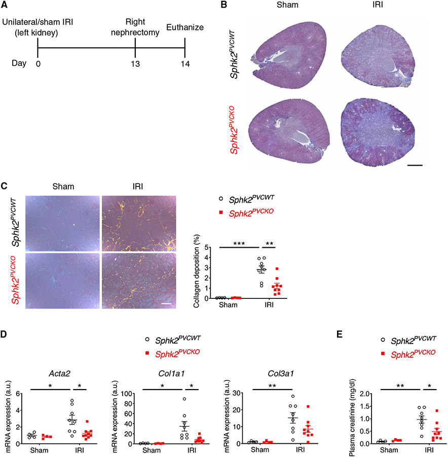
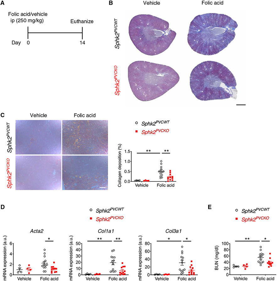
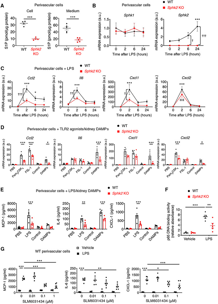
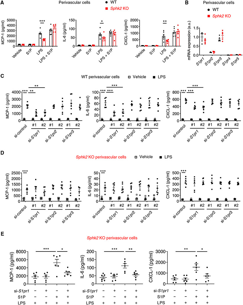
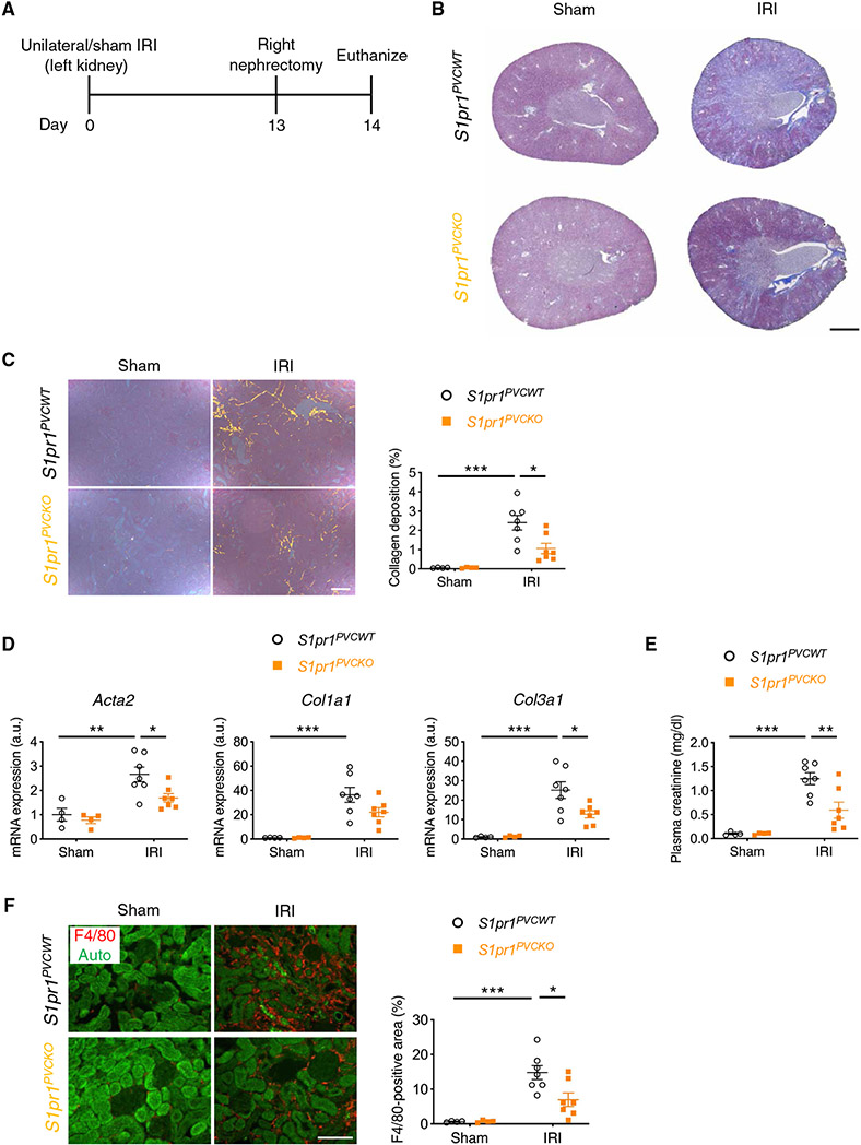
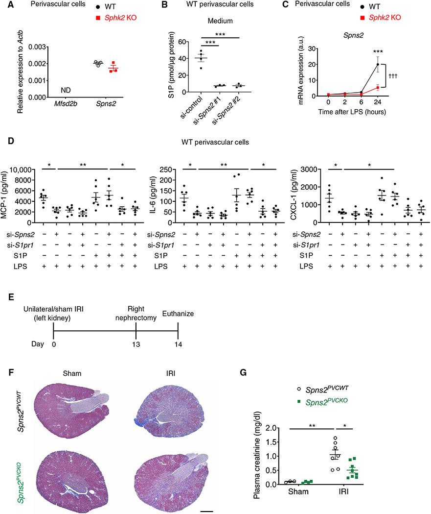
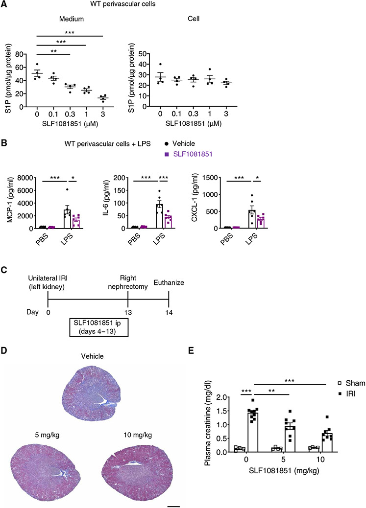
Chronic kidney disease (CKD), characterized by sustained inflammation and progressive fibrosis, is highly prevalent and can eventually progress to end-stage kidney disease. However, current treatments to slow CKD progression are limited. Sphingosine 1-phosphate (S1P), a product of sphingolipid catabolism, is a pleiotropic mediator involved in many cellular functions, and drugs targeting S1P signaling have previously been studied particularly for autoimmune diseases. The primary mechanism of most of these drugs is functional antagonism of S1P receptor-1 (S1P1) expressed on lymphocytes and the resultant immunosuppressive effect. Here, we documented the role of local S1P signaling in perivascular cells in the progression of kidney fibrosis using primary kidney perivascular cells and several conditional mouse models. S1P was predominantly produced by sphingosine kinase 2 in kidney perivascular cells and exported via spinster homolog 2 (Spns2). It bound to S1P1 expressed in perivascular cells to enhance production of proinflammatory cytokines/chemokines upon injury, leading to immune cell infiltration and subsequent fibrosis. A small-molecule Spns2 inhibitor blocked S1P transport, resulting in suppression of inflammatory signaling in human and mouse kidney perivascular cells in vitro and amelioration of kidney fibrosis in mice. Our study provides insight into the regulation of inflammation and fibrosis by S1P and demonstrates the potential of Spns2 inhibition as a treatment for CKD and potentially other inflammatory and fibrotic diseases that avoids the adverse events associated with systemic modulation of S1P receptors.


Figures

Fig. 1.
Fig. 1.. Sphk2 deletion in renal perivascular cells ameliorates kidney fibrosis in the unilateral IRI mouse model.
(A) Protocol for unilateral IRI to induce kidney fibrosis (for B to E). Sphk2PVCWT and Sphk2PVCKO mice underwent left renal pedicle clamp for 30 min (day 0) and right nephrectomy at day 13 and were euthanized at day 14. (B) Representative Masson’s trichrome staining of collagen in kidney sections at day 14. (C) Picrosirius red staining of kidney (polarized microscopy, right) with quantification of red/yellow birefringence of mature collagen fibers as a percentage of the total surface area of kidney section at day 14 (left). n = 4 to 9 per group. (D) Acta2, Col1a1, and Col3a1 transcript expression (from whole kidney) at day 14. n = 4 to 9 per group. a.u., arbitrary units. (E) Plasma creatinine concentrations at day 14. n = 4 to 9 per group. Scale bars, 1 mm (B) and 200 μm (C). Data are represented as means ± SEM. *P < 0.05, **P < 0.01, and ***P < 0.001; two-way ANOVA followed by post hoc multiple comparisons test (Tukey’s). a.u., arbitrary units.
Fig. 2.
Fig. 2.. Sphk2 deletion in renal perivascular cells ameliorates kidney fibrosis in the folic acid mouse model.
(A) Protocol for inducing kidney fibrosis by injecting folic acid (for B to E). Sphk2PVCWT and Sphk2PVCKO mice were given folic acid (250 mg/kg, ip) and euthanized at day 14. (B) Representative Masson’s trichrome staining of collagen in kidney sections at day 14. (C) Picrosirius red staining of kidney (polarized microscopy) with quantification of red/yellow birefringence of mature collagen fibers as a percentage of the total surface area of kidney section at day 14. n = 4 to 13 per group. (D) Acta2, Col1a1, and Col3a1 transcript expression (from whole kidney) at day 14. n = 4 to 13 per group. (E) Blood urea nitrogen (BUN) concentrations at day 14. n = 4 to 13 per group. Scale bars, 1 mm (B) and 200 μm (C). Data are represented as means ± SEM. *P < 0.05 and **P < 0.01; two-way ANOVA followed by post hoc multiple comparisons test (Tukey’s).
Fig. 3.
Fig. 3.. SphK2 inhibition suppresses inflammatory signaling in mouse perivascular cells.
(A to G) Experiments were performed using primary kidney perivascular cells isolated from Sphk2PVCWT (WT) and Sphk2PVCKO [Sphk2 knockout (KO)] mice. (A) S1P concentrations in perivascular cells and in media. (B and C) Time course of transcript expression of Sphk1/Sphk2 (B) and proinflammatory cytokines and chemokines (Ccl2, Il6, Cxcl1, and Cxcl2) after LPS treatment. (D) Transcript expression of Ccl2, Il6, Cxcl1, and Cxcl2 at 2 hours after treatment with TLR2 agonists (Pam3CSK4 and FSL-1) or kidney DAMPs. (E) MCP-1, IL-6, and CXCL-1 concentrations in supernatants of kidney perivascular cells treated with LPS or kidney DAMPs for 24 hours. (F) NF-κB activity at 1 hour after treatment with LPS. (G) Dose response of a selective SphK2 inhibitor (SLM6031434) on MCP-1, IL-6, and CXCL-1 concentrations in supernatants of wild-type kidney perivascular cells at 24 hours after LPS treatment. n = 4 to 5 (A), n = 5 (B to F), or n = 6 (G) per group. Data are represented as means ± SEM. *P < 0.05, **P < 0.01, and ***P < 0.001 by unpaired two-sided Student’s t test (A) or two-way ANOVA followed by post hoc multiple comparisons test (Tukey’s) (D to G). *P < 0.05 and ***P < 0.001 (versus WT at 0 hours); †P < 0.05, ††P < 0.01, and †††P < 0.001 (WT versus Sphk2 KO at the same time point); ‡‡‡P < 0.001 (versus Sphk2 KO at 0 hours) by two-way ANOVA followed by post hoc multiple comparisons test (Tukey’s) (B and C).
Fig. 4.
Fig. 4.. S1P produced by SphK2 is exported to the extracellular space and binds to S1P1 to enhance inflammatory signaling in perivascular cells.
(A to E) Experiments were performed using primary kidney perivascular cells isolated from Sphk2PVCWT [wild-type (WT)] and Sphk2PVCKO (Sphk2 KO) mice. (A) MCP-1, IL-6, and CXCL-1 concentrations in supernatants of primary kidney perivascular cells treated with LPS (100 ng/ml) for 24 hours with or without S1P supplementation (100 nM). (B) S1pr1-5 transcript expression at baseline. (C and D) MCP-1, IL-6, and CXCL-1 concentrations in supernatants of WT (C) and Sphk2 KO (D) kidney perivascular cells at 24 hours after treatment with LPS (100 ng/ml). Cells were treated with control, S1pr1, S1pr2, or S1pr3 siRNA before LPS stimulation. (E) MCP-1, IL-6, and CXCL-1 concentrations in supernatants of Sphk2 KO kidney perivascular cells treated with LPS (100 ng/ml) for 24 hours with or without S1P supplementation (100 nM). Cells were treated with control or S1pr1 siRNA before LPS stimulation. n = 6 (A, C, D, and E), n = 5 (B) per group. Data are represented as means ± SEM. *P < 0.05, **P < 0.01, and ***P < 0.001 by two-way (A to D) or one-way (E) ANOVA followed by post hoc multiple comparisons test (Tukey’s).
Fig. 5.
Fig. 5.. S1pr1 deletion in renal perivascular cells ameliorates kidney fibrosis in the unilateral IRI mouse model.
(A) Protocol for unilateral IRI to induce kidney fibrosis in mice (for B to F). S1pr1PVCKO (Foxd1-Cre;S1pr1fl/fl) mice, in which S1pr1 is deleted in kidney perivascular cells, and Cre-negative littermate control (S1pr1PVCWT) mice underwent left renal pedicle clamp for 30 min (day 0) and right nephrectomy at day 13 and were euthanized at day 14. (B) Representative Masson’s trichrome staining of collagen in kidney sections at day 14. (C) Picrosirius red staining of kidney (polarized microscopy, left) with quantification of red/yellow birefringence of mature collagen fibers as a percentage of the total surface area of kidney section at day 14 (right). (D) Acta2, Col1a1, and Col3a1 transcript expression (from whole kidney) at day 14. (E) Plasma creatinine concentrations at day 14. (F) Macrophage staining (F4/80, red, left) of kidney with quantification of F4/80-positive area as a percentage of the total surface area of kidney section at day 14 (right). Auto, autofluorescence (green). Scale bars, 1 mm (B), 200 μm (C), and 100 μm (F). n = 4 to 7 per group. Data are represented as means ± SEM. *P < 0.05, **P < 0.01, and ***P<0.001; two-way ANOVA followed by post hoc multiple comparisons test (Tukey’s).
Fig. 6.
Fig. 6.. Spns2 is an S1P transporter expressed in perivascular cells and enhances inflammatory signaling and fibrosis on injury.
(A) Mfsd2b and Spns2 transcript expression at baseline in primary kidney perivascular cells isolated from Sphk2PVCWT (WT) and Sphk2PVCKO (Sphk2 KO) mice. ND, not detected. (B) S1P concentrations in the supernatant of wild-type kidney perivascular cells treated with control or Spns2 siRNA. (C) Spns2 transcript expression over time after LPS treatment in primary kidney perivascular cells isolated from Sphk2PVCWT(WT) and Sphk2PVCKO (Sphk2 KO) mice. (D) MCP-1, IL-6, and CXCL-1 concentrations in supernatants of wild-type kidney perivascular cells treated with LPS (100 ng/ml) for 24 hours with or without S1P supplementation (100 nM). Spns2 and/or S1pr1 were knocked down before LPS stimulation. (E) Protocol for unilateral IRI to induce kidney fibrosis (for F and G). Spns2PVCKO (Foxd1-Cre;Spns2fl/fl) mice, in which Spns2 is deleted in kidney perivascular cells, and Cre-negative littermate control (Spns2PVCWT) mice underwent left renal pedicle clamp for 34 min (day 0), right nephrectomy at day 13, and were euthanized at day 14. (F) Representative Masson’s trichrome staining of collagen in kidney sections at day 14. (G) Plasma creatinine concentrations at day 14. Scale bar, 1 mm. n = 3 (A), n = 3 to 4 (B), n = 5 (C), n = 6 (D), or n = 3 to 8 (E to G) per group. Data are represented as means ± SEM. *P < 0.05, **P < 0.01, and ***P < 0.001 by one-way ANOVA followed by post hoc multiple comparisons test (Tukey’s) (B and D). ***P < 0.001 (versus WT at 0 hour); †††P < 0.001 (WT versus Sphk2 KO) by two-way ANOVA followed by post hoc multiple comparisons test (Tukey’s) (C). *P < 0.05 and **P < 0.01 by two-way ANOVA followed by post hoc multiple comparisons test (Tukey’s) (G).
Fig. 7.
Fig. 7.. Pharmacological inhibition of Spns2 suppresses S1P transport and inflammatory signaling in perivascular cells and ameliorates kidney fibrosis.
(A) Dose response of a selective Spns2 inhibitor (SLF1081851) on S1P concentrations in wild-type kidney perivascular cells and media. (B) MCP-1, IL-6, and CXCL-1 concentrations in supernatants of wild-type kidney perivascular cells treated with LPS (100 ng/ml) for 24 hours. Cells were treated with 3 μM SLF1081851 or vehicle (0.1% fatty acid–free BSA) before stimulation. (C) Timeline of experiments (D and E) to investigate the effect of SLF1081851 on kidney fibrosis in wild-type mice. Mice underwent unilateral IRI (day 0) and were administered SLF1081851 (5 or 10 mg/kg, ip) or vehicle (5% hydroxypropyl-β-cyclodextrin) once daily from day 4 to day 13, followed by right nephrectomy at day 13 and euthanasia at day 14. (D) Representative Masson’s trichrome staining of collagen in kidney sections at day 14. (E) Plasma creatinine concentrations at day 14. Scale bar, 1 mm. n = 4 (A), n = 6 (B), or n = 4 to 9 per group (C to E). Data are represented as means ± SEM. *P < 0.05, **P < 0.01, and ***P < 0.001 by one-way (A) or two-way (B and E) ANOVA followed by post hoc multiple comparisons test (Tukey’s).

Comment in

References

    1. Webster AC, Nagler EV, Morton RL, Masson P, Chronic kidney disease. Lancet 389, 1238–1252 (2017). - PubMed
    1. Eckardt KU, Coresh J, Devuyst O, Johnson RJ, Kottgen A, Levey AS, Levin A, Evolving importance of kidney disease: From subspecialty to global health burden. Lancet 382, 158–169 (2013). - PubMed
    1. Keith DS, Nichols GA, Gullion CM, Brown JB, Smith DH, Longitudinal follow-up and outcomes among a population with chronic kidney disease in a large managed care organization. Arch. Intern. Med 164, 659–663 (2004). - PubMed
    1. Go AS, Chertow GM, Fan D, McCulloch CE, Hsu CY, Chronic kidney disease and the risks of death, cardiovascular events, and hospitalization. N. Engl. J. Med 351, 1296–1305 (2004). - PubMed
    1. National Kidney F, KDOQI clinical practice guideline for diabetes and CKD: 2012 update. Am. J. Kidney Dis. 60, 850–886 (2012). - PubMed

Publication types