. 2022 Oct 13;140(15):1723-1734.
doi: 10.1182/blood.2022015883.

The use of pluripotent stem cells to generate diagnostic tools for transfusion medicine

Affiliations

The use of pluripotent stem cells to generate diagnostic tools for transfusion medicine

Hyun Hyung An et al. Blood. .

Abstract

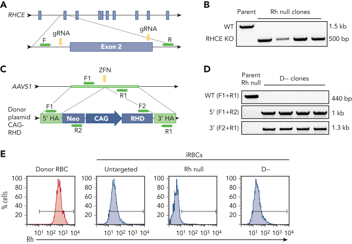
Red blood cell (RBC) transfusion is one of the most common medical treatments, with more than 10 million units transfused per year in the United States alone. Alloimmunization to foreign Rh proteins (RhD and RhCE) on donor RBCs remains a challenge for transfusion effectiveness and safety. Alloantibody production disproportionately affects patients with sickle cell disease who frequently receive blood transfusions and exhibit high genetic diversity in the Rh blood group system. With hundreds of RH variants now known, precise identification of Rh antibody targets is hampered by the lack of appropriate reagent RBCs with uncommon Rh antigen phenotypes. Using a combination of human-induced pluripotent stem cell (iPSC) reprogramming and gene editing, we designed a renewable source of cells with unique Rh profiles to facilitate the identification of complex Rh antibodies. We engineered a very rare Rh null iPSC line lacking both RHD and RHCE. By targeting the AAVS1 safe harbor locus in this Rh null background, any combination of RHD or RHCE complementary DNAs could be reintroduced to generate RBCs that express specific Rh antigens such as RhD alone (designated D--), Goa+, or DAK+. The RBCs derived from these iPSCs (iRBCs) are compatible with standard laboratory assays used worldwide and can determine the precise specificity of Rh antibodies in patient plasma. Rh-engineered iRBCs can provide a readily accessible diagnostic tool and guide future efforts to produce an alternative source of rare RBCs for alloimmunized patients.


Conflict of interest statement

Conflict-of-interest disclosure: US Patent Application No. 16/757,815, entitled “Engineered Red Blood Cells Having Rare Antigen Phenotypes” by S.T.C. and C.M.W. in the name of The Children’s Hospital of Philadelphia and New York Blood Center, Inc (national stage entry of International Application No. PCT/US2018/057932); manuscript describes engineered red blood cells covered in patent application. The remaining authors declare no competing financial interests.

Figures

Image 1
Graphical abstract
Figure 1
Figure 1.
Generation of Rh null and D-- iPSCs. (A) Schematic for CRISPR-Cas9–mediated disruption of RHCE in RhD-negative iPSC to generate Rh null iPSCs. (B) PCR products amplified with primers targeting intron 1 and intron 2 identifies iPSC clones with Cas9-mediated large deletion of ∼1 kb. (C) Schematic for ZFN-mediated insertion of RHD cDNA into the safe harbor AAVS1 locus of Rh null iPSCs to generate D-- iPSCs. (D) PCR products amplified using primers indicated in panel C to identify clones with successful integration of the RHD cDNA cassette. (E) Cell surface Rh protein visualized by flow cytometry using a pan-Rh antibody in control donor RBCs (red), and untargeted parent, Rh null, and D-- iRBCs (blue). CAG, CAG promoter; F, forward primer; gRNA, guide RNA; HA, homology arm; KO, knockout; Neo, neomycin resistance cassette; R, reverse primer.
Figure 2
Figure 2.
Generation of iRBCs. (A) Schematic of 12-day erythroid liquid culture of iPSC-derived HPCs into mature iRBCs. (B) Representative flow cytometric analysis of common cell surface erythroid maturation markers on day 12 of ce (rr) iRBC culture compared with control dRBCs. (C) Morphology of control dRBCs and day 12 ce iRBCs (May-Grunwald Giemsa stain). (D) Size distribution of day 12 ce iRBCs compared with control dRBCs measured by Countess Automated Cell Counter (n = 205 and 1435, respectively, Welch’s t test). (E) Fold expansion of iRBC per fresh HPC in erythroid-specific culture on days 3, 6, 9, and 12 (n = 22 independent assays). Ery, erythroid; IMDM, Iscove’s modified Dulbecco medium; pen strep, penicillin-streptomycin; Epo, erythropoietin, SCF, stem cell factor.
Figure 3
Figure 3.
Detection of Rh antigen expression on iRBCs by gel column agglutination. (A) Rh antigen typing of DCe/DCe (R1R1), DcE/cE (R2R2), and ce/ce (rr) esRBCs or iRBCs using commercial monoclonal RBC typing reagents for the 5 common Rh antigens (anti-D, anti-C, anti-c, anti-E, and anti-e indicated above) on buffered gel cards. Agglutination prevents cells from traveling through the gel matrix to the bottom of each column upon centrifugation and indicates cell surface expression of the corresponding antigen. (B-D) (B) Rh typing for common Rh antigens of Rh null, D--, D+ Goa+, and D+ DAK+ iRBCs, and (C) D+c+e+, VVS+ iRBCs, and (D) C+c+e+, hrB− and D+c+e+, hrS− ficin-treated iRBCs.
Figure 4
Figure 4.
Detection of Rh antibodies in patient plasma using iRBCs and gel column agglutination. (A-E) Plasma containing (A) anti-D, (B) anti-C, (C) anti-c, (D) anti-E, and (E) anti-e were tested against a panel of control dRBCs and ficin-treated iRBCs or esRBCs. Each assay included control dRBCs expressing or lacking the corresponding Rh antigen (left) along with Rh null and D-- iRBCs, and iRBCs/esRBCs negative or positive for the corresponding Rh antigen (right). Agglutination prevents cells from traveling through the gel matrix to the bottom of each column upon centrifugation and indicates the presence of antibody in the plasma sample.
Figure 5
Figure 5.
iRBCs identify antibodies against high-prevalence or low-prevalence Rh antigens in patient plasma. (A-B) Patient plasma containing antibody against (A) the high-prevalence antigen hrS or (B) low-prevalence antigens V or Goa were tested against a panel of control dRBCs and ficin-treated iRBCs. Each assay included the 3 control dRBCs routinely used for antibody screening: DCe (R1R1), DcE (R2R2), and ce (rr) phenotypes (left). In panel A, iRBCs that were Rh null, D--, e+ hrS− hrB+, and e+ hrS+ hrB− were selected to show hrS specificity (right). In panel B, patient plasma was tested against iRBCs that were Rh null, D--, or expressing low-prevalence Goa, DAK, or VVS antigens. Agglutination of cells prevents them from traveling through the gel matrix to the bottom of each column upon centrifugation and indicates the presence of antibody in the plasma sample.
Figure 6
Figure 6.
Rh null iRBCs have comparable O2 binding capacity and deformability compared with WT iRBCs. (A) Representative and (B) quantification of alpha-like and beta-like globin chain analysis of day 12 iRBCs by HPLC (n = 5 independent assays). (C) Representative and (D) quantification of hemoglobin tetramer analysis of day 12 iRBCs by HPLC. (n = 5). (E) O2 dissociation curve of adult dRBCs, PB-RBCs, WT and Rh null iRBCs. (F) The partial pressure of O2 at 50% oxyhemoglobin (p50) interpolated from Sigmoidal 4PL curves of each type of RBC (n = 3 each, one-way ANOVA), ∗P < .05; ∗∗P < .01. (G) Average elongation indices of dRBCs, PB-RBCs, and WT or Rh null iRBCs determined by Lineweaver-Burk fitting for ektacytometry (n = 6 dRBCs, n = 3 for all others). (H) Elongation indices at 5 Pa, 10 Pa, and maximum elongation (n = 6 dRBCs, n = 3 for all others, two-way ANOVA), ∗∗∗∗P < .0001. (I) Representative images of fully “stretched” dRBCs, pbRBCs, and iRBCs obtained on the RheoScan AnD system. ANOVA, analysis of variance; HbF, fetal hemoglobin; O2, oxygen; Pa, Pascal; PB-RBCs, peripheral blood CD34+ cultured RBCs; WT, wild type.

Comment in

References

    1. Nickel RS, Hendrickson JE, Fasano RM, et al. Impact of red blood cell alloimmunization on sickle cell disease mortality: a case series. Transfusion. 2016;56(1):107–114. - PubMed
    1. Linder GE, Chou ST. Red blood cell transfusion and alloimmunization in sickle cell disease. Haematologica. 2021;106(7):1805–1815. - PMC - PubMed
    1. Chou ST, Alsawas M, Fasano RM, et al. American Society of Hematology 2020 guidelines for sickle cell disease: transfusion support. Blood Advances. 2020;4(2):327–355. - PMC - PubMed
    1. Noizat-Pirenne F, Tournamille C. Relevance of RH variants in transfusion of sickle cell patients. Transfus Clin Biol. 2011;18(5):527–535. - PubMed
    1. Chou ST, Jackson T, Vege S, Smith-Whitley K, Friedman DF, Westhoff CM. High prevalence of red blood alloimmunization in sickle cell disease despite transfusion from Rh-matched minority donors. Blood. 2013;122(6):1062–1071. - PubMed

Publication types

Substances