

Suppressor mutations that make the essential transcription factor Spn1/Iws1 dispensable in Saccharomyces cerevisiae
- PMID: 35977387
- PMCID: PMC9526074
- DOI: 10.1093/genetics/iyac125
Suppressor mutations that make the essential transcription factor Spn1/Iws1 dispensable in Saccharomyces cerevisiae
Abstract
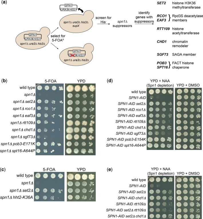
Spn1/Iws1 is an essential eukaryotic transcription elongation factor that is conserved from yeast to humans as an integral member of the RNA polymerase II elongation complex. Several studies have shown that Spn1 functions as a histone chaperone to control transcription, RNA splicing, genome stability, and histone modifications. However, the precise role of Spn1 is not understood, and there is little understanding of why it is essential for viability. To address these issues, we have isolated 8 suppressor mutations that bypass the essential requirement for Spn1 in Saccharomyces cerevisiae. Unexpectedly, the suppressors identify several functionally distinct complexes and activities, including the histone chaperone FACT, the histone methyltransferase Set2, the Rpd3S histone deacetylase complex, the histone acetyltransferase Rtt109, the nucleosome remodeler Chd1, and a member of the SAGA coactivator complex, Sgf73. The identification of these distinct groups suggests that there are multiple ways in which Spn1 bypass can occur, including changes in histone acetylation and alterations in other histone chaperones. Thus, Spn1 may function to overcome repressive chromatin by multiple mechanisms during transcription. Our results suggest that bypassing a subset of these functions allows viability in the absence of Spn1.
Keywords: Iws1; Spn1; chromatin; histone chaperone; transcription.
© The Author(s) 2022. Published by Oxford University Press on behalf of Genetics Society of America. All rights reserved. For permissions, please email: journals.permissions@oup.com.
Figures
Similar articles
-
Insights into Spt6: a histone chaperone that functions in transcription, DNA replication, and genome stability.Trends Genet. 2023 Nov;39(11):858-872. doi: 10.1016/j.tig.2023.06.008. Epub 2023 Jul 20. Trends Genet. 2023. PMID: 37481442 Free PMC article. Review.
-
The conserved elongation factor Spn1 is required for normal transcription, histone modifications, and splicing in Saccharomyces cerevisiae.Nucleic Acids Res. 2020 Oct 9;48(18):10241-10258. doi: 10.1093/nar/gkaa745. Nucleic Acids Res. 2020. PMID: 32941642 Free PMC article.
-
Essential histone chaperones collaborate to regulate transcription and chromatin integrity.Genes Dev. 2021 May 1;35(9-10):698-712. doi: 10.1101/gad.348431.121. Epub 2021 Apr 22. Genes Dev. 2021. PMID: 33888559 Free PMC article.
-
Combinatorial Genetic Control of Rpd3S Through Histone H3K4 and H3K36 Methylation in Budding Yeast.G3 (Bethesda). 2018 Nov 6;8(11):3411-3420. doi: 10.1534/g3.118.200589. G3 (Bethesda). 2018. PMID: 30158320 Free PMC article.
-
Changing the DNA landscape: putting a SPN on chromatin.Curr Top Microbiol Immunol. 2003;274:171-201. doi: 10.1007/978-3-642-55747-7_7. Curr Top Microbiol Immunol. 2003. PMID: 12596908 Review.
Cited by
-
Inherent asymmetry of Rpd3S coordinates its nucleosome engagement and association with elongating RNA polymerase II.Nat Struct Mol Biol. 2025 Apr;32(4):687-697. doi: 10.1038/s41594-024-01453-w. Epub 2025 Jan 8. Nat Struct Mol Biol. 2025. PMID: 39779918
-
ERC 2.0 - evolutionary rate covariation update improves inference of functional interactions across large phylogenies.bioRxiv [Preprint]. 2025 Feb 28:2025.02.24.639970. doi: 10.1101/2025.02.24.639970. bioRxiv. 2025. Update in: Genome Res. 2025 Aug 7. doi: 10.1101/gr.280586.125. PMID: 40060623 Free PMC article. Updated. Preprint.
-
Insights into Spt6: a histone chaperone that functions in transcription, DNA replication, and genome stability.Trends Genet. 2023 Nov;39(11):858-872. doi: 10.1016/j.tig.2023.06.008. Epub 2023 Jul 20. Trends Genet. 2023. PMID: 37481442 Free PMC article. Review.
-
IWS1 positions downstream DNA to globally stimulate Pol II elongation.Nat Commun. 2025 Aug 20;16(1):7747. doi: 10.1038/s41467-025-62913-5. Nat Commun. 2025. PMID: 40835814 Free PMC article.
-
Spt6-Spn1 interaction is required for RNA polymerase II association and precise nucleosome positioning along transcribed genes.J Biol Chem. 2025 May;301(5):108436. doi: 10.1016/j.jbc.2025.108436. Epub 2025 Mar 22. J Biol Chem. 2025. PMID: 40127868 Free PMC article.
References
-
- Bergkessel M, Guthrie C, Abelson J.. Yeast-gene replacement using PCR products. Methods Enzymol. 2013;533:43–55. - PubMed
Publication types
MeSH terms
Substances
Grants and funding
LinkOut - more resources
Full Text Sources
Molecular Biology Databases
