

The Nuclear-to-Cytoplasmic Ratio: Coupling DNA Content to Cell Size, Cell Cycle, and Biosynthetic Capacity
- PMID: 35977407
- PMCID: PMC10165727
- DOI: 10.1146/annurev-genet-080320-030537
The Nuclear-to-Cytoplasmic Ratio: Coupling DNA Content to Cell Size, Cell Cycle, and Biosynthetic Capacity
Abstract
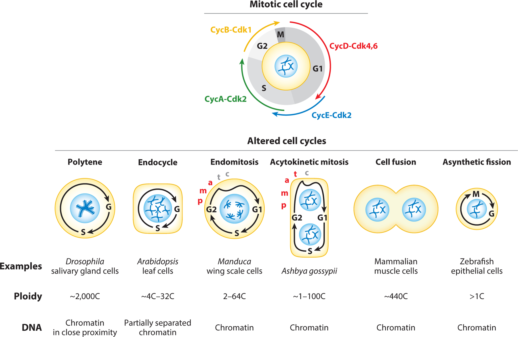
Though cell size varies between different cells and across species, the nuclear-to-cytoplasmic (N/C) ratio is largely maintained across species and within cell types. A cell maintains a relatively constant N/C ratio by coupling DNA content, nuclear size, and cell size. We explore how cells couple cell division and growth to DNA content. In some cases, cells use DNA as a molecular yardstick to control the availability of cell cycle regulators. In other cases, DNA sets a limit for biosynthetic capacity. Developmentally programmed variations in the N/C ratio for a given cell type suggest that a specific N/C ratio is required to respond to given physiological demands. Recent observations connecting decreased N/C ratios with cellular senescence indicate that maintaining the proper N/C ratio is essential for proper cellular functioning. Together, these findings suggest a causative, not simply correlative, role for the N/C ratio in regulating cell growth and cell cycle progression.
Keywords: DNA-to-cytoplasmic ratio; biosynthetic activity; cell cycle; cell size; nuclear-to-cytoplasmic ratio; polyploidy.
Figures
Similar articles
-
A mechanism of intracellular timing and its cooperation with extracellular signals in controlling cell proliferation and differentiation, an amended hypothesis.J Theor Biol. 2001 Aug 7;211(3):239-51. doi: 10.1006/jtbi.2001.2342. J Theor Biol. 2001. PMID: 11444955
-
Simultaneous analysis of cell cycle kinetics at two different DNA ploidy levels based on DNA content and cyclin B measurements.Cancer Res. 1993 Nov 1;53(21):5096-9. Cancer Res. 1993. PMID: 8221643
-
Both Nuclear Size and DNA Amount Contribute to Midblastula Transition Timing in Xenopus laevis.Sci Rep. 2017 Aug 11;7(1):7908. doi: 10.1038/s41598-017-08243-z. Sci Rep. 2017. PMID: 28801588 Free PMC article.
-
Plant cell size: Links to cell cycle, differentiation and ploidy.Curr Opin Plant Biol. 2024 Apr;78:102527. doi: 10.1016/j.pbi.2024.102527. Epub 2024 Mar 13. Curr Opin Plant Biol. 2024. PMID: 38484440 Review.
-
On the Molecular Mechanisms Regulating Animal Cell Size Homeostasis.Trends Genet. 2020 May;36(5):360-372. doi: 10.1016/j.tig.2020.01.011. Epub 2020 Feb 20. Trends Genet. 2020. PMID: 32294416 Free PMC article. Review.
Cited by
-
A Travel through Landscapes of Seed Dormancy.Plants (Basel). 2023 Nov 24;12(23):3963. doi: 10.3390/plants12233963. Plants (Basel). 2023. PMID: 38068600 Free PMC article. Review.
-
Impact of ionizing radiation on cell-ECM mechanical crosstalk in breast cancer.Front Bioeng Biotechnol. 2024 Jun 6;12:1408789. doi: 10.3389/fbioe.2024.1408789. eCollection 2024. Front Bioeng Biotechnol. 2024. PMID: 38903185 Free PMC article.
-
When one nucleus is not enough: Intestinal polyploidy fuels healthier progeny in C. elegans.J Cell Biol. 2025 Mar 3;224(3):e202412192. doi: 10.1083/jcb.202412192. Epub 2025 Feb 11. J Cell Biol. 2025. PMID: 39932557
-
Lipid Priming of Adipose Mesenchymal Stromal Cells with Docosahexaenoic Acid: Impact on Cell Differentiation, Senescence and the Secretome Neuroregulatory Profile.Tissue Eng Regen Med. 2025 Jan;22(1):113-128. doi: 10.1007/s13770-024-00679-5. Epub 2024 Nov 4. Tissue Eng Regen Med. 2025. PMID: 39495459 Free PMC article.
-
RCC1 depletion drives protein transport defects and rupture in micronuclei.bioRxiv [Preprint]. 2024 Sep 5:2024.09.04.611299. doi: 10.1101/2024.09.04.611299. bioRxiv. 2024. PMID: 39282444 Free PMC article. Preprint.
References
-
- Adams MD, Celniker SE, Holt RA, Evans CA, Gocayne JD, et al. 2000. The genome sequence of Drosophila melanogaster. Science 287(5461):2185–95 - PubMed
-
- Adamson ED, Woodland HR. 1977. Changes in the rate of histone synthesis during oocyte maturation and very early development of Xenopus laevis. Dev. Biol 57(1):136–49 - PubMed
Publication types
MeSH terms
Grants and funding
LinkOut - more resources
Full Text Sources
