. 2022 Aug 16;3(8):100715.
doi: 10.1016/j.xcrm.2022.100715.

A single-cell transcriptional gradient in human cutaneous memory T cells restricts Th17/Tc17 identity

Affiliations

A single-cell transcriptional gradient in human cutaneous memory T cells restricts Th17/Tc17 identity

Christopher P Cook et al. Cell Rep Med. .

Abstract

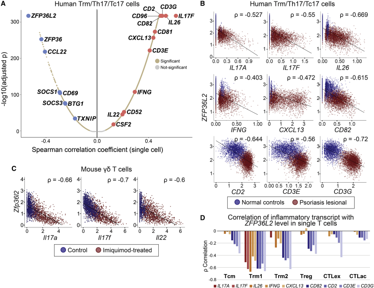
The homeostatic mechanisms that fail to restrain chronic tissue inflammation in diseases, such as psoriasis vulgaris, remain incompletely understood. We profiled transcriptomes and epitopes of single psoriatic and normal skin-resident T cells, revealing a gradated transcriptional program of coordinately regulated inflammation-suppressive genes. This program, which is sharply suppressed in lesional skin, strikingly restricts Th17/Tc17 cytokine and other inflammatory mediators on the single-cell level. CRISPR-based deactivation of two core components of this inflammation-suppressive program, ZFP36L2 and ZFP36, replicates the interleukin-17A (IL-17A), granulocyte macrophage-colony-stimulating factor (GM-CSF), and interferon gamma (IFNγ) elevation in psoriatic memory T cells deficient in these transcripts, functionally validating their influence. Combinatoric expression analysis indicates the suppression of specific inflammatory mediators by individual program members. Finally, we find that therapeutic IL-23 blockade reduces Th17/Tc17 cell frequency in lesional skin but fails to normalize this inflammatory-suppressive program, suggesting how treated lesions may be primed for recurrence after withdrawal of treatment.

Keywords: ZFP36; ZFP36L2; cytokine; inflammation; psoriasis; resident-memory T cell; tristetraprolin scRNA-seq.


Conflict of interest statement

Declaration of interests A.M. is a cofounder of Arsenal Biosciences, Spotlight Therapeutics, and Survey Genomics. A.M. serves on the boards of directors at Spotlight Therapeutics and Survey Genomics and is board observer (and former member of the board of directors) at Arsenal Biosciences. A.M. is a member of the scientific advisory boards of Arsenal Biosciences, Spotlight Therapeutics, Survey Genomics, and NewLimit. A.M. owns stock in Arsenal Biosciences, Spotlight Therapeutics, NewLimit, Survey Genomics, PACT Pharma, and Merck. A.M. has received fees from Arsenal Biosciences, Spotlight Therapeutics, NewLimit, 23andMe, PACT Pharma, Juno Therapeutics, Trizell, Vertex, Merck, Amgen, Genentech, AlphaSights, Rupert Case Management, Bernstein, and ALDA. A.M. is an investor in and informal advisor to Offline Ventures and a client of EPIQ.

Figures

None
Graphical abstract
Figure 1
Figure 1
Psoriatic T cells expressing IL17A and IL17F harbor skin-resident memory markers and classify into both Th17 and Tc17 subpopulations (A) Single-cell RNA sequencing (scRNA-seq) and cellular indexing of transcriptomes and epitopes sequencing (CITE-seq; protein epitope) marker expression defining major T cell subpopulations (>5% of all T cells) from 8 psoriasis and 7 normal skin samples. (B) Frequency of six major T cell subpopulations in each donor sample. (C) Differentially expressed transcripts in the Trm1 subclass in lesional psoriatic skin versus healthy controls (Table S4). The x axis denotes average log2FC in transcript counts between disease and healthy controls (increases in red, decreases in blue). The y axis denotes negative log10 of the adjusted p value. Key cytokines (red) and inflammation-suppressive genes (blue) are labeled. (D) Psoriatic cytokine expression in both 800 CD4 transcript-positive cells (Th17) and 1,573 CD8 transcript-positive cells (Tc17) from 8 psoriasis samples.
Figure 2
Figure 2
scRNA-seq identifies ZFP36L2 as the transcript most anti-correlated with psoriatic inflammatory gene expression in skin-resident T cells (A) Positive correlation of IL17A expression in single Trm/Th17/Tc17 cells with expression of pro-inflammatory transcripts and cytokines (right half of graph, labeled in red). Deficiency in IL17A-expressing single T cells of inflammation-suppressive genes such as ZFP36L2, ZFP36, and BTG1 is shown as negative Spearman correlations (left half of graph, labeled in blue). Data from 8 psoriasis and 7 normal samples. An analogous correlation graph for IL17F is shown in Figure S2. (B) ZFP36L2 suppression predicts induction of numerous other pro-inflammatory mediator transcripts from Th17/Tc17 in lesional psoriatic skin from this study (red, all 8 samples pooled) versus healthy controls (blue, all 7 samples pooled). The y axis shows ZFP36L2 expression, the x axis shows imputed cytokine transcript levels, and each point represents a single T cell. (C) scRNA-seq from prior studies of CD45+ cutaneous immune cells isolated from imiquimod-provoked (red, 3 samples pooled) versus control treated mice (blue, 3 samples pooled) show maximal expression of IL17A, IL17F, and IL22 with highest ZFP36L2 suppression. The y axis shows ZFP36L2 expression, the x axis shows imputed cytokine transcript levels, and each point represents a single γδ T cell. For both axes, the standard imputed expression value (iCellR) has been normalized and log-transformed (Seurat; STAR Methods). (D) Relationship between ZFP36L2 loss and inflammatory mediator expression seen in all major skin T cell classes but strongest in skin-resident memory T cells.
Figure 3
Figure 3
ZFP36L2 and ZFP36 knockout increases the intracellular concentration of 3′ ARE-containing cytokines Frequency of CD4+ (top rows) and CD8+ (bottom rows) T cell staining for the cytokines indicated on the y axis. (A) Frequency of cytokine-positive cells from ZFP36L2 knockout T cells shown in green for each of two gRNAs (L2g1 and L2g2, biological triplicate experiments). (B) Frequency of cytokine-positive cells from ZFP36 knockout T cells shown in orange for two gRNAs (36g1 and 36g2, biological triplicate experiments).AAVS1 and NT are AAVS1 safe-harbor-targeting and non-targeting negative control gRNAs. ∗p < 0.05, ∗∗p < 0.01, ∗∗∗p < 0.001, Student’s two-sample, two-tailed t tests. Error bars: SEM. Sanger gene-inactivation percentages displayed at bottom. Selected cytokine assessments, including the L2g1 ZFP36L2 guide for IL-17A, were performed using an additional donor (Figure S5).
Figure 4
Figure 4
Loss of a coordinated inflammation-suppressive program centered on ZFP36L2 defines inflamed psoriatic skin-resident memory cells (A) ZFP36L2 expression coordinates with that of numerous other global inflammatory suppressors in Th17/Tc17 single cells in lesional psoriatic skin (red, all 8 samples pooled) compared with healthy controls (blue, all 7 samples pooled), a program we term the ZFP36L2 inflammation suppressive transcript program (ZIST). The y axis shows ZFP36L2 expression, the x axis shows transcript levels for the specified suppressor gene, and each point represents a single T cell. (B) Correlation between most ZIST genes is statistically significant in different T cell subclasses but is strongest in Trm1. (C) Combinatoric analysis shows top six ZIST gene groups whose combined downregulation best predicts individual inflammatory transcript expression (Spearman correlation coefficient displayed at right for gene named at left, different ZIST transcript subsets denoted by filled red circles in each row), revealing predictive associations of ZFP36L2 for IL17A, as well as of BTG1 for CD82.
Figure 5
Figure 5
The ZIST program gradient mirrors an inherent molecular trajectory organizing the transition from uninflamed to psoriatic skin-resident memory T cells (A) A pseudotime constructed from a batch-corrected manifold shows distributed representation of all samples. (B–D) Uninflamed Trms and psoriatic Th17/Tc17 cells are clearly distinguished in this pseudotime, distinct from (C) CD4/CD8 or (D) central memory marker expression (e.g., CCR7/SELL). (E–G) ZFP36L2 and ZIST expression reflects the natural pseudotime, in inverse relation to (G) a mapping of inflammatory genes IL17A, IL17F, IL26, IFNG, CXCL13, CD2, CD82, and CD3E/G. (H) A rooted trajectory shows a single dominant path from uninflamed cells (root node, white circle 1), with linearly arrayed transitional states (black circles) and divergent development sinks/end states (gray circles).
Figure 6
Figure 6
IL-23 blockade with tildrakizumab eliminates most Th17/Tc17 T cells in three treated patients, but residual Trms show persistent suppression of the ZIST program (A) Inferred IL17A+, IL17F+, and IL26+ cell numbers per 6 mm biopsy shown for normal, psoriatic, and mid-IL-23 blockade (>8 weeks) for three individual patients (see Table S6 for CD4+/CD8+ ratios). (B) ZIST member suppression persists in residual, IL-23-blockaded skin-resident T cells (brown) versus normal, uninflamed Trms (mauve), similar to untreated psoriatic Th17/Tc17 cells (blue) despite nearly complete clinical resolution. Three asterisks (∗∗∗) denote differences in displayed violin plots of p < 2 × 10−16 (Kruskal-Wallis test). (C) Scatterplots illustrate ZIST program members are co-suppressed with ZFP36L2 in Trm1 class single cells isolated from mid-treatment biopsies, showing persistence of the gradient despite the context of IL-23 blockade.

References

    1. Lowes M.A., Kikuchi T., Fuentes-Duculan J., Cardinale I., Zaba L.C., Haider A.S., Bowman E.P., Krueger J.G. Psoriasis vulgaris lesions contain discrete populations of Th1 and Th17 T cells. J. Invest. Dermatol. 2008;128:1207–1211. - PubMed
    1. Cheuk S., Wikén M., Blomqvist L., Nylén S., Talme T., Ståhle M., Eidsmo L. Epidermal Th22 and Tc17 cells Form a Localized disease memory in clinically healed psoriasis. J. Immunol. 2014;192:3111–3120. - PMC - PubMed
    1. Liu Y., Wang H., Taylor M., Cook C., Martínez-Berdeja A., North J.P., Harirchian P., Hailer A.A., Zhao Z., Ghadially R., et al. Classification of human chronic inflammatory skin disease based on single-cell immune profiling. Sci. Immunol. 2022;7:eabl9165. - PMC - PubMed
    1. Reynolds G., Vegh P., Fletcher J., Poyner E.F.M., Stephenson E., Goh I., Botting R.A., Huang N., Olabi B., Dubois A., et al. Developmental cell programs are co-opted in inflammatory skin disease. Science. 2021;371:eaba6500. - PMC - PubMed
    1. Hughes T.K., Wadsworth M.H., Gierahn T.M., Do T., Weiss D., Andrade P.R., Ma F., de Andrade Silva B.J., Shao S., Tsoi L.C., et al. Second-strand synthesis-based Massively Parallel scRNA-seq reveals cellular states and molecular features of human inflammatory skin Pathologies. Immunity. 2020;53:878–894.e7. - PMC - PubMed

Publication types

Substances