. 2022 Sep 12;57(17):2127-2139.e6.
doi: 10.1016/j.devcel.2022.07.015. Epub 2022 Aug 16.

The spatiotemporal dynamics of microglia across the human lifespan

Affiliations

The spatiotemporal dynamics of microglia across the human lifespan

David A Menassa et al. Dev Cell. .

Abstract

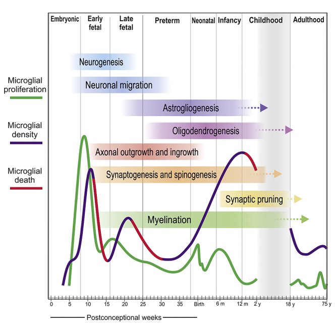
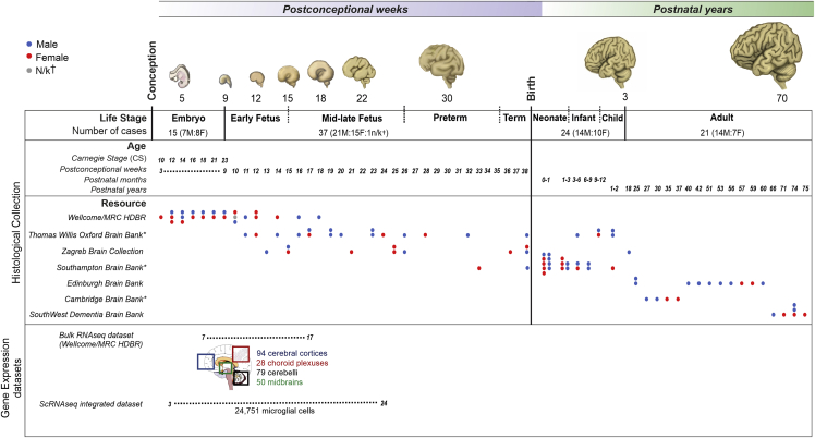
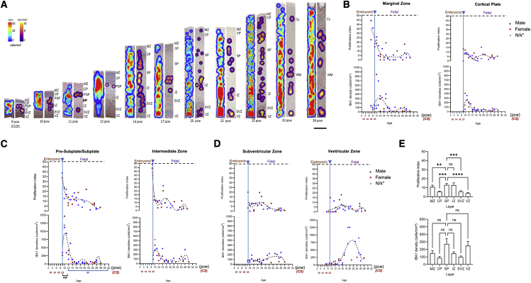
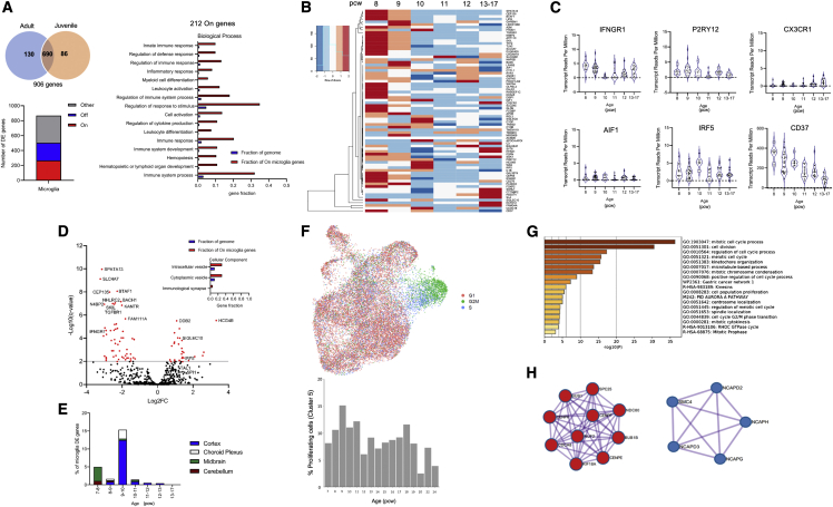
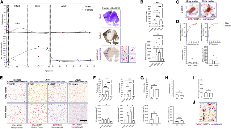
Microglia, the brain's resident macrophages, shape neural development and are key neuroimmune hubs in the pathological signatures of neurodevelopmental disorders. Despite the importance of microglia, their development has not been carefully examined in the human brain, and most of our knowledge derives from rodents. We aimed to address this gap in knowledge by establishing an extensive collection of 97 post-mortem tissues in order to enable quantitative, sex-matched, detailed analysis of microglia across the human lifespan. We identify the dynamics of these cells in the human telencephalon, describing waves in microglial density across gestation, infancy, and childhood, controlled by a balance of proliferation and apoptosis, which track key neurodevelopmental milestones. These profound changes in microglia are also observed in bulk RNA-seq and single-cell RNA-seq datasets. This study provides a detailed insight into the spatiotemporal dynamics of microglia across the human lifespan and serves as a foundation for elucidating how microglia contribute to shaping neurodevelopment in humans.

Keywords: RNA-seq; apoptosis; neurodevelopment; proliferation; single-cell RNA-seq.


Conflict of interest statement

Declaration of interests The authors declare no competing interests.

Figures

None
Graphical abstract
Figure 1
Figure 1
Overview of the study Post-mortem collection of human tissues across the lifespan with 52 prenatal and 45 postnatal cases, which adds up to n = 97. The overall number of cases collected, sampled, and analyzed was in excess of 130 (see STAR Methods section and Table S2). Temporal windows mapped onto key human neurodevelopmental milestones across the lifespan were defined consistent with existing classifications (Carroll et al., 2021; Silbereis et al., 2016; Kostović et al., 2002): the embryonic period (3–8 pcw), the early fetal period (9–15 pcw), the mid-late fetal period (16–25 pcw), the preterm period (26–35 pcw), the term period (36 pcw–birth), the neonatal period (0–1 month), the infancy period (1–12 months), childhood (1–3 years), and adulthood (>18 years). Gene expression datasets: bulk RNA-seq of 251 samples from the Wellcome MRC/HDBR resource in 4 anatomical regions between 7 and 17 postconceptional weeks and an integrated dataset of 24,751 microglial cells from 4 single-cell RNA-seq studies (Cao et al., 2020; Kracht et al., 2020; Bian et al., 2020; Fan et al., 2020) between 3 and 24 postconceptional weeks. F, female; M, male; MRC/HDBR, Medical Research Council/Human Developmental Biology Resource; † indicates not known, and indicates brain banks that are part of the BRAINUK network. Embryonic brain drawings were based on our own samples, and other model brains were redrawn and colored based on illustrations from Haymaker and Adams (1982).
Figure 2
Figure 2
Early colonization of the human embryo (A) Representative sagittal cross-section through a CS10 (late 3rd pcw) human embryo. Immunolabeling was done on consecutive 8-μm sections identifying the presence of PU.1 cells in organs apart from the brain and the absence of IBA1+ cells overall. Scale bars: 500 μm (left); 40 μm (right). (B) Representative sagittal sections through whole human embryos from CS12, the age of appearance of IBA1+ cells across the entire embryo, through to CS21. Insets show the location of proliferating IBA1+ cells in both the liver and the dorsal telencephalon. Scale bars: 1 mm (top); 100 μm (bottom). (C) IBA1 proliferative dynamics and cell densities in the developing brain (n = 14) and the liver (n = 11). Black arrows represent the two most relevant time points for microglial densities: CS12 (4th pcw) for the first colonization of the brain rudiment and CS23 (9th pcw), the transition from embryonic to early fetal life. (D) Mean IBA1 proliferation in the liver (n = 11) and the developing brain (n = 11) across the embryonic period. All data are shown as mean ± SEM. (E) Cumulative frequency distribution plots of densities and proliferative indices by sex in the developing brain (7F:7M) (top panel); mean differences between sexes in microglial densities and proliferative indices across the embryonic age in the developing brain (7F:7M) (bottom panel, data are shown as mean ± SEM). (F) Representative migratory profiles of IBA1+ cells in the bilaminar telencephalon at 5 pcw (n = 2). Data are shown as mean ± SEM. (G) IBA1+ cell distributions in the CS12 embryo showing very few cells in the forebrain and a much higher density of cells in the hindbrain and midbrain. Scale bars: 500 μm. (H) Entry routes of IBA1+ cells into the brain rudiment in the forebrain and the hindbrain. Scale bars: 100 μm. C, cardiac muscle; CP, cortical plate; GE, ganglionic eminence; H, hindbrain; L, liver; M, meninges; MB, midbrain; Me, mesenchyme; ML, mantle layer; SC, spinal cord; T, telencephalon; VZ, ventricular zone. For all panels, asterisks represent adjusted p value significance as follows: p < 0.05, ∗∗p < 0.001, ∗∗∗p < 0.0001, and ns p > 0.05.
Figure 3
Figure 3
Developmental dynamics of microglia in the cortex (A) Representative laminar structure of the developing cortex with its transient zones observed from CS23 (9th pcw) in humans and representative cortical columns from 10 to 12 pcw showing (1) the development of the pre-subplate to the subplate at 12.5 pcw and the alignment of microglial cells at the CP-PSP/SP boundary and (2) the distribution of microglial cells across transient zones. Scale bars: 2 mm (left); 100 μm (right). (B) Corrected microglial densities (against fold change in frontal telencephalic wall thickness with age) and proliferative dynamics in the telencephalon during development (CS10 [late 3rd/early 4th pcw]) until term (38 pcw) (n = 50). n/k, not known. Embryonic and early fetal temporal windows are most significant for proliferation against other temporal windows, while the early fetal window is the most significant against other temporal windows. (C) Equally spaced temporal windows for proliferation levels (top) and densities (bottom) around the most significant first wave. Data are represented as mean ± SEM. (D) Mean apoptotic index around the first peak of densities (n = 15, 8 cases between 7 and 11 pcw and 7 cases between 12 and 16 pcw) (top panel). Data are represented as mean ± SEM. Representative microglial cell death photomicrographs observed in wave 1 following the decrease in densities (bottom panel, black arrows in B). Scale bars: 20 μm. (E) Migratory profile of microglia and type of migration in representative cases from the telencephalon (n = 12). (F) Representative profile (top panel) of TMEM119+ and IBA1+ cell densities around the first significant density wave (10–16 pcw) (n = 6). Mean TMEM119+ and IBA1+ cell densities across the prenatal period (10–28 pcw, n = 10) (bottom panel). Data are shown as mean ± SEM. (G) Ratio of labeled TMEM119+/IBA1+ cells during the prenatal period (10–28 pcw, n = 10). Data are presented as mean ±SEM. (H) Representative confocal images of TMEM119+ cells in the MZ and the VZ of a 13-pcw case (left) and double labeling of TMEM119/IBA1 in bright field in a neocortical column at 13 pcw observed in the MZ (right). Scale bars (left): 50 μm; scale bars (right): 100 μm. (I) Wave 2 microglial cell death at 18 and 23 pcw. Scale bars: 20 μm. (J) Non-microglial death observed in the GE and in cortical transient zones at CS23 (9th pcw) (top, scale bars: 100 μm) and at 24 pcw (bottom, scale bars: 50 μm). (K) Proliferative dynamics and densities by sex across human development shown as relative cumulative frequency distribution plots (top panel) and mean values between the sexes (27M:23F) (bottom panel). Data are shown as mean ± SEM. R, right; L, left; CP, cortical plate; GE, ganglionic eminence; IZ, intermediate zone; M, meninges; MZ, marginal zone; PSP, pre-subplate; SP, subplate; SVZ, subventricular zone; VZ, ventricular zone. For all panels, asterisks represent adjusted p value significance as follows: p < 0.05, ∗∗p < 0.001, ∗∗∗p < 0.0001, and ns p > 0.05.
Figure 4
Figure 4
Spatiotemporal microglial dynamics within the developing cortex (A) Representative heatmaps showing microglial dynamics throughout development in the human cortex. Heatmaps were constructed based on the density of immunoreactive cells. Density of all IBA1 immunoreactive cells are represented by blue-red spectral heatmaps (left column of each stage), while IBA1+/Ki67+ double-positive cells are represented by purple-yellow spectral heatmaps (right column of each stage). Approximate densities are shown on the color bar with minimal density being blue/purple, maximum density being red/yellow. All cortical columns are 450-μm wide. (B–D) (B) Proliferation and densities of microglia in the marginal zone (n = 35), cortical plate (n = 31); (C) the subplate (n = 31) and the intermediate zone (n = 31); (D) the subventricular (n = 31) and the ventricular zones (n = 35). (E) Proliferation and densities in the subplate, the intermediate zone, and remaining cortical layers. Data are shown as mean ±SEM. CP, cortical plate; GM, gray matter; IZ, intermediate zone; MZ, marginal zone; PSP, pre-subplate; SP, subplate; SVZ, subventricular zone; VZ, ventricular zone; WM, white matter. For all panels, asterisks represent adjusted p value significance as follows: p < 0.05, ∗∗p < 0.001, ∗∗∗p < 0.0001, and ns p > 0.05.
Figure 5
Figure 5
Microglial gene signature during development (A) 906 genes chosen from two published microglial-specific cortical adult and juvenile gene lists (Gosselin et al., 2017; Galatro et al., 2017). Bar plot shows the number of constitutively expressed microglial genes (“On”), unexpressed genes (“Off”) and those with sporadic expression in the developing cortex (“Other”) with gene ontology analysis of constitutively expressed genes only. (B) Heatmap of highly enriched microglial genes during development (n = 75). (C) Transcript reads per million expression levels of a selection of homeostatic and immune modulatory microglial markers during development. (D) Volcano plot of upregulated and downregulated microglial genes at the peak of differential gene expression between 9 and 10 pcw, with gene ontology analysis significant for cellular component. (E) Regional differential expression between time points of the cortical adult microglial genes. (F) Actively cycling and proliferating microglia in the scRNA-seq dataset were identified across the gestational age. Only ages with a minimum of 50 cells were selected (7–24 pcw). Cycling and proliferating cell signatures display a bimodal pattern of proliferation consistent with histological findings. (G) Metascape analysis of conserved cycling and proliferating microglia markers. (H) Gene ontology and protein-protein interaction enrichment analysis marking key mitotic cell-cycle processes. Protein-protein interaction enrichment identifies mitotic spindle checkpoint and amplification of signals from the kinetochores (in red), as well as mitotic chromosome condensation and condensing I complex (in blue). DE, differentially expressed genes.
Figure 6
Figure 6
Postnatal dynamics of microglia in the cortex (A) Microglial densities and proliferative dynamics in early postnatal life (n = 24; 14M:10F) and adulthood (n = 21; 14M:7F) and a representative cross-section of frontal cortex with anatomical histochemistry and photomicrographs of homeostatic and proliferating microglial morphologies in gray and white matters. Neonatal and infant proliferation is most significant against other postnatal windows, while microglial densities are most significant in the child against other temporal windows. Scale bars: 2 mm (left); 40 μm (right). (B) Mean proliferation and mean densities in the neonate, the infant, child, and adult. Data are shown as mean ± SEM. (C) Microglial cell death in gray and white matters at 2 years (black arrow in A). Scale bars: 15 μm. (D) Cumulative relative frequency distribution plots of microglial densities and proliferation by sex (top panel) and mean proliferation and densities by sex (28M:17F) (bottom panel, data are shown as mean ± SEM). (E) Representative photomicrographs of cell densities in gray and white matters from birth to 2 years. Scale bars: 500 μm. (F) Regional differences of microglial densities and proliferation in gray (left) and white matters (right) between the neonate, infant, child, and adult (n = 45). Data are shown as mean ± SEM. (G) Proliferation rates and densities in early postnatal life in gray and white matters (n = 24). Data are shown as mean ± SEM. (H) Regional differences in the adult in gray and white matters in proliferation (top) and density (bottom). Data are shown as mean ± SEM. (I) Mean TMEM119 and IBA1 cell densities postnatally (n = 5) (left) and ratio of TMEM119/IBA1 between early life (3–6 months, n = 2) and adult life (40–70 years, n = 3). Data are represented as mean ± SEM. (J) Representative photomicrograph of TMEM119/IBA1 labeling showing colocalization of both markers (black arrows). Scale bars: 25 μm. CC, cingulate cortex; GM, gray matter; WM, white matter. For all panels, asterisks represent adjusted p value significance as follows: p < 0.05, ∗∗p < 0.001, ∗∗∗p < 0.0001, and ns p > 0.05.

References

    1. Ameijeiras-Alonso J., Crujeiras R.M., Rodríguez-Casal A. Mode testing, critical bandwidth and excess mass. Test. 2019;28:900–919.
    1. Anders S., Pyl P.T., Huber W. HTSeq—a Python framework to work with high-throughput sequencing data. Bioinformatics. 2015;31:166–169. - PMC - PubMed
    1. Askew K., Li K., Olmos-Alonso A., Garcia-Moreno F., Liang Y., Richardson P., Tipton T., Chapman M.A., Riecken K., Beccari S., et al. Coupled proliferation and apoptosis maintain the rapid turnover of microglia in the adult brain. Cell Rep. 2017;18:391–405. - PMC - PubMed
    1. Badimon A., Strasburger H.J., Ayata P., Chen X., Nair A., Ikegami A., Hwang P., Chan A.T., Graves S.M., Uweru J.O., et al. Negative feedback control of neuronal activity by microglia. Nature. 2020;586:417–423. - PMC - PubMed
    1. Bayer S.A., Altman J. CRC Press; 2006. The human brain during the late first trimester. Atlas of Human Central Nervous System Development v 4.

Publication types

LinkOut - more resources