. 2022 Dec;81(12):1712-1721.
doi: 10.1136/ard-2022-222795. Epub 2022 Aug 17.

Loss-of-function variants in SAT1 cause X-linked childhood-onset systemic lupus erythematosus

Affiliations

Loss-of-function variants in SAT1 cause X-linked childhood-onset systemic lupus erythematosus

Lingxiao Xu et al. Ann Rheum Dis. 2022 Dec.

Abstract

Objectives: Families that contain multiple siblings affected with childhood onset of systemic lupus erythematosus (SLE) likely have strong genetic predispositions. We performed whole exome sequencing (WES) to identify familial rare risk variants and to assess their effects in lupus.

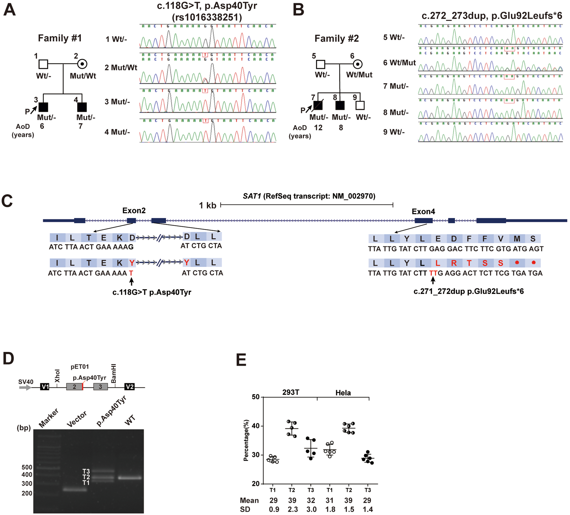
Methods: Sanger sequencing validated the two ultra-rare, predicted pathogenic risk variants discovered by WES and identified additional variants in 562 additional patients with SLE. Effects of a splice site variant and a frameshift variant were assessed using a Minigene assay and CRISPR/Cas9-mediated knock-in (KI) mice, respectively.

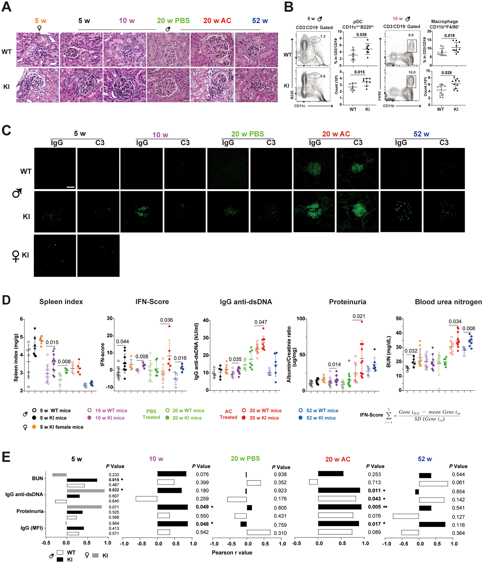
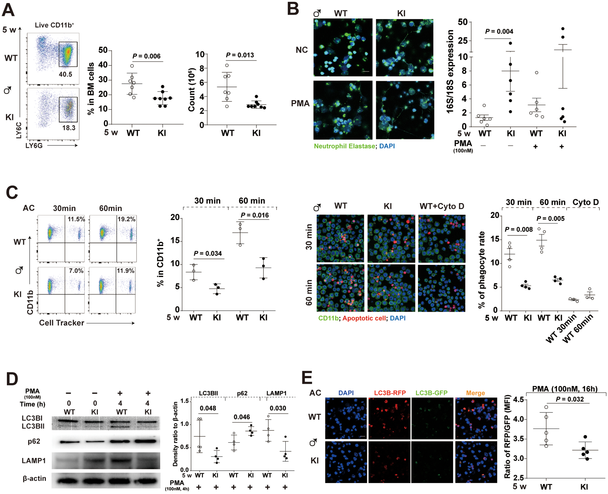
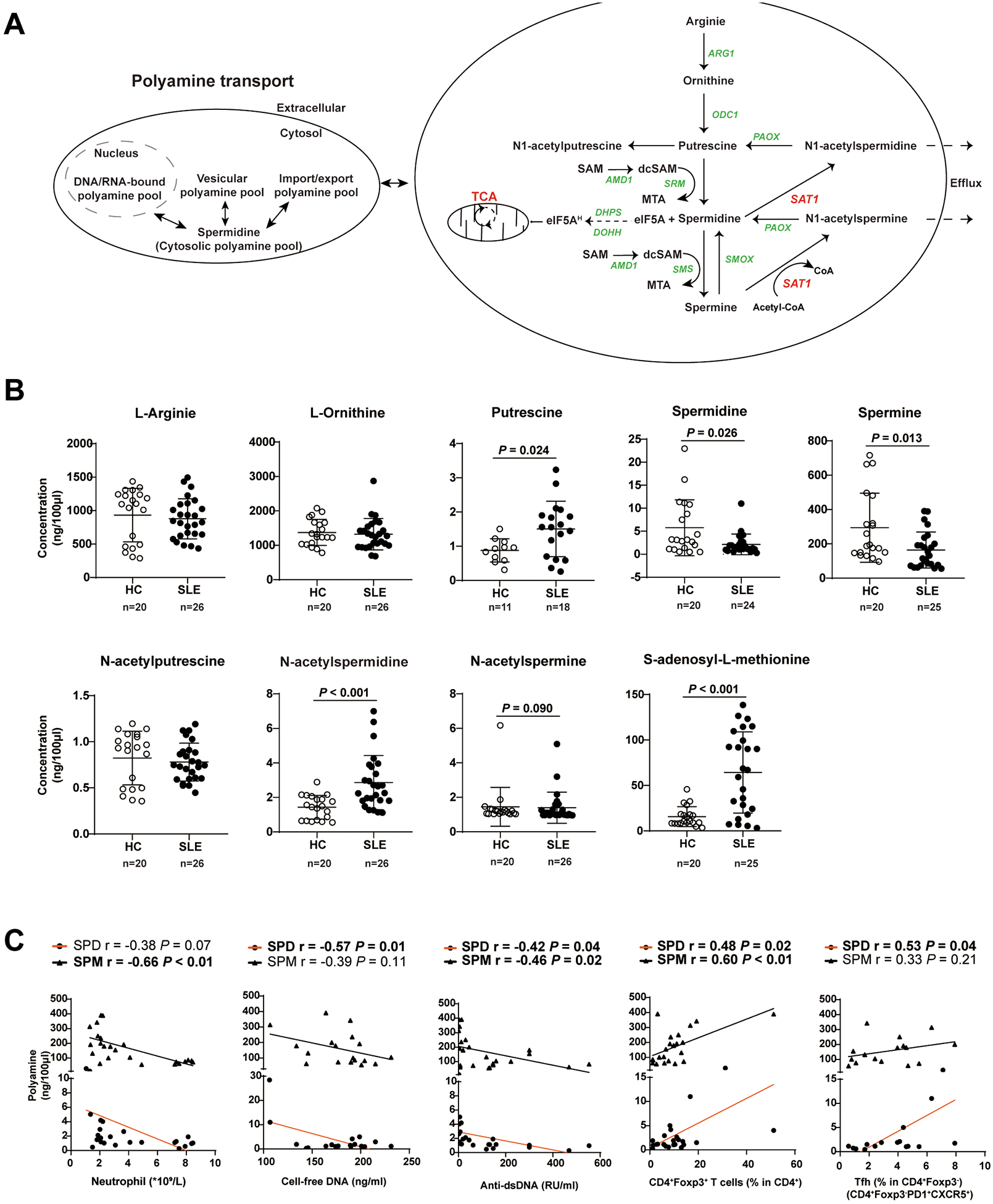
Results: The two familial ultra-rare, predicted loss-of-function (LOF) SAT1 variants exhibited X-linked recessive Mendelian inheritance in two unrelated African-American families. Each LOF variant was transmitted from the heterozygous unaffected mother to her two sons with childhood-onset SLE. The p.Asp40Tyr variant affected a splice donor site causing deleterious transcripts. The young hemizygous male and homozygous female Sat1 p.Glu92Leufs*6 KI mice spontaneously developed splenomegaly, enlarged glomeruli with leucocyte infiltration, proteinuria and elevated expression of type I interferon-inducible genes. SAT1 is highly expressed in neutrophils and encodes spermidine/spermine-N1-acetyltransferase 1 (SSAT1), a rate-limiting enzyme in polyamine catabolism. Young male KI mice exhibited neutrophil defects and decreased proportions of Foxp3 +CD4+ T-cell subsets. Circulating neutrophil counts and proportions of Foxp3 +CD4+ T cells correlated with decreased plasma levels of spermine in treatment-naive, incipient SLE patients.

Conclusions: We identified two novel SAT1 LOF variants, showed the ability of the frameshift variant to confer murine lupus, highlighted the pathogenic role of dysregulated polyamine catabolism and identified SAT1 LOF variants as new monogenic causes for SLE.

Keywords: Autoimmune Diseases; Immune System Diseases; Lupus Erythematosus, Systemic.


Conflict of interest statement

Competing interests: None declared.

Figures

Figure 1.
Figure 1.. Identification of p.Asp40Tyr and p.Glu92Leufs*6 SAT1 variants segregating with disease status in each family.
(A and B) Pedigree information of two unrelated African-American families containing SLE-affected sibpairs and Sanger sequencing data that confirmed Mendelian inheritance of either p.Glu92Leufs*6 or p.Asp40Tyr variant of X-linked SAT1 in each family. AoD, age of diagnosis. P, proband. p.Asp40Tyr is identified as rs1016338251 by Human Longevity company, but no annotation of any individual is available. (C) Locations of the p.Asp40Tyr (NM_002970: c.118G>T, ChrX(GRCh38):g.23783709G>T) and p.Glu92Leufs*6 (NM_002970: c.272_273dup, ChrX(GRCh38):g.23785397–23785398dup) SAT1 variants and the corresponding sequences based on human reference genome build GRCh38/hg38. (D and E) The genomic segment containing p.Asp40Tyr cloned into the Minigene assay vector resulted in aberrantly spliced transcripts in transfected 293T and HeLa cell lines. A schematic of the p.Asp40Tyr minigene plasmid and electrophoresed RT-PCR products from 293T cells transfected with a minigene plasmid with either Asp40- or Tyr40- containing genomic segment (D). The percentage of each spliced transcript from three independent transfection experiments is depicted. Data are mean ± SD (E).
Figure 2.
Figure 2.. The young Sat1p.Glu92Leufs*6 KI male and female mice spontaneously develop lupus-like autoimmune disorder.
(A) Hematoxylin-eosin-stained kidney sections from female (5 wks-old) and male Sat1p.Glu92Leufs*6 KI mice and WT littermates that were naïve (5, 10 or 52 wks-old), or injected with either PBS (phosphate buffer saline) or apoptotic cells (AC) starting at 10 wks-old and sacrificed at 20 wks-old. Bar: 50μm. (B) Gating strategy and percentage of plasmacytoid dendritic cells (pDC)(CD3CD19CD11cintB220hi) and macrophages (CD3CD19CD11bhiF4/80+) in spleen cells from 5- and 10-week-old naive male mice, respectively. Open circle, WT mice; closed circle, KI littermates. Data are mean ± SD. Mann-Whiney U test. (C) Immunofluorescent staining of mouse Immunoglobulin G (IgG) and complement 3 (C3) depositions in the frozen kidney sections of Sat1p.Glu92Leufs*6 female KI mice, male KI and WT littermates. Bar: 50μm. (D) Levels of Spleen index, type I IFN-Scores, serum IgG anti-dsDNA, proteinuria, and blood urea nitrogen (BUN) in 5 to 52-week-old female KI mice, male KI and WT littermates. Open circle, male WT mice; closed circle, male KI mice. Black, 5-week-old mice; purple, 10-week-old mice, green, 20-week-old mice injected with PBS; red, 20-week-old mice injected with apoptotic cells; blue, 52-week-old mice. Data are mean ± SD. Yellow closed circle, 5-week-old female KI mice. Mann-Whiney U test. (E) Correlation analysis of levels of IgG deposition in the kidney, proteinuria, serum anti-dsDNA and BUN with type I IFN-Scores in splenocytes of 5 to 52-week-old KI mice. Closed box depicts statistically significant correlation. White box, male WT mice; Black box, male KI mice; Gray box, female KI mice.
Figure 3.
Figure 3.. Decreased levels and defective functions of bone marrow (BM)-isolated neutrophils from young male Sat1p.Glu92Leufs*6 mice.
(A) Gating strategy, decreased percentage, and cell numbers of BM-isolated neutrophils (CD11b+LY6G+LY6Cint) in 5-week-old male KI mice compared to their WT littermates. Data are mean ± SD. Mann-Whiney U test. (B) (Left) Representative images of the Neutrophil extracellular traps (NET) induced by PMA in BM-isolated neutrophils. (Right) Quantitation of mitochondrial (16S; officially known as MT-RNR2) and chromosomal (18S; officially known as RNA18S5) DNA in the immuno-precipitated total oxidized DNA from overnight culture supernatants of BM-isolated neutrophils of either WT or KI littermates incubated in the absence (spontaneous NETosis) or presence of PMA (induced NETosis). Green immunofluorescence represents neutrophil elastase and blue represents DNA (Hoechst_33342) of confocal images. Bar: 10μm; PMA, phorbol myristate acetate, 100nM; Incubation time, 24 hours. Data are mean ± SD. Mann-Whiney U test. (C) Defective engulfment of Cell Tracker-labeled apoptotic cells (AC) by BM-isolated neutrophils from 5-week-old male KI mice after 30- or 60- min co-cultures assessed by either flow cytometry or confocal microscopy; BM-isolated neutrophils: AC=1:5. Cyto D, Cytochalasin D, an inhibitor of actin polymerization, at 10μM; Bar: 10μm. Data are mean ± SD. Unpaired t-test. (D) Representative Western blot of LC3B, p62 (an autophagosome cargo protein), LAMP1 and β-actin, and quantification of relative levels of LC3B-II to LC3B-I, and relative levels of p62 or LAMP1 to β-actin in PMA-stimulated groups. PMA, 100nM. Bar: 10μm. Incubation time, 4 hours. Data are mean ± SD. Unpaired t-test. (E) Decreased levels of autophagic flux in PMA-treated BM-isolated neutrophils from 5-week-old male KI mice. The left panel depicts representative fluorescence images of autophagic flux assays using an RFP-GFP-LC3B tandem construct that only the GFP signal could be quenched by the acidic lysosomal pH, and the right panel depicts relative ratios of RFP: GFP in each group. Bar: 10μm; PMA, 100nM; Incubation time, 16 hours. Data are mean ± SD. Unpaired t-test.
Figure 4.
Figure 4.. Plasma concentration of nine metabolites related to polyamine homeostasis in treatment-naïve patients affected with SLE and in age- and sex- matched healthy controls (HCs).
(A) The polyamine metabolism pathway. ARG1, arginase 1; ODC1, ornithine decarboxylase 1; PAOX, peroxisomal N(1)-acetyl-spermine/spermidine oxidase; SAT1, spermine/spermidine N1-acetyltransferase 1; SMS, spermine synthase; SMOX, Spermine oxidase; DHPS, deoxyhypusine synthase; DOHH, deoxyhypusine hydroxylase; eIF5A, eukaryotic initiation factor 5A; eIF5AH, eukaryotic initiation factor 5A hypusination; CoA, coenzyme A; dcSAM, decarboxylated SAM, S-adenosylmethionine; dcSAM, decarboxylated S-adenosylmethionine; SRM, spermidine synthase. (B) Quantification of nine polyamine metabolites (ng/100μL) found in fasting plasma samples of treatment-naïve, newly diagnosed SLE patients (n = 26) and age-and sex-matched HCs (n = 20). Open circle, health control; closed circle, SLE patient. Data are mean ± SD. Mann-Whiney U test. (C) Pearson correlations of polyamine concentrations (SPM and SPD, Spermine and Spermidine, respectively) with counts of blood neutrophils, percentages of Tfh and CD4+Foxp3+ T cells, serum levels of cell-free DNA and IgG anti-double-strand DNA antibodies in treatment-naïve SLE patients (n=26). Closed circle and orange line, spermidine; triangle and black line, spermine. Each symbol represents a sample from one individual subject.

References

    1. Ha E, Bae S-C, Kim K. Recent advances in understanding the genetic basis of systemic lupus erythematosus. Semin Immunopathol 2022;44:29–46. - PubMed
    1. Harley ITW, Sawalha AH. Systemic lupus erythematosus as a genetic disease. Clin Immunol 2022;236:108953. - PMC - PubMed
    1. Deng Y, Tsao BP. Updates in lupus genetics. Curr Rheumatol Rep 2017;19:1–13. - PubMed
    1. Cui Y, Sheng Y, Zhang X. Genetic susceptibility to SLE: recent progress from GWAS. J Autoimmun 2013;41:25–33. - PubMed
    1. Truedsson L Classical pathway deficiencies - a short analytical review. Mol Immunol 2015;68:14–19. - PubMed

Publication types