Transient splenial lesions of the corpus callosum and infectious diseases
- PMID: 35977887
- PMCID: PMC9475166
- DOI: 10.4266/acc.2022.00864
Transient splenial lesions of the corpus callosum and infectious diseases
Abstract
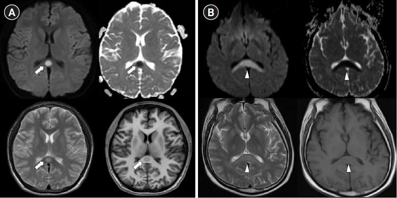
Transient splenial lesion of the corpus callosum can be observed in various diseases such as cancer, drug use, metabolic disorders, and cerebrovascular disorders, as well as in patients with infectious diseases. During the coronavirus disease 2019 (COVID-19) pandemic, there were increasing reports of these lesions being detected on brain imaging tests performed in patients with neurological symptoms. On brain magnetic resonance imaging, findings suggestive of cytotoxic edema are observed in the splenium; these are known to disappear with improvement of clinical symptoms. Cytokinopathy caused by infection increases the permeability of the blood-brain barrier and activates the glial cells of the brain to induce cytotoxic edema. Most patients have a good prognosis. The causes, mechanism, diagnosis, treatment and prognosis of transient splenial lesions of the corpus callosum will be summarized in this review.
Keywords: COVID-19; corpus callosum; infections; magnetic resonance imaging.
Conflict of interest statement
Dong-Ick Shin is an editorial board member of the journal but was not involved in the peer reviewer selection, evaluation, or decision process of this article. No other potential conflicts of interest relevant to this article were reported.
Figures
Similar articles
-
Transient Splenial Lesion of the Corpus Callosum Related to Migraine with Aura in a Pediatric Patient.Acta Medica (Hradec Kralove). 2016;59(2):64-6. doi: 10.14712/18059694.2016.91. Acta Medica (Hradec Kralove). 2016. PMID: 27526308
-
Cytotoxic lesions of the corpus callosum in children: Etiology, clinical and radiological features, and prognosis.Brain Dev. 2021 Oct;43(9):919-930. doi: 10.1016/j.braindev.2021.05.001. Epub 2021 Jun 11. Brain Dev. 2021. PMID: 34120800
-
Lesions in the Splenium of the Corpus Callosum on MRI in Children: A Review.J Neuroimaging. 2017 Nov;27(6):549-561. doi: 10.1111/jon.12455. Epub 2017 Jun 27. J Neuroimaging. 2017. PMID: 28654166 Review.
-
Transient Splenial Lesion of the Corpus Callosum After Cabergoline Treatment.World Neurosurg. 2018 Jun;114:257-258. doi: 10.1016/j.wneu.2018.03.166. Epub 2018 Mar 31. World Neurosurg. 2018. PMID: 29614359
-
Transient blindness associated with mild encephalitis/encephalopathy with a reversible splenial lesion (MERS): a case report and review of literature.Ital J Pediatr. 2020 Oct 12;46(1):152. doi: 10.1186/s13052-020-00918-0. Ital J Pediatr. 2020. PMID: 33046117 Free PMC article. Review.
Cited by
-
Legionella-induced dysarthria and rhabdomyolysis with acute renal failure achieving recovery.Clin Case Rep. 2024 Mar 7;12(3):e8628. doi: 10.1002/ccr3.8628. eCollection 2024 Mar. Clin Case Rep. 2024. PMID: 38464574 Free PMC article.
-
Contrasting association pattern of plasma low-density lipoprotein with white matter integrity in APOE4 carriers versus non-carriers.Neurobiol Aging. 2024 Nov;143:41-52. doi: 10.1016/j.neurobiolaging.2024.08.005. Epub 2024 Aug 22. Neurobiol Aging. 2024. PMID: 39213809
-
Tuberculous Meningitis in a Child: A Rare Presentation of Cytotoxic Lesion of the Corpus Callosum.Trop Med Infect Dis. 2025 Apr 4;10(4):96. doi: 10.3390/tropicalmed10040096. Trop Med Infect Dis. 2025. PMID: 40278769 Free PMC article.
-
COVID-19-Associated Cytotoxic Lesions of the Corpus Callosum in Chinese Patients: A Retrospective Study.Brain Behav. 2025 Jun;15(6):e70547. doi: 10.1002/brb3.70547. Brain Behav. 2025. PMID: 40437871 Free PMC article.
-
Case report: Re-evaluating reversibility of cytotoxic lesions of the corpus callosum.Front Neuroimaging. 2025 Feb 17;4:1436931. doi: 10.3389/fnimg.2025.1436931. eCollection 2025. Front Neuroimaging. 2025. PMID: 40034189 Free PMC article.
References
-
- Raybaud C. The corpus callosum, the other great forebrain commissures, and the septum pellucidum: anatomy, development, and malformation. Neuroradiology. 2010;52:447–77. - PubMed
-
- Takanashi J, Hirasawa K, Tada H. Reversible restricted diffusion of entire corpus callosum. J Neurol Sci. 2006;247:101–4. - PubMed
-
- Starkey J, Kobayashi N, Numaguchi Y, Moritani T. Cytotoxic lesions of the corpus callosum that show restricted diffusion: mechanisms, causes, and manifestations. Radiographics. 2017;37:562–76. - PubMed
-
- Garcia-Monco JC, Cortina IE, Ferreira E, Martínez A, Ruiz L, Cabrera A, et al. Reversible splenial lesion syndrome (RESLES): what's in a name? J Neuroimaging. 2011;21:e1–14. - PubMed
LinkOut - more resources
Full Text Sources