. 2022 Aug 17;13(1):4843.
doi: 10.1038/s41467-022-32535-2.

Halogenation of tyrosine perturbs large-scale protein self-organization

Affiliations

Halogenation of tyrosine perturbs large-scale protein self-organization

Huan Sun et al. Nat Commun. .

Abstract

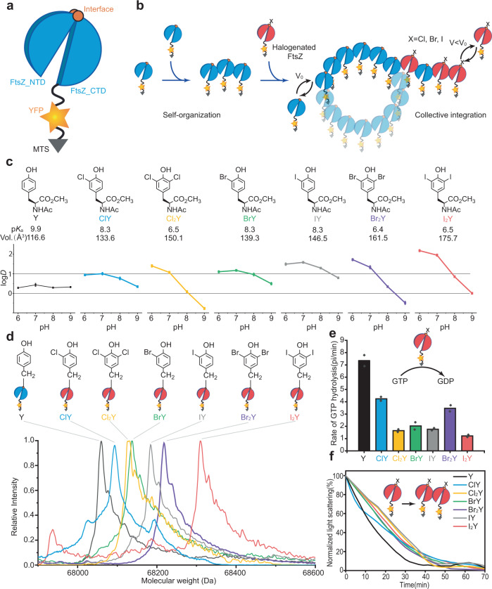
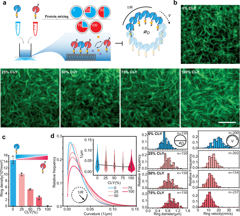
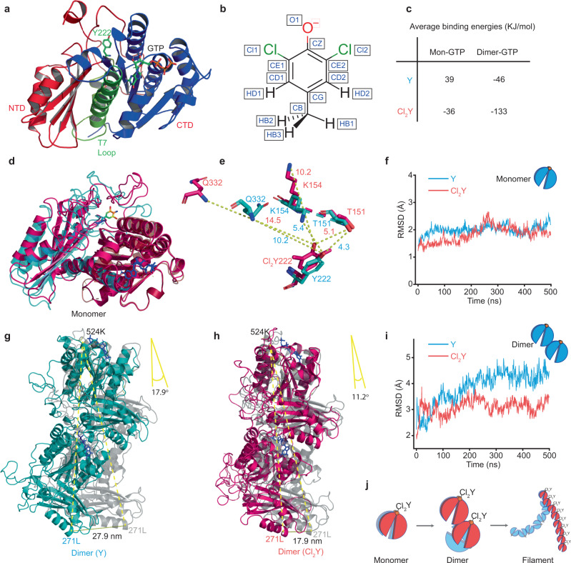
Protein halogenation is a common non-enzymatic post-translational modification contributing to aging, oxidative stress-related diseases and cancer. Here, we report a genetically encodable halogenation of tyrosine residues in a reconstituted prokaryotic filamentous cell-division protein (FtsZ) as a platform to elucidate the implications of halogenation that can be extrapolated to living systems of much higher complexity. We show how single halogenations can fine-tune protein structures and dynamics of FtsZ with subtle perturbations collectively amplified by the process of FtsZ self-organization. Based on experiments and theories, we have gained valuable insights into the mechanism of halogen influence. The bending of FtsZ structures occurs by affecting surface charges and internal domain distances and is reflected in the decline of GTPase activities by reducing GTP binding energy during polymerization. Our results point to a better understanding of the physiological and pathological effects of protein halogenation and may contribute to the development of potential diagnostic tools.


Conflict of interest statement

The authors declare no competing interests.

Figures

Fig. 1
Fig. 1. Genetic incorporation of halogenated tyrosine analogues in FtsZ based constructs.
a Schematic illustration of the FtsZ construct (FtsZ-YFP-mts): FtsZ (1–366, split blue cylinder), yellow fluorescent protein (YFP, yellow star) and a membrane target sequence (mts) (grey triangle). V and V0 indicate the treadmilling speed. b Schematic model for inhibition of FtsZ ring formation by halogenated residues. c Molecular properties of the tyrosine analogues as dependent from halogenation. Experimental lipophilicity was determined in methyl N-acetylaminoacetates against buffers at different pH values, pKa of the phenolic side-chain was experimentally determined in free amino acids, molecular volume is calculated for free amino acids. d Chemical structures of the halogenated tyrosine analogues incorporated into FtsZ and the deconvoluted ESI-MS spectra. The expected and observed masses are listed in Supplementary Table 3. e GTPase activities of wild type FtsZ-YFP-mts (Y) and halogenated FtsZ(Y222X)-YFP-mts (X = ClY, BrY, Br2Y, I2Y, Cl2Y, and IY). Data from two independent replicates are shown as means with standard deviations. f Dynamic light scattering of wild type FtsZ-YFP-mts and halogenated FtsZ-YFP-mts variants. Source data of cf are provided as a Source Data file.
Fig. 2
Fig. 2. Halogenation affects FtsZ patterning and treadmilling dynamics.
a Schematic illustrating FtsZ self-organization on model membrane monitored by TIRF. b Representative cytoskeletal pattern of wild type FtsZ-YFP-mts (Y) and halogenated FtsZ(Y222X)-YFP-mts (X = ClY, BrY, Br2Y, I2Y, ClY, and IY) on supported membrane (0.5 μM proteins, 4 mM GTP and 1 mM Mg2+). Scale bar: 3 μm. The experiment was performed three times under identical conditions. c Ring size distributions of wild type FtsZ-YFP-mts and halogenated FtsZ-YFP-mts. D indicates the ring diameter. Analysis of Variance (ANOVA) one-way statistical test (p1 = 6.16 × 10−18; p2 = 4.91 × 10−5, p3 = 1.28 × 10−7; p4 = 1.18 × 10−20). d Representative kymograph along the circumference of the vortices formed by wild type FtsZ-YFP-mts and halogenated FtsZ-YFP-mts. The respective slopes (red lines) correspond to the treadmilling velocity (V) of the vortices. e Velocity distributions for wild type FtsZ-YFP-mts and halogenated FtsZ-YFP-mts. V indicates the ring velocity. Analysis of Variance (ANOVA) one-way statistical test (p1 = 9.57 × 10−18; p2 = 2.01 × 10−13, p3 = 6.08 × 10−21; p4 = 4.92 × 10−39). f Filament curvature distributions for wild type FtsZ-YFP-mts (n = 1700 filaments) and FtsZ(Y222 Cl2Y/IY)-YFP-mts (n = 700 filaments). Wild type FtsZ-YFP-mts (Y) is shown in black, FtsZ(Y222Cl2Y)-YFP-mts (Cl2Y) is red and FtsZ(Y222IY)-YFP-mts is blue (IY). 1/R represents the filament curvature. Analysis of Variance (ANOVA) one-way statistical test (p1 = 6.00 × 10−25; p2 = 3.19 × 10−4). Box plots in f: the lines represent medians, box limits represent quartiles 1 and 3, whiskers represent 1.5 × interquartile range and points are outliers. g Snapshots and h fluorescence recovery curves for wild type FtsZ-YFP-mts and FtsZ(Y222Cl2Y)-YFP-mts after photo-bleaching. Scale bar: 3 μm. Inset: The half-life of fluorescence recovery. Data from three independent replicates are shown as means with standard deviations. Analysis of Variance (ANOVA) one-way statistical test (p1 = 1.28 × 10−5). The solid curves in c and e represent the Gaussian fit. The solid curves in f represent the extreme fit. Source data of c, f and h are provided as a Source Data file.
Fig. 3
Fig. 3. Partial halogenated FtsZ disrupts dynamic pattern formations of wild type FtsZ.
a Schematic representation of the self-organization assay with wild type FtsZ-YFP-mts (Y) and FtsZ(Y222Cl2Y)-YFP-mts (Cl2Y) at certain proportions. b Representative cytoskeleton images of wild type FtsZ-YFP-mts and FtsZ(Y222Cl2Y)-YFP-mts at certain proportions on supported membrane (0.5 µM proteins, 4 mM GTP and 1 mM Mg2+). Scale bar: 5 μm. The experiment was performed three times under identical conditions. c Ring densities for wild type FtsZ-YFP-mts mixed with FtsZ(Y222Cl2Y)-YFP-mts. Data from three independent replicates are shown as means with standard deviations. d Distributions of curvature (left), ring size (middle), and ring velocity (right) of wild type FtsZ-YFP-mts mixed with FtsZ(Y222Cl2Y)-YFP-mts in certain proportions. Curvatures in d. were calculated for the mixture of ring and filaments, representing the overall curvatures of the bulk reaction. The solid curves in d (left panel) represent the extreme fit for the histograms (n100 = 829, n75 = 781, n50 = 312, n25 = 217, n0 = 1475). The histograms of the curvatures are not shown. Box plots in d (inset of left panel): the lines represent medians, box limits represent quartiles 1 and 3, whiskers represent 1.5 × interquartile range and points are outliers. The solid curves in the middle and right panels represent the Gaussian fit. Source data of c, d are provided as a Source Data file. D, V and 1/R in Fig. 3 indicate the ring diameter, velocity and curvature separately.
Fig. 4
Fig. 4. Structure simulations of halogenated FtsZ.
a Monomer structure of wild type FtsZ. The main protein body is shown in cartoon representation, GTP and Tyr222 are shown in licorice representation. The NTD is colored red, CTD is blue and the central H7 helix is shown in green. b Charge calculation of 3,5-dichloro-tyrosine. c Average binding energies calculated according to the protocol described in Methods section using the Adaptive Poisson-Boltzmann Solver (APBS) software with time frames from the last 100 ns of MD simulations. Two sets of interactions were analyzed: Mon-GTP describes the interaction between monomer body and Mg2+-GTP; Di-GTP illustrates the interactions between dimer body and Mg2+-GTP at dimer interface. d Structural alignment of FtsZ(Y222Cl2Y) (Red) with the wild type FtsZ (cyan). e Overlay of the mutant sites of wild type FtsZ(cyan) and FtsZ(Y222Cl2Y) (red), showing the conformational changes induced by the Y222Cl2Y mutation. f Time evolution of the backbone RMSD of FtsZ monomer (backbone atoms only) after alignment to the energy-minimized FtsZ(Y222Cl2Y) model. Residues 13 to 316 are shown, excluding the disordered regions of sequence (head and C-terminal tail). g, h The bending motion of dimer models. Dimer alignments of g wild type FtsZ (cyan) and h FtsZ(Y222Cl2Y) (red) with their energy-minimized models (grey). i Time evolution of the backbone RMSD of the FtsZ dimer when aligned to same selection of reference dimer structures, after energy minimization using AMBER software in the dielectric constant (ε = 4). Residues 13–316 are shown for each monomer. j The scheme illustrates protein self-organization amplify the changes of structure by site-specific halogenation. Source data of f and i are provided as a Source Data file.

Similar articles

Cited by

References

    1. Harmel R, Fiedler D. Features and regulation of non-enzymatic post-translational modifications. Nat. Chem. Biol. 2018;14:244–252. doi: 10.1038/nchembio.2575. - DOI - PubMed
    1. Pimviriyakul P, Wongnate T, Tinikul R, Chaiyen P. Microbial degradation of halogenated aromatics: molecular mechanisms and enzymatic reactions. Microb. Biotechnol. 2020;13:67–86. doi: 10.1111/1751-7915.13488. - DOI - PMC - PubMed
    1. Feeney MB, Schöneich C. Tyrosine modifications in aging. Antioxid. Redox Signal. 2012;17:1571–1579. doi: 10.1089/ars.2012.4595. - DOI - PMC - PubMed
    1. Olaniran AO, Igbinosa EO. Chlorophenols and other related derivatives of environmental concern: properties, distribution and microbial degradation processes. Chemosphere. 2011;83:1297–1306. doi: 10.1016/j.chemosphere.2011.04.009. - DOI - PubMed
    1. Santos, A. L. & Lindner, A. B. Protein posttranslational modifications: roles in aging and age-related disease. Oxid. Med. Cell. Longev. 2017, 1–19 (2017). - PMC - PubMed

Publication types

MeSH terms