. 2022 Aug 17;13(1):4829.
doi: 10.1038/s41467-022-31859-3.

A nanoengineered topical transmucosal cisplatin delivery system induces anti-tumor response in animal models and patients with oral cancer

Affiliations

A nanoengineered topical transmucosal cisplatin delivery system induces anti-tumor response in animal models and patients with oral cancer

Manijeh Goldberg et al. Nat Commun. .

Erratum in

Abstract

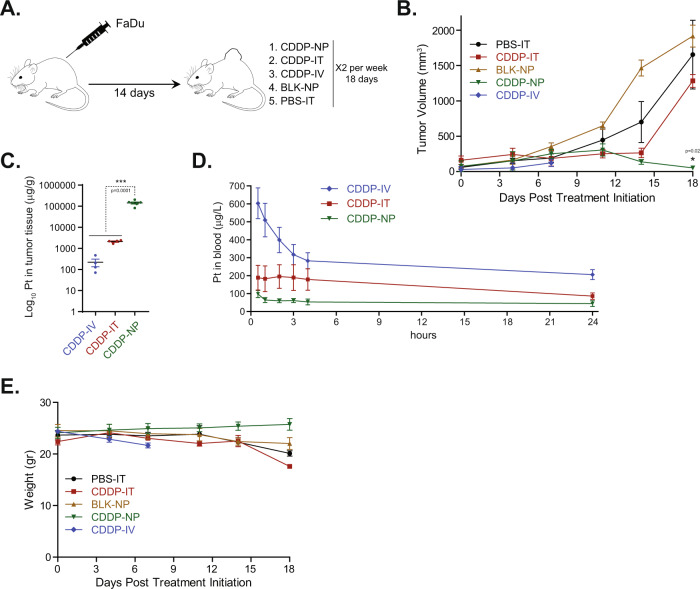
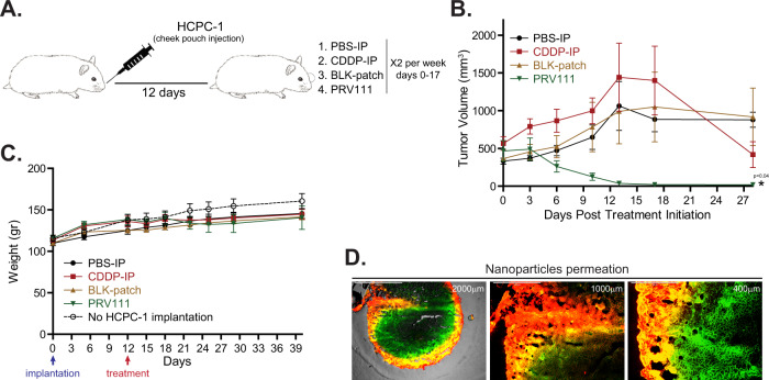
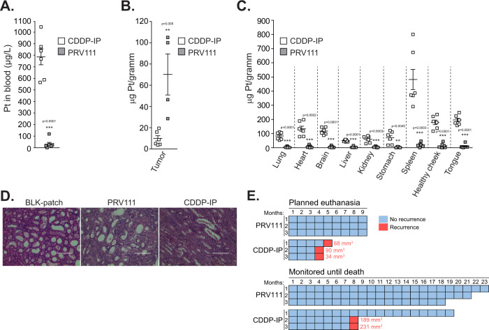
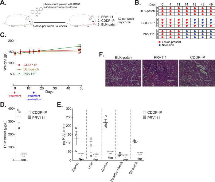
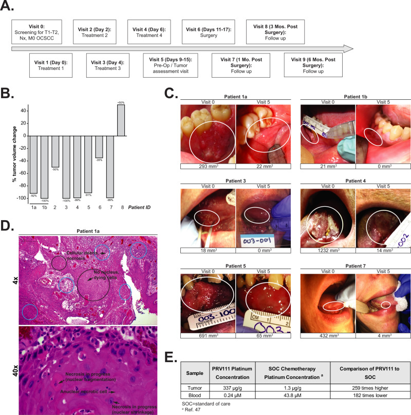
Despite therapeutic advancements, oral cavity squamous cell carcinoma (OCSCC) remains a difficult disease to treat. Systemic platinum-based chemotherapy often leads to dose-limiting toxicity (DLT), affecting quality of life. PRV111 is a nanotechnology-based system for local delivery of cisplatin loaded chitosan particles, that penetrate tumor tissue and lymphatic channels while avoiding systemic circulation and toxicity. Here we evaluate PRV111 using animal models of oral cancer, followed by a clinical trial in patients with OCSCC. In vivo, PRV111 results in elevated cisplatin retention in tumors and negligible systemic levels, compared to the intravenous, intraperitoneal or intratumoral delivery. Furthermore, PRV111 produces robust anti-tumor responses in subcutaneous and orthotopic cancer models and results in complete regression of carcinogen-induced premalignant lesions. In a phase 1/2, open-label, single-arm trial (NCT03502148), primary endpoints of efficacy (≥30% tumor volume reduction) and safety (incidence of DLTs) of neoadjuvant PRV111 were reached, with 69% tumor reduction in ~7 days and over 87% response rate. Secondary endpoints (cisplatin biodistribution, loco-regional control, and technical success) were achieved. No DLTs or drug-related serious adverse events were reported. No locoregional recurrences were evident in 6 months. Integration of PRV111 with current standard of care may improve health outcomes and survival of patients with OCSCC.


Conflict of interest statement

M.G., A.M., A.B., B.L., P.C., S.C., B.F. and E.R.G. are affiliated with Privo Technologies. N.A. and S.G. serve as advisors for Privo Technologies. The remaining authors declare no competing interests.

Figures

Fig. 1
Fig. 1. Cisplatin chitosan nanoparticles (CDDP-NP) demonstrate improved local drug retention and induce potent anti-tumor response in FaDu tumor bearing mice xenografts.
A Schematic representation of the experiment. B Nude mice xenografts bearing subcutaneous human FaDu HNSCC cell line induced tumors were treated with either PBS-IT, CDDP-IT, CDDP-IV, BLK-NP, or CDDP-NP. Graphs show the average tumor volume for 6 animals per group ± SEM. P-value was performed by a 1-sided Wilcoxon Rank-sum Test. Asterisk represents statistical significance between the CDDP-IT and CDDP-NP groups (p < 0.05). C Residual tumors were harvested after 4 treatments (day 14) for CDDP-NP (n = 6) or CDDP-IT (n = 4) and 2 treatments (day 7) for CDDP-IV (n = 4). Samples were weighted, homogenized and cisplatin level was quantified by ICP-MS, shown as average ± SEM (p < 0.0001; Student’s unpaired t-test). D Blood was collected from 4 animals per group after administration of the first dose at indicated time points and level of cisplatin was quantified by ICP-MS, shown as average ± SEM. E Graph shows the average body weights for 6 animals per group ± SEM. Source data are provided as a Source Data file.
Fig. 2
Fig. 2. Local application of PRV111 induced robust anti-tumor response in hamster orthotopic oral cancer model.
A Schematic representation of the experiment. B Golden Syrian hamsters bearing orthotopic tumors induced by injection of HCPC-1 cell line into the cheek pouch were treated with either PBS-IP, CDDP-IP, BLK-patch or PRV111. Graphs show the average tumor volume for 6 animals per group ± SEM. P-value was performed by a 1-sided Wilcoxon Rank-sum method. Asterisk represents statistical significance between the CDDP-IP and PRV111 groups (p < 0.05). C Graph shows the average body weights for 6 animals per group ± SEM. Body weights of 5 tumor free animals without treatment were measure along the tumor bearing counterparts (dashed line). D Representative fluorescence images of tumor sections at indicated magnification taken after treatment of the hamster with PRV111 patch containing chitosan particles labeled with Cy5 (red) and encapsulating FITC (green). Yellow areas display dual-labeling, NPs with encapsulated FITC. Permeation experiment was repeated in 6 tumors. Source data are provided as a Source Data file.
Fig. 3
Fig. 3. Topical administration of PRV111 reduced cisplatin associated toxicities and prevented tumor recurrence in vivo.
A Blood was collected after administration of the first dose of CDDP-IP or PRV111 (6 animals per group), and level of cisplatin was quantified by ICP-MS, shown as average ± SEM (p = 0.0001; Student’s unpaired t-test). B, C Hamsters treated with CDDP-IP or PRV111 (6 animals per group) were sacrificed and residual tumors as well as lungs, heart, brain, liver, kidneys, stomach, spleen, healthy contralateral cheek pouch, and tongue were harvested. Samples were weighted, processed, and biodistribution of cisplatin was assessed by ICP-MS, shown as average ± SEM (p = 0.005 for tumor tissue all body organs analyzed; Student’s unpaired t-test). Note, residual tumors were collected from four animals treated with PRV111. D Representative H&E stained histopathological images of kidneys (×200; scale bar—100 μm). Three tumors per group were stained. E Animals that were tumor free after treatment with either PRV111 or CDDP-IP (6 per group) were monitored for recurrence and survival during the indicated time period (months) post treatment cessation. Animals were evaluated and weighted weekly until day 45, and once a month thereafter. Cyan squares indicate a month when animal remained tumor-free. Pink squares indicate a month when recurrence was detected. Tumor volume upon recurrence is provided next to the each pink square. Source data are provided as a Source Data file.
Fig. 4
Fig. 4. PRV111 induced rapid and sustainable regression of carcinogen-induced oral premalignant lesions.
A Schematic representation of the experiment. B Golden Syrian hamsters bearing DMBA-induced oral dysplastic lesion were treated with either BLK-patch, CDDP-IP or PRV111 (3 animals per group). Red circle indicates presence of the premalignant lesion, whereas blue circle indicates that the premalignant lesions was undetected. C Graph shows the average body weights for the 3 hamsters in each indicated group  ± SEM. D Blood was collected from 3 animals per group after administration of the first dose of CDDP-IP or PRV111, and level of cisplatin was quantified by ICP-MS, shown as average ± SEM (p < 0.0019; Student’s unpaired t-test). E Hamsters treated with CDDP-IP or PRV111 (n = 3) were sacrificed and indicated organs were harvested, weighted, processed, and biodistribution of cisplatin was assessed by ICP-MS, shown as average ± SEM (p < 0.05 for all body organs analyzed; Student’s unpaired t-test). F Representative H&E stained histopathological images of kidneys (200×; scale bar—100 μm). Three tumors per group were stained. Source data are provided as a Source Data file.
Fig. 5
Fig. 5. PRV111 induced a rapid anti-tumor response in patients with locally advanced OCSCC.
A Schematic representation of the clinical trial timeline. B Tumor volume changes from baseline and end of study for each patient treated with PRV111. C Tumor photographs and volume measurements for subjects which achieved >90% tumor volume reduction post RPV111 treatment. D Representative H&E image of post-PRV111 treatment tumor sample. Top: Background of residual cancer cells (black circles) and evidence of treatment effect in the form of necrosis/ulceration, cellular debris, and acute/chronic inflammation (blue circles). 4×; scale bar – 1000um. Bottom: Area of minimal residual cancer cells and evidence of treatment effect in the form of necrosis/ulceration and cellular debris, as indicated by black arrows. 40×; scale bar—100μm. E Biodistribution of platinum detected in the blood, and tumor tissue following PRV111 therapy compared with standard of care cisplatin treatment.
Fig. 6
Fig. 6. PRV111 increased the number of TILs.
A Fold change increase in TILs after treatment of 8 patients with PRV111 relative to pretreatment biopsies, shown as average ± SEM (p < 0.001; Student’s unpaired t-test). Four slides were analyzed for each specimen and average was used for fold change calculation. B Representative images (10×; scale bar—1000μm) of pre- and post-treatment sections stained for CD3, CD8, and CD4 T cells. Changes in brown staining indicates increase in tumor-infiltrating lymphocytes following PRV111 treatment. Source data are provided as a Source Data file.

Similar articles

Cited by

References

    1. Howlader N, N. A. et al. (eds). SEER Cancer Statistics Review, 1975-2017, National Cancer Institute. Bethesda, MD, https://seer.cancer.gov/csr/1975_2017/, based on November 2019 SEER data submission, posted to the SEER web site, April 2020. (2020).
    1. Kramer S, et al. Combined radiation-therapy and surgery in the management of advanced head and neck-cancer - final report of study 73-03 of the radiation-therapy-oncology-group. Head. Neck Surg. 1987;10:19–30. doi: 10.1002/hed.2890100105. - DOI - PubMed
    1. Brizel DM, et al. Hyperfractionated irradiation with or without concurrent chemotherapy for locally advanced head and neck cancer. N. Engl. J. Med. 1998;338:1798–1804. doi: 10.1056/NEJM199806183382503. - DOI - PubMed
    1. Cohen EE, Lingen MW, Vokes EE. The expanding role of systemic therapy in head and neck cancer. J. Clin. Oncol. 2004;22:1743–1752. doi: 10.1200/JCO.2004.06.147. - DOI - PubMed
    1. Pignon JP, Bourhis J, Domenge C, Designe L, Grp MNC. Chemotherapy added to locoregional treatment for head and neck squamous-cell carcinoma: three meta-analyses of updated individual data. Lancet. 2000;355:949–955. doi: 10.1016/S0140-6736(00)90011-4. - DOI - PubMed

Publication types

MeSH terms

Associated data