Working correlates of protection predict SchuS4-derived-vaccine candidates with improved efficacy against an intracellular bacterium, Francisella tularensis
- PMID: 35977964
- PMCID: PMC9385090
- DOI: 10.1038/s41541-022-00506-9
Working correlates of protection predict SchuS4-derived-vaccine candidates with improved efficacy against an intracellular bacterium, Francisella tularensis
Abstract
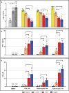
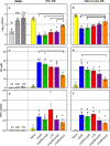
Francisella tularensis, the causative agent of tularemia, is classified as Tier 1 Select Agent with bioterrorism potential. The efficacy of the only available vaccine, LVS, is uncertain and it is not licensed in the U.S. Previously, by using an approach generally applicable to intracellular pathogens, we identified working correlates that predict successful vaccination in rodents. Here, we applied these correlates to evaluate a panel of SchuS4-derived live attenuated vaccines, namely SchuS4-ΔclpB, ΔclpB-ΔfupA, ΔclpB-ΔcapB, and ΔclpB-ΔwbtC. We combined in vitro co-cultures to quantify rodent T-cell functions and multivariate regression analyses to predict relative vaccine strength. The predictions were tested by rat vaccination and challenge studies, which demonstrated a clear relationship between the hierarchy of in vitro measurements and in vivo vaccine protection. Thus, these studies demonstrated the potential power a panel of correlates to screen and predict the efficacy of Francisella vaccine candidates, and in vivo studies in Fischer 344 rats confirmed that SchuS4-ΔclpB and ΔclpB-ΔcapB may be better vaccine candidates than LVS.
© 2022. This is a U.S. Government work and not under copyright protection in the US; foreign copyright protection may apply.
Conflict of interest statement
The authors declare competing interests. Patent application and Patent applicants: National Research Council of Canada. Name of inventor(s): Joseph Wayne Conlan and Anders Sjostedt. Application numbers: US8993302B2 (granted), EP2424974B1 (granted), CA2760098C (granted), ES2553763T3 (granted), US20210008191A1 (pending), CA3094404A1 (pending), EP3768820A1 (pending). Specific aspect of manuscript covered in the patent application: “The present invention relates to a mutant
Figures


Similar articles
-
In vivo and in vitro immune responses against Francisella tularensis vaccines are comparable among Fischer 344 rat substrains.Front Microbiol. 2023 Jul 13;14:1224480. doi: 10.3389/fmicb.2023.1224480. eCollection 2023. Front Microbiol. 2023. PMID: 37547680 Free PMC article.
-
A Francisella tularensis live vaccine strain (LVS) mutant with a deletion in capB, encoding a putative capsular biosynthesis protein, is significantly more attenuated than LVS yet induces potent protective immunity in mice against F. tularensis challenge.Infect Immun. 2010 Oct;78(10):4341-55. doi: 10.1128/IAI.00192-10. Epub 2010 Jul 19. Infect Immun. 2010. PMID: 20643859 Free PMC article.
-
Development of functional and molecular correlates of vaccine-induced protection for a model intracellular pathogen, F. tularensis LVS.PLoS Pathog. 2012 Jan;8(1):e1002494. doi: 10.1371/journal.ppat.1002494. Epub 2012 Jan 19. PLoS Pathog. 2012. PMID: 22275868 Free PMC article.
-
Vaccination strategies for Francisella tularensis.Adv Drug Deliv Rev. 2005 Jun 17;57(9):1403-14. doi: 10.1016/j.addr.2005.01.030. Adv Drug Deliv Rev. 2005. PMID: 15919131 Review.
-
Progress, challenges, and opportunities in Francisella vaccine development.Expert Rev Vaccines. 2016 Sep;15(9):1183-96. doi: 10.1586/14760584.2016.1170601. Epub 2016 May 3. Expert Rev Vaccines. 2016. PMID: 27010448 Review.
Cited by
-
Analyses of human immune responses to Francisella tularensis identify correlates of protection.Front Immunol. 2023 Sep 15;14:1238391. doi: 10.3389/fimmu.2023.1238391. eCollection 2023. Front Immunol. 2023. PMID: 37781364 Free PMC article.
-
In vivo and in vitro immune responses against Francisella tularensis vaccines are comparable among Fischer 344 rat substrains.Front Microbiol. 2023 Jul 13;14:1224480. doi: 10.3389/fmicb.2023.1224480. eCollection 2023. Front Microbiol. 2023. PMID: 37547680 Free PMC article.
-
The rLVS ΔcapB/iglABC vaccine provides potent protection in Fischer rats against inhalational tularemia caused by various virulent Francisella tularensis strains.Hum Vaccin Immunother. 2023 Dec 15;19(3):2277083. doi: 10.1080/21645515.2023.2277083. Epub 2023 Nov 17. Hum Vaccin Immunother. 2023. PMID: 37975637 Free PMC article.
-
Oropharyngeal Tularemia: A Case From Rural Armenia Highlighting Diagnostic Challenges and Treatment Outcomes.Cureus. 2025 May 19;17(5):e84439. doi: 10.7759/cureus.84439. eCollection 2025 May. Cureus. 2025. PMID: 40539185 Free PMC article.
-
Current vaccine strategies and novel approaches to combatting Francisella infection.Vaccine. 2024 Apr 2;42(9):2171-2180. doi: 10.1016/j.vaccine.2024.02.086. Epub 2024 Mar 8. Vaccine. 2024. PMID: 38461051 Free PMC article. Review.
References
-
- Eigelsbach HT, Downs CM. Prophylactic effectiveness of live and killed tularemia vaccines. J. Immunol. 1961;87:415–425. - PubMed
Grants and funding
LinkOut - more resources
Full Text Sources

