. 2022 Aug 17;12(1):13955.
doi: 10.1038/s41598-022-17716-9.

Low pH structure of heliorhodopsin reveals chloride binding site and intramolecular signaling pathway

Affiliations

Low pH structure of heliorhodopsin reveals chloride binding site and intramolecular signaling pathway

Jessica E Besaw et al. Sci Rep. .

Abstract

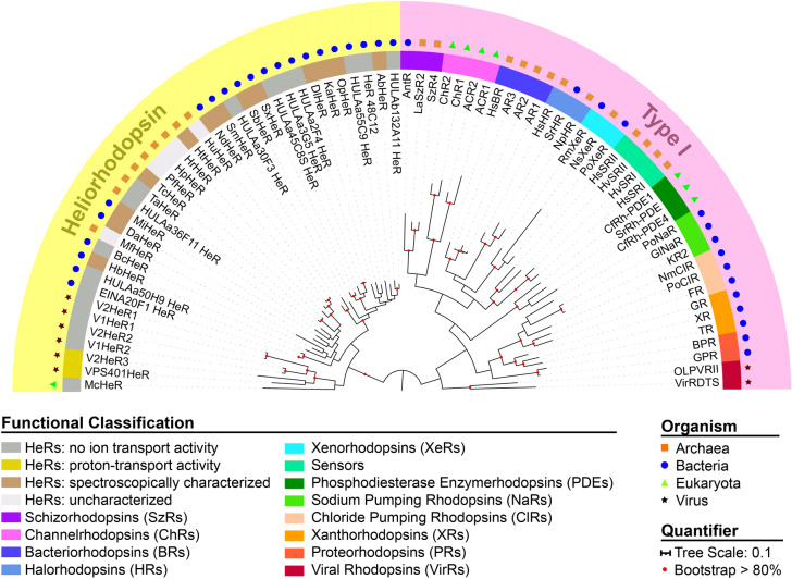
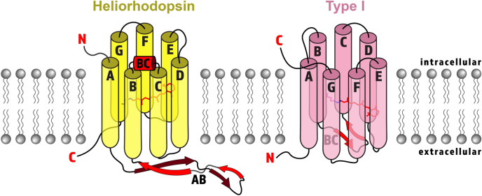
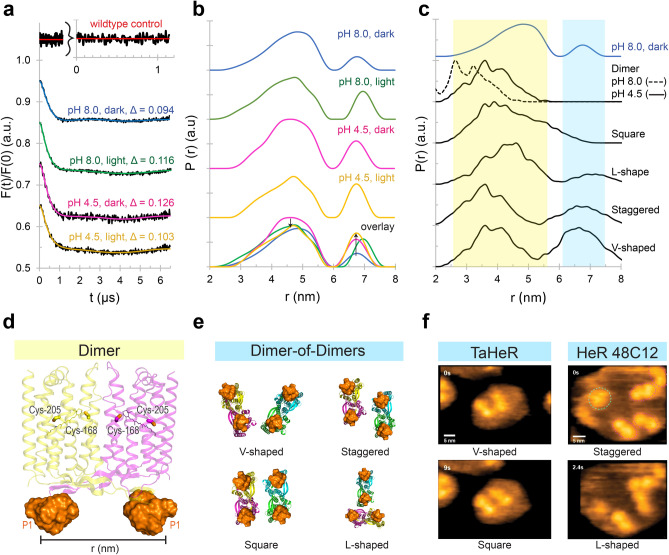
Within the microbial rhodopsin family, heliorhodopsins (HeRs) form a phylogenetically distinct group of light-harvesting retinal proteins with largely unknown functions. We have determined the 1.97 Å resolution X-ray crystal structure of Thermoplasmatales archaeon SG8-52-1 heliorhodopsin (TaHeR) in the presence of NaCl under acidic conditions (pH 4.5), which complements the known 2.4 Å TaHeR structure acquired at pH 8.0. The low pH structure revealed that the hydrophilic Schiff base cavity (SBC) accommodates a chloride anion to stabilize the protonated retinal Schiff base when its primary counterion (Glu-108) is neutralized. Comparison of the two structures at different pH revealed conformational changes connecting the SBC and the extracellular loop linking helices A-B. We corroborated this intramolecular signaling transduction pathway with computational studies, which revealed allosteric network changes propagating from the perturbed SBC to the intracellular and extracellular space, suggesting TaHeR may function as a sensory rhodopsin. This intramolecular signaling mechanism may be conserved among HeRs, as similar changes were observed for HeR 48C12 between its pH 8.8 and pH 4.3 structures. We additionally performed DEER experiments, which suggests that TaHeR forms possible dimer-of-dimer associations which may be integral to its putative functionality as a light sensor in binding a transducer protein.


Conflict of interest statement

The authors declare no competing interests.

Figures

Figure 1
Figure 1
Phylogenetic tree of microbial rhodopsins with representative HeRs and type I rhodopsins. HeRs and type I rhodopsins form distinct branches arising from < 15% sequence identity. Microbial rhodopsins are widely distributed among archaea (orange squares), bacteria (blue circles), eukaryota (green triangles), and viruses (black stars). Type I rhodopsins have numerous diverse functions including outward H+ pumps (BRs, XRs, PRs, VirRs), inward H+ pumps (XeRs, SzRs), outward Cl pumps (HRs, ClRs), outward Na+ pumps (NaRs), anion and cation channels (ChRs), sensors, and enzymerhodopsins (including PDEs). A small group of viral HeRs show proton-transport activity (gold). Many HeRs have no ion transport activity (grey). Some HeRs that have been spectroscopically characterized only (brown) or are uncharacterized (white) are also shown. The tree scale bar shows the average number of amino acid substitutions per site. Red colored circles indicate bootstrap values > 80% for 100 replicates. See Supplementary Table S1 for the full names of proteins and sequence (NCBI searchable codes).
Figure 2
Figure 2
Cartoon structure representation of HeRs (yellow) and type I rhodopsins (pink) with major structural differences emphasized in red. Both HeRs and type I rhodopsins have seven transmembrane α-helices labelled A–G (except enzymerhodopsin, which has eight helices), with a covalently linked retinal chromophore (red sticks) bound to a lysine (pink or yellow sticks) by a Schiff base linkage (blue sticks) on helix G. HeRs have an inverted orientation in the membrane with the N-terminus (red N) facing inward and C-terminus (red C) facing outward, which consequently flips the retinal orientation. HeRs possess a long extracellular A–B loop (AB) composed of a twisted β-sheet (red arrows), and an intracellular B–C loop forming an α-helix (red cylinder labelled BC). In contrast, type I rhodopsins typically have an extracellular B–C loop forming a β-sheet.
Figure 3
Figure 3
Electron density map and model of TaHeR crystallized at pH 4.5. (a) Asymmetric unit of TaHeR with electron density of water molecules (red spheres), chloride ions (green spheres) and retinal (yellow sticks). A red box highlights a single water molecule present in the highly hydrophobic extracellular half of TaHeR. (b) Electron density map of the SBC, with black dashed lines revealing a water-mediated hydrogen bonding network. (c) Electron density map of the protein backbone with the corresponding residue numbers listed above. Conserved residues in the suggested intramolecular signal transduction pathway have been labelled. For all images, the 2Fo − Fc map is contoured at 1 σ (blue mesh), while Fo–Fc is contoured at − 3 σ (red mesh) and in green at + 3 σ (green mesh).
Figure 4
Figure 4
Schiff base cavity (SBC). SBC (grey surface) of (a) TaHeR and (b) HeR 48C12 under acidic and basic conditions. Under slightly basic conditions, the SBC of both TaHeR (PDB ID 6is6) and HeR 48C12 (PDB ID 6su3) contain only water molecules (red spheres) and are capped by two ionic interactions: (1) the RSBH+/Glu counterion pair and (2) the Arg/Glu pair. Under acidic conditions, the Glu counterion accepts a proton and is neutral. The SBC of TaHeR accommodates a negatively charged chloride ion (green sphere) while HeR 48C12 (PDB ID 6su4) accommodates a negatively charged acetate ion to maintain charge balance with the RSBH+. The negative charges are indicated by red dashes, while the positive charges are shown by blue pluses. ‡ Weak electron density adjacent to Ser-76 in the pH 4.3 HeR 48C12 structure has been modeled as a water molecule, but this molecule may also be absent, as in TaHeR.
Figure 5
Figure 5
A putatively conserved intramolecular signaling pathway for TaHeR and HeR 48C12. (a) TaHeR dimer structure at pH 8 (cyan, PDB ID 6is6) and pH 4.5 (yellow, PDB ID 7u55). (b) HeR 48C12 dimer structure at pH 8.8 (green, PDB ID 6su3) and pH 4.3 (pink, PDB ID 6su4). The conserved intramolecular signaling mechanism involves a negatively charged anion (chloride or acetate) occupying the SBC (grey surface), switching the orientation of His-23, displacing water molecules (red arrows), disordering Gln-26 and disordering Trp-243 (in TaHeR) or Trp-246 (in HeR 48C12). The overlay reveals there is a 1 Å movement of the intracellular B–C loop. TaHeR has an additional 6 Å shift of the A − B tail from the electrostatic attraction of Tyr-48 and Asn-53 to an external chloride ion. Waters are shown as small spheres colored to match protein, and chloride ions as large green spheres (belonging to pH 4.5 TaHeR only). The 2Fo − Fc maps (blue mesh) are contoured at 1 σ, and Fo − Fc is contoured at + 3 σ for positive (green mesh) and − 3 σ for negative (red mesh) electron density, respectively. ‡ In HeR 48C12 at pH 4.3, there is no electron density to account for this water in the chain A protomer.
Figure 6
Figure 6
Allosteric transmission and conformational dynamics modelling in TaHeR and HeR 48C12 dimers. (a, c) The allosteric network within HeR dimers is revealed through rigidity theory allostery analysis. Allosteric transmission is measured by changes in conformational degrees of freedom (red/blue gradient bar) experienced upon rigidification of the negatively charged ion (chloride or acetate, green) occupying the SBC (grey surface) of the acidic pH structure. Amino acids within 3 Å of the charged ion (grey portions of the helix) were omitted from the coloring scheme to prevent skewed coloring biases, as neighbouring residues would naturally have high allosteric effects. (b, d) Constrained geometric Monte Carlo-based dynamics analysis predicts individual residue dynamics measured as the root mean squared fluctuation (RMSF). The extracellular β-sheet (A–B loop) that was further on investigated by DEER is highlighted in pink.
Figure 7
Figure 7
DEER of TaHeR-I51P1 suggests HeR can adopt dimer-of-dimer assemblies that are pH- and light-dependent. (a) The DEER dipolar evolution function of TaHeR-I51P1 with indicated modulation depth (Δ) at pH 8 and pH 4.5 in dark and light conditions. The inset (red) shows data from wildtype TaHeR incubated with IAP spin label. Here, the lack of dipolar interactions indicated that native cysteines could not be sufficiently spin-labelled. (b) The DEER distance distribution, P(r), revealed two spatially separated peaks at 4.7 nm and 6.6 nm. The experimental parameters allow for accurate distance distributions up to 5.9 nm and mean distances up to 7.4 nm. Relative to the pH 8 dark sample, light or acidification increases the population of the 6.6 nm peak (up arrow). Relative to the pH 4.5 dark sample, illumination slightly decreases the population of the 4.7 nm peak (down arrow). DEER experiments were performed in duplicate. (c) MMM distance distributions of dimer and dimer-of-dimer assemblies compared to the pH 8 dark sample. Dimer-of-dimer assemblies were generated from AlphaFold (square, L-shaped) or PyMOL (staggered, V-shaped), with corresponding models shown in panel e. (d) Model of the TaHeR-I51P1 dimer with the P1 rotamer cloud (orange surface) from the pH 4.5 structure. The two native cysteines, Cys-168 and Cys-205, are inaccessible to spin-labelling. (e) Dimer-of-dimer models with P1 rotamer clouds (orange surface). (f) HS-AFM images of TaHeR and HeR 48C12 from the supplementary videos of ref. showing similar dimer-of-dimer assemblies. Reprinted by permission from CCC: Springer Nature Shihoya et al. (2019).

Similar articles

  • Internal Proton Transfer in the Activation of Heliorhodopsin.
    Singh M, Hashimoto M, Katayama K, Furutani Y, Kandori H. Singh M, et al. J Mol Biol. 2024 Mar 1;436(5):168273. doi: 10.1016/j.jmb.2023.168273. Epub 2023 Sep 12. J Mol Biol. 2024. PMID: 37709010
  • High-resolution structural insights into the heliorhodopsin family.
    Kovalev K, Volkov D, Astashkin R, Alekseev A, Gushchin I, Haro-Moreno JM, Chizhov I, Siletsky S, Mamedov M, Rogachev A, Balandin T, Borshchevskiy V, Popov A, Bourenkov G, Bamberg E, Rodriguez-Valera F, Büldt G, Gordeliy V. Kovalev K, et al. Proc Natl Acad Sci U S A. 2020 Feb 25;117(8):4131-4141. doi: 10.1073/pnas.1915888117. Epub 2020 Feb 7. Proc Natl Acad Sci U S A. 2020. PMID: 32034096 Free PMC article.
  • Specific zinc binding to heliorhodopsin.
    Hashimoto M, Miyagawa K, Singh M, Katayama K, Shoji M, Furutani Y, Shigeta Y, Kandori H. Hashimoto M, et al. Phys Chem Chem Phys. 2023 Jan 27;25(4):3535-3543. doi: 10.1039/d2cp04718g. Phys Chem Chem Phys. 2023. PMID: 36637167
  • Microbial Rhodopsins: The Last Two Decades.
    Rozenberg A, Inoue K, Kandori H, Béjà O. Rozenberg A, et al. Annu Rev Microbiol. 2021 Oct 8;75:427-447. doi: 10.1146/annurev-micro-031721-020452. Epub 2021 Aug 3. Annu Rev Microbiol. 2021. PMID: 34343014 Review.
  • Coupling of protonation switches during rhodopsin activation.
    Vogel R, Sakmar TP, Sheves M, Siebert F. Vogel R, et al. Photochem Photobiol. 2007 Mar-Apr;83(2):286-92. doi: 10.1562/2006-06-19-IR-937. Photochem Photobiol. 2007. PMID: 17576345 Review.

Cited by

References

    1. Pushkarev A, et al. A distinct abundant group of microbial rhodopsins discovered using functional metagenomics. Nature. 2018;558:595–599. doi: 10.1038/s41586-018-0225-9. - DOI - PMC - PubMed
    1. Rozenberg A, Inoue K, Kandori H, Beja O. Microbial rhodopsins: The last two decades. Annu. Rev. Microbiol. 2021;75:427–447. doi: 10.1146/annurev-micro-031721-020452. - DOI - PubMed
    1. Shibukawa A, et al. Photochemical characterization of a new heliorhodopsin from the gram-negative eubacterium Bellilinea caldifistulae (BcHeR) and comparison with heliorhodopsin-48C12. Biochemistry. 2019;58:2934–2943. doi: 10.1021/acs.biochem.9b00257. - DOI - PubMed
    1. Chazan A, et al. Diverse heliorhodopsins detected via functional metagenomics in freshwater Actinobacteria, Chloroflexi and Archaea. Environ. Microbiol. 2022;24:110–121. doi: 10.1111/1462-2920.15890. - DOI - PubMed
    1. Kovalev K, et al. High-resolution structural insights into the heliorhodopsin family. Proc. Natl. Acad. Sci. USA. 2020;117:4131–4141. doi: 10.1073/pnas.1915888117. - DOI - PMC - PubMed

Publication types