. 2022 Aug 17;13(1):4846.
doi: 10.1038/s41467-022-32599-0.

MARTX toxin of Vibrio vulnificus induces RBC phosphatidylserine exposure that can contribute to thrombosis

Affiliations

MARTX toxin of Vibrio vulnificus induces RBC phosphatidylserine exposure that can contribute to thrombosis

Han Young Chung et al. Nat Commun. .

Abstract

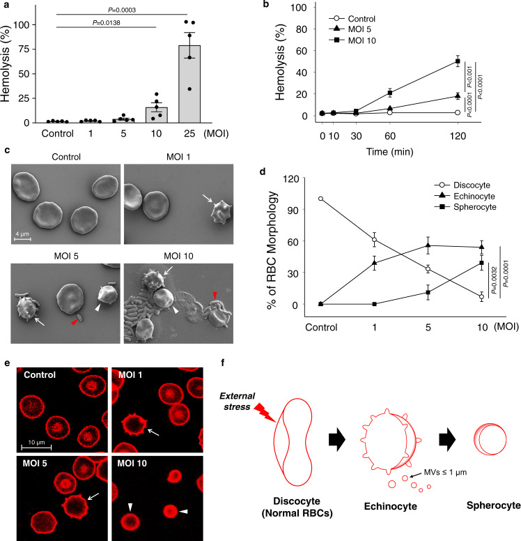
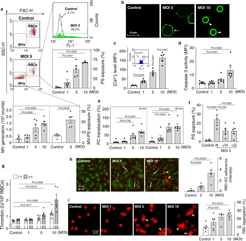
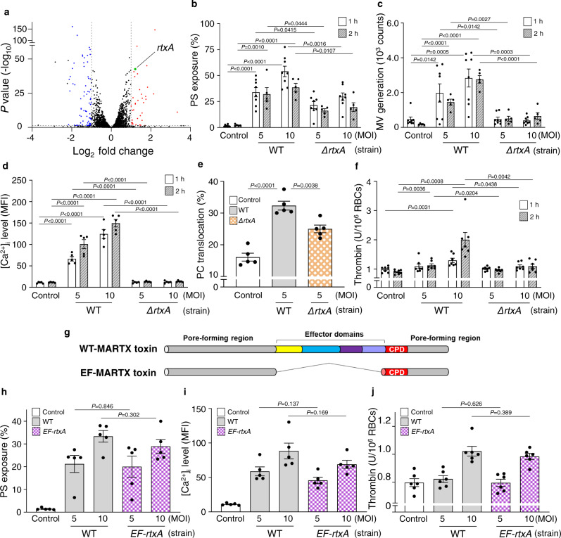
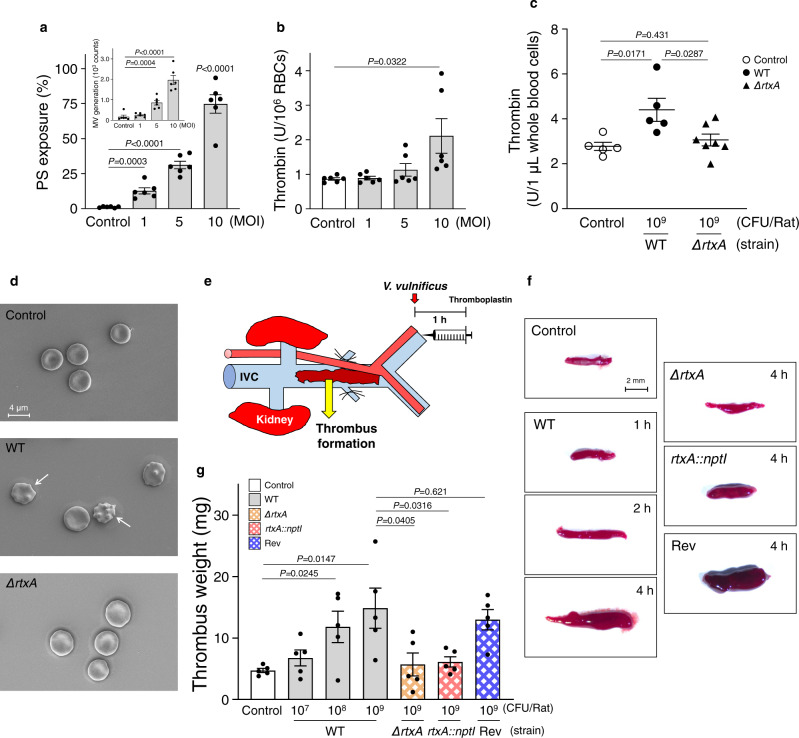
V. vulnificus-infected patients suffer from hemolytic anemia and circulatory lesions, often accompanied by venous thrombosis. However, the pathophysiological mechanism of venous thrombosis associated with V. vulnificus infection remains largely unknown. Herein, V. vulnificus infection at the sub-hemolytic level induced shape change of human red blood cells (RBCs) accompanied by phosphatidylserine exposure, and microvesicle generation, leading to the procoagulant activation of RBCs and ultimately, acquisition of prothrombotic activity. Of note, V. vulnificus exposed to RBCs substantially upregulated the rtxA gene encoding multifunctional autoprocessing repeats-in-toxin (MARTX) toxin. Mutant studies showed that V. vulnificus-induced RBC procoagulant activity was due to the pore forming region of the MARTX toxin causing intracellular Ca2+ influx in RBCs. In a rat venous thrombosis model triggered by tissue factor and stasis, the V. vulnificus wild type increased thrombosis while the ΔrtxA mutant failed to increase thrombosis, confirming that V. vulnificus induces thrombosis through the procoagulant activation of RBCs via the mediation of the MARTX toxin.


Conflict of interest statement

The authors declare no competing interests.

Figures

Fig. 1
Fig. 1. Infection of human RBCs with V. vulnificus induces hemolysis and shape change.
a, b RBCs were infected with V. vulnificus at various MOIs and incubated for 1 h (a) (n = 5, two-tailed Student’s t-test) or for different times (b) (n = 6, two-way analysis of variance (ANOVA) followed by Duncan’s multiple range test) as indicated. c RBCs were infected with V. vulnificus at various MOIs and incubated for 1 h and shape changes were analyzed by SEM. The red arrowhead indicates V. vulnificus; white arrow, echinocyte; and the white arrowhead, spherocyte. The scale bar is presented. d Different shapes of RBCs were quantified from the SEM images obtained from six independent experiments and were expressed as the percent of the specific shapes per total RBCs (n = 6, two-tailed Student’s t-test). e RBCs were infected with V. vulnificus at various MOIs and incubated for 1 h. The representative images by confocal microscopy among five independent experiments were presented. Different shapes of RBCs are marked with the same symbols used for Fig. 1c. Scale bar is presented. f A schematic diagram represents a typical morphological change of RBCs under external stress. The means ± SE were calculated from at least five independent experiments. Control, uninfected; MOI multiplicity of infection, SEM scanning electron microscopy, and MV microvesicle.
Fig. 2
Fig. 2. Infection of human RBCs with V. vulnificus increases MV generation, PS exposure, and procoagulant and prothrombotic activity.
a, b RBCs were infected with V. vulnificus at various MOIs and incubated for 1 h. a RBCs and MV were separately analyzed based on forward-scatter height (FSC-H) and side-scatter height (SSC-H) by flow cytometry (top left). The extents of PS exposures of the RBCs are determined and presented as a histogram with fluorescence FL-1 (top right) and as bar graphs (middle right) (n = 5, two-tailed Student’s t-test). Consistently, MV generated from RBC membranes (bottom left) and PS exposures of the MVs (bottom right) (n = 5, two-tailed Student’s t-test) are presented. b PS exposures of the RBCs stained with Annexin V-FITC were analyzed by confocal microscopy and different shapes of RBCs are marked with the same symbols used for Fig. 1c. The representative images among five independent experiments were presented. The scale bar is presented. c Intracellular Ca2+ [Ca2+]i level in the RBCs infected with V. vulnificus for 1 h are presented as bar graphs and as a histogram with fluorescence FL-1 (c, insert) (n = 5, two-tailed Student’s t-test). dg RBCs were infected with V. vulnificus and then caspase-3 activity (d) (n = 7, two-tailed Student’s t-test), PC translocation to assess scramblase activity (e) (n = 6, two-tailed Student’s t-test), PS exposures in the presence of various caspase inhibitors (f) (n = 5, two-tailed Student’s t-test), and thrombin generation (g) (n = 5, two-tailed Student’s t-test) were determined. h RBCs pre-infected with V. vulnificus for 30 min were moved to ECs (green fluorescence), and then RBCs (red fluorescence) adhered to ECs (white arrows) and aggregation (yellow arrowheads) were analyzed by fluorescence microscopy. The relative intensity of the red fluorescence adhered to ECs was presented as a bar graph (n = 5, two-tailed Student’s t-test). i RBCs were infected with V. vulnificus for 1 h and self-aggregation of the RBCs (yellow arrowheads) were analyzed by fluorescence microscopy. Percentages of the RBC aggregation were presented as bar graphs (n = 5, two-tailed Student’s t-test). The means ± SE were calculated from at least five independent experiments. Control, uninfected; PC phosphatidylcholine, N no inhibitor, VI Z-DEVD-FMK, and Q Q-VD-OPh.
Fig. 3
Fig. 3. The V. vulnificus MARTX toxin is responsible for the procoagulant and prothrombotic activity of human RBCs.
a A volcano plot representing differentially expressed genes in V. vulnificus exposed to RBCs. The genes significantly upregulated or downregulated (|log2 fold change| ≥1.0, and P value ≤0.05) are shown in red or blue dots, respectively. A rtxA gene is indicated by a green dot. bf RBCs were infected with either V. vulnificus WT or ΔrtxA at various MOIs for 1 or 2 h as indicated. Then, PS exposure and MV generation (b, c, respectively) (n = 8 in 1 h, n = 5 in 2 h, two-tailed Student’s t-test), intracellular Ca2+ [Ca2+]i level (d) (n = 5, two-tailed Student’s t-test), PC translocation to assess scramblase activity (e) (n = 5, two-tailed Student’s t-test), and thrombin generation (f) (n = 7, two-tailed Student’s t-test) of the RBCs were determined. g Schematic diagrams of the WT-MARTX toxin and the EF-MARTX toxin. Different colors represent the pore-forming region and each effector domain. hj RBCs were infected with either V. vulnificus WT or EF-rtxA strains at the indicated MOIs for 1 h. Then, PS exposure (h) (n = 5, two-tailed Student’s t-test), [Ca2+]i level (i) (n = 5, two-tailed Student’s t-test), and thrombin generation (j) (n = 6, two-tailed Student’s t-test) were determined. The means ± SE were calculated from at least five independent experiments. WT wild type, EF effector-free, ΔrtxA a mutant producing no MARTX toxin, EF-rtxA a mutant producing the EF-MARTX toxin, control uninfected, MOI multiplicity of infection, PS phosphatidylserine, MV microvesicle, PC phosphatidylcholine, and CPD cysteine protease domain.
Fig. 4
Fig. 4. The V. vulnificus MARTX toxin is responsible for rat venous thrombosis.
a, b Rat RBCs were infected with V. vulnificus at the indicated MOIs and incubated for 10 min, and then PS exposure and MV generation (a, insert) (a) (n = 6, two-tailed Student’s t-test), and thrombin generation (b) (n = 6, two-tailed Student’s t-test) were determined. c Rats were infected IV with either V. vulnificus WT or ΔrtxA mutant (109 CFU/Rat) for 1 h, and then whole blood cells were collected and used to determine thrombin generation ex vivo (n = 5 in control, n = 5 in WT, n = 7 in ΔrtxA mutant, two-tailed Student’s t-test). d Rats were infected IV with either V. vulnificus WT or ΔrtxA mutant (108 CFU/Rat) for 1 h, and then whole blood cells were collected and removed platelet-rich plasma and buffy coat to observe RBCs morphology using SEM. The representative images by SEM images among five independent experiments were presented. The white arrow indicates echinocyte. The scale bar is presented. e A schematic diagram presents the isolation of thrombus formed in 16 mm of inferior vena cava (IVC) exposed after 1 h IV infection with V. vulnificus and subsequent injection with thromboplastin. f Rats were infected IV with V. vulnificus WT, ΔrtxA, rtxA::nptI mutant, or revertant strain (107 CFU/Rat) as described above and then incubated for different times as indicated, and then thrombus formed in the IVC were analyzed by stereomicroscopy. The scale bar is presented. g Rats were infected IV with V. vulnificus WT, ΔrtxA, rtxA::nptI mutant, or revertant strain for 1 h at various doses as indicated, and then weights of the formed thrombus in the IVC were determined (n = 5, two-tailed Student’s t-test). The means ± SE were calculated from at least five independent experiments. WT wild type, ΔrtxA a mutant producing no MARTX toxin, rtxA::nptI a mutant producing no MARTX toxin, nptI aminoglycoside 3’-phosphotransferase gene, Rev a revertant producing the WT-MARTX toxin, SEM scanning electron microscopy, control, uninfected, MOI multiplicity of infection, and IVC inferior vena cava.
Fig. 5
Fig. 5. Summary: effects of the V. vulnificus MARTX toxin on the procoagulant and prothrombotic activity of human RBCs leading to thrombosis.
V. vulnificus infects RBCs at the sub-hemolytic level and produces the pore-forming MARTX toxin that induces procoagulant activity accompanied by the elevation of the [Ca2+]i level, caspase-3, and scramblase activity, then leading to PS exposure and MV generation. Procoagulant activity is responsible for the shape changes from discocyte to spherocyte, enhancing the prothrombotic activity such as thrombin generation, adherence to EC, and self-aggregation of the RBCs, which ultimately promotes thrombosis. Different colors in the MARTX toxin represent each effector domain. PS phosphatidylserine, MV microvesicle, and EC endothelial cell.

Similar articles

Cited by

References

    1. Baker-Austin C, Oliver JD. Vibrio vulnificus: new insights into a deadly opportunistic pathogen. Environ. Microbiol. 2018;20:423–430. doi: 10.1111/1462-2920.13955. - DOI - PubMed
    1. Jones MK, Oliver JD. Vibrio vulnificus: disease and pathogenesis. Infect. Immun. 2009;77:1723–1733. doi: 10.1128/IAI.01046-08. - DOI - PMC - PubMed
    1. Hong GL, et al. Surgical treatment of 19 cases with Vibrio necrotising fasciitis. Burns. 2012;38:290–295. doi: 10.1016/j.burns.2011.04.013. - DOI - PubMed
    1. Sun, Y., Lin, Y. Z. & Chen, Z. G. An uncommon case of necrotizing fasciitis and septic shock caused by Vibrio vulnificus infection–related freshwater shrimp stung. Int. J. Low. Extrem. Wounds 1534734620973992 (2020). - PubMed
    1. Centers for Disease Control and Prevention (CDC Vibrio vulnificus infections associated with eating raw oysters–Los Angeles, 1996. MMWR. 1996;45:621–624. - PubMed

Publication types