. 2022 Aug 17;12(1):13970.
doi: 10.1038/s41598-022-18232-6.

LINE-1 and Alu methylation signatures in autism spectrum disorder and their associations with the expression of autism-related genes

Affiliations

LINE-1 and Alu methylation signatures in autism spectrum disorder and their associations with the expression of autism-related genes

Thanit Saeliw et al. Sci Rep. .

Abstract

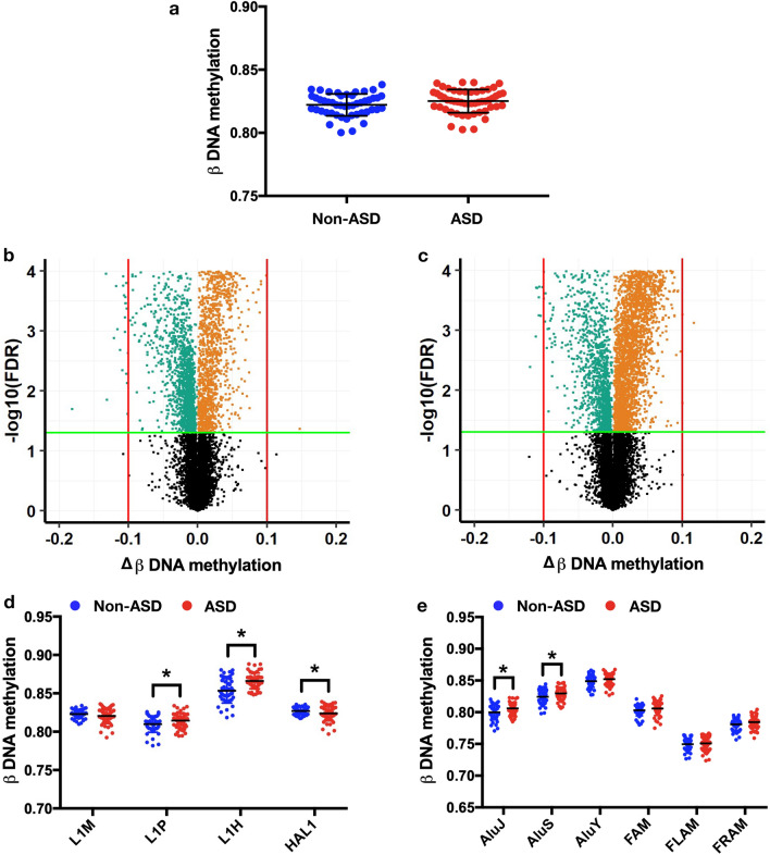
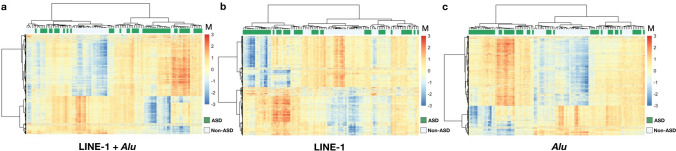
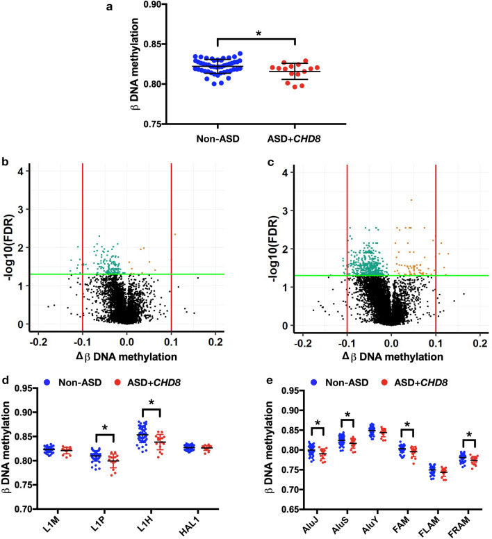
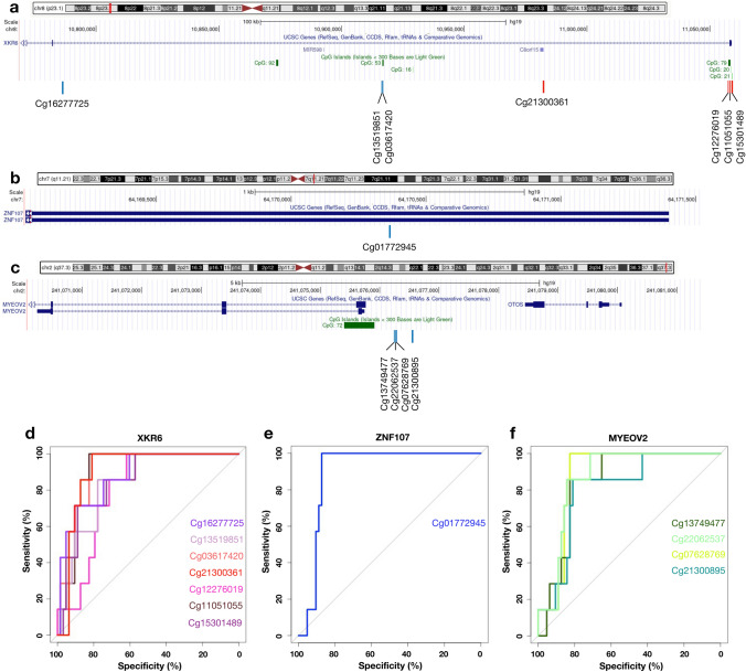
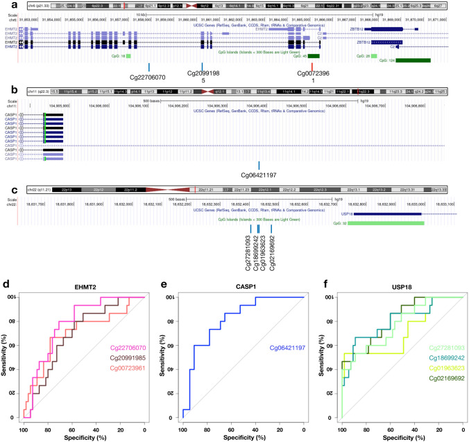
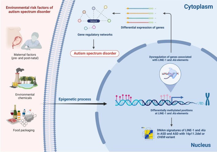
Long interspersed nucleotide element-1 (LINE-1) and Alu elements are retrotransposons whose abilities cause abnormal gene expression and genomic instability. Several studies have focused on DNA methylation profiling of gene regions, but the locus-specific methylation of LINE-1 and Alu elements has not been identified in autism spectrum disorder (ASD). Here we interrogated locus- and family-specific methylation profiles of LINE-1 and Alu elements in ASD whole blood using publicly-available Illumina Infinium 450 K methylation datasets from heterogeneous ASD and ASD variants (Chromodomain Helicase DNA-binding 8 (CHD8) and 16p11.2del). Total DNA methylation of repetitive elements were notably hypomethylated exclusively in ASD with CHD8 variants. Methylation alteration in a family-specific manner including L1P, L1H, HAL, AluJ, and AluS families were observed in the heterogeneous ASD and ASD with CHD8 variants. Moreover, LINE-1 and Alu methylation within target genes is inversely related to the expression level in each ASD variant. The DNA methylation signatures of the LINE-1 and Alu elements in ASD whole blood, as well as their associations with the expression of ASD-related genes, have been identified. If confirmed in future larger studies, these findings may contribute to the identification of epigenomic biomarkers of ASD.


Conflict of interest statement

The authors declare no competing interests.

Figures

Figure 1
Figure 1
Methylation of repetitive elements (LINE-1 and Alu) in non-ASD (n = 48) and ASD (n = 52). Total methylation of REs (a). Volcano plots of mean change in methylation (Δβ) of LINE-1 (b) and Alu (c) against − log10 FDR-adjusted p value (PFDR) of ASD compared with non-ASD; the green line represents PFDR = 0.05, the red line represents 10% of methylation changes, green dots represent hypomethylation loci, and orange dots represent hypermethylation loci. Changes in DNA methylation (Δβ) of ASD compared with non-ASD by a subfamily of LINE-1 (d) and Alu (e). Mean ± SD. *p < 0.05.
Figure 2
Figure 2
Unsupervised hierarchical cluster heatmap of the significant differentially methylated loci of the repetitive elements in non-ASD and ASD. Clustering of the 7165 significant differentially methylated loci (combining of LINE-1 and Alu loci) (a). Clustering of the 2802 significant differentially methylated loci (LINE-1) (b). Clustering of the 4363 significant differentially methylated loci (Alu) (c). The color scale indicates methylation level (M value), from low (blue) to high (red). Green color represents ASD and white color represents non-ASD.
Figure 3
Figure 3
Methylation of repetitive elements (LINE-1 and Alu) in non-ASD (n = 48) and ASD patients who carry CHD8 variants (n = 15). DNA methylation (a). Volcano plots of mean change in methylation (Δβ) of LINE-1 (b) and Alu (c) against − log10 FDR-adjusted p value (PFDR) of ASD compared with non-ASD; the green line represents PFDR = 0.05, the red line represents 10% of methylation changes, green dots represent hypomethylation loci, and orange dots represent hypermethylation loci. Changes in DNA methylation (Δβ) of ASD with CHD8 compared with non-ASD by a subfamily of LINE-1 (d) and Alu (e). Mean ± SD. *p < 0.05.
Figure 4
Figure 4
Genomic location and specificity of the unique differentially methylated regions in ASD with 16p11.2 deletion. Genomic location on XK related 6 (XKR6) (a), Zinc Finger Protein 107 (ZNF107) (b) and Myeloma overexpressed 2 (MYEOV2) (c): blue line represents hypomethylation, red line represents hypermethylation. The receiver operating characteristic (ROC) analysis of the unique differentially methylated regions of ASD with 16p11.2 deletion (n = 7) was performed against non-ASD (n = 48) and ASD with CHD8 variants (n = 15). Specificity and sensitivity of the unique differentially methylated regions for XKR6 (d), ZNF107 (e) and MYEOV2 (f).
Figure 5
Figure 5
Genomic location and specificity of the unique differentially methylated regions in ASD with CHD8 variants. Genomic location on Euchromatic Histone Lysine Methyltransferase 2 (EHMT2) (a), Caspase 1 (CASP1) (b) and Ubiquitin Specific Peptidase 18 (USP18) (c): blue line represents hypomethylation, the red line represents hypermethylation. The receiver operating characteristic (ROC) analysis of the unique differentially methylated regions of ASD with CHD8 variants (n = 15) was performed against non-ASD (n = 48) and ASD with 16p11.2 deletion (n = 7). Specificity and sensitivity of the unique differentially methylated regions for EHMT2 (d), CASP1 (e) and USP18 (f).
Figure 6
Figure 6
Schematic diagram illustrating a possible mechanism of LINE-1 and Alu elements in ASD (created with https://biorender.com/).

References

    1. Association AP. Diagnostic and Statistical Manual of Mental Disorders 5th edn (2013).
    1. Maenner MJ, Shaw KA, Bakian AV, Bilder DA, Durkin MS, Esler A, et al. Prevalence and characteristics of autism spectrum disorder among children aged 8 years—Autism and developmental disabilities monitoring network, 11 sites, United States, 2018. MMWR Surveill. Summ. 2021;70(11):1–16. doi: 10.15585/mmwr.ss7011a1. - DOI - PMC - PubMed
    1. Schaefer GB, Mendelsohn NJ. Genetics evaluation for the etiologic diagnosis of autism spectrum disorders. Genet. Med. 2008;10(1):4–12. doi: 10.1097/GIM.0b013e31815efdd7. - DOI - PubMed
    1. Bolte S, Girdler S, Marschik PB. The contribution of environmental exposure to the etiology of autism spectrum disorder. Cell Mol. Life Sci. 2019;76(7):1275–1297. doi: 10.1007/s00018-018-2988-4. - DOI - PMC - PubMed
    1. Stamou M, Streifel KM, Goines PE, Lein PJ. Neuronal connectivity as a convergent target of gene x environment interactions that confer risk for Autism Spectrum Disorders. Neurotoxicol. Teratol. 2013;36:3–16. doi: 10.1016/j.ntt.2012.12.001. - DOI - PMC - PubMed

Publication types

MeSH terms