Review
. 2022 Dec;44(6):2623-2653.
doi: 10.1007/s11357-022-00620-5. Epub 2022 Aug 18.

Microbiota mitochondria disorders as hubs for early age-related macular degeneration

Affiliations
Review

Microbiota mitochondria disorders as hubs for early age-related macular degeneration

János Fehér et al. Geroscience. 2022 Dec.

Abstract

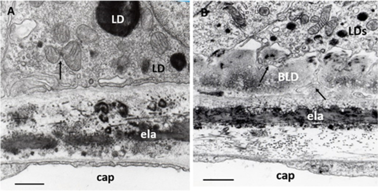
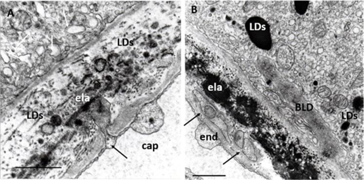
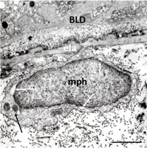
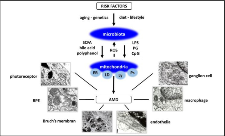
Age-related macular degeneration (AMD) is a progressive neurodegenerative disease affecting the central area (macula lutea) of the retina. Research on the pathogenic mechanism of AMD showed complex cellular contribution governed by such risk factors as aging, genetic predisposition, diet, and lifestyle. Recent studies suggested that microbiota is a transducer and a modifier of risk factors for neurodegenerative diseases, and mitochondria may be one of the intracellular targets of microbial signaling molecules. This review explores studies supporting a new concept on the contribution of microbiota-mitochondria disorders to AMD. We discuss metabolic, vascular, immune, and neuronal mechanism in AMD as well as key alterations of photoreceptor cells, retinal pigment epithelium (RPE), Bruch's membrane, choriocapillaris endothelial, immune, and neuronal cells. Special attention was paid to alterations of mitochondria contact sites (MCSs), an organelle network of mitochondria, endoplasmic reticulum, lipid droplets (LDs), and peroxisomes being documented based on our own electron microscopic findings from surgically removed human eyes. Morphometry of Bruch's membrane lipids and proteoglycans has also been performed in early AMD and aged controls. Microbial metabolites (short-chain fatty acids, polyphenols, and secondary bile acids) and microbial compounds (lipopolysaccharide, peptidoglycan, and bacterial DNA)-now called postbiotics-in addition to local effects on resident microbiota and mucous membrane, regulate systemic metabolic, vascular, immune, and neuronal mechanisms in normal conditions and in various common diseases. We also discuss their antioxidant, anti-inflammatory, and metabolic effects as well as experimental and clinical observations on regulating the main processes of photoreceptor renewal, mitophagy, and autophagy in early AMD. These findings support an emerging concept that microbiota-mitochondria disorders may be a crucial pathogenic mechanism of early AMD; and similarly, to other age-related neurodegenerative diseases, new treatment approaches should be targeted at these disorders.

Keywords: Age-related macular degeneration; Bruch’s membrane; Choriocapillaris; Electron microscopy; Ferroptosis; Innate immunity; Lipid droplets; Microbiota; Microglia; Mitochondria; Mitochondria contact sites; Morphometry; Photoreceptor; Retinal pigment epithelium.


Conflict of interest statement

The authors declare no competing interests.

Figures

Fig. 1
Fig. 1
Photoreceptor inner segment. A Normal aged cone and rods. Ellipsoid (el) contains numerous, densely packet, uniformly long mitochondria with well-preserved cristae and matrix. Myoid (my) contains a few endoplasmic reticula (ER) and normal cytoplasm free from inclusions, similarly to neighboring rods (R). olm: outer limiting membrane; 76 years; male; ×12,000; bar: 1 μm. B Early AMD cone. Ellipsoid (el) contains mitochondria of various shapes and sizes with focal loss of cristae and matrix. Myoid (my) is shorter and contains some endoplasmic reticulum (ER), lysosome (Ly), electrodense, and lipid-containing inclusions (LDs) next to the mitochondria. olm: outer limiting membrane; 62 years; male; ×20,000; bar: 0.1 μm
Fig. 2
Fig. 2
MCSs in the RPE. A MCSs in young RPE (normal). Typical organization of contact sites between mitochondria, endoplasmic reticulum (ER), and LDs (black arrows). ER show dilatation at some places, phenomena of ER-stress (white arrows), one of these dilatations is continuous with a small LD (widened white arrow). Eight years; male; ×32,000; 1 μm. B MCSs in adult RPE (normal): Several contact sites between mitochondria and ER (black arrows), ER show dilatation at some places, phenomena of ER-stress (white arrows), one of these dilatations is continuous with a small LD (widened white arrow). At the center of the pictures, multiple contact sites and presumed mitophagy can be seen (double black arrows). Thirty-eight years; male; ×24,000; 1 μm
Fig. 3
Fig. 3
RPE alterations. A Normal aged RPE. Characteristic ultrastructural features: sparsely distributed mitochondria in the cytoplasm, which contains numerous small vacuoles features of the lysosome (Ly) and electrodense LDs of various sizes (LDs); mitochondrial cristae and matrix mostly with normal appearance, one of them forms a contact site with small peroxisome (white arrow); two of them show mitochondrial fission (black arrows); some small round-form mitochondria with confluent electron-dense cristae characteristic for iron overload seen in ferroptosis (fp); basal lamina (BL) of RPE shows normal appearance. Eighty-one years; female; ×22,000;1 μm. B Early AMD. Characteristic ultrastructural features: a few sparsely distributed mitochondria in the cytoplasm which contains numerous (Ly); focal loss of mitochondrial cristae and matrix, one of them forms a MCSs with LDs and lysosome (black arrows) and one contact site between a small LDs and lysosome (white arrow); some small round-form mitochondria with confluent electron-dense cristae suggesting seen in ferroptosis (fp); LDs in the cytoplasm (LDs) one in the upper right angel, some smaller in the basal cytoplasm of RPE; BLD with some round-form electron-translucent areas characteristic to non-membrane-bound cholesterol. n: nucleus. Seventy-two years; male; ×22,000; bar: 1 μm
Fig. 4
Fig. 4
Drusen in early AMD. A LDs in early AMD. LDs of different electron densities and sizes are located under the well-circumscribed elevation of the RPE basal lamina (LDs). All other layers of BM show normal aged features. Note a MCS with ER in the endothelial cell (arrow) and several pinocytotic or exocytotic vesicles next to the BM (white arrow). Seventy-one years; female; ×22,000; bar 1 μm. B Drusen in early AMD. Electron microscopy of drusen containing LDs of various sizes and electron-density, membrane-fragments, and electron-translucent vacuoles surrounded by appearance homogeneous material. Endothelial cell shows normal fenestration and a dome-shaped basal laminar thickening (star). Eighty-one years; female; ×22,000; bar: 1 μm
Fig. 5
Fig. 5
BLD in early AMD. A Normal aged Bruch’s membrane. RPE contains some small mitochondria, two of them show fission (arrow) and electrodense LDs; inner and outer collagenous layers contain electrodense inclusions of various shapes and sizes, as well as some of them are round and single membrane-bound; elastic layer, basal lamina of RPE, and capillary endothelium are normal. ela: elastic layer, cap: choriocapillaris. Seventy-four years; male; ×28,000; bar: 1 μm. B BLD in early AMD. A longitudinal section of BLD between the basal lamina and basal cytoplasm of RPE. In some places, it is amorphous in appearance with small electron-translucent vacuoles, while in other palaces it shows filamentary structures with electron-dense patches due to lipids. Basal in-foldings of the RPE in some places may form deep protrusions into the BLD, reaching the basal lamina of the RPE (black arrows). The cytoplasm of RPE contains numerous small LDs and only a few mitochondria of various sizes but well-preserved cristae and matrix. ela: elastic layer, cap: choriocapillaris. Seventy-one years; female; ×25,000; bar:1 μm
Fig. 6
Fig. 6
Collagen, lipids, and proteoglycans in Bruch’s membrane of aged normal eyes. A Light microscopy showed PAS-positive staining in the Bruch’s membrane (arrows), as well as in the intercapillary and pericapillary connective tissue, indicating their high carbohydrate content. PAS alcian-blue hematoxylin staining, 72 years; male; ×350; bar: 10 μm. B Collagen fibrils in Bruch’s membrane. After the collagen-specific phenol reaction, two layers of Bruch’s membrane showed intense anisotropy with polarization microscopy. They were prominent just beneath the RPE and much less in the capillary wall (arrows). Seventy-six years; male; ×350; bar: 10 μm. C Lipids in the basal lamina of RPE and capillary walls. Polarization microscopy of unstained sections showed anisotropy just beneath the RPE and in the choriocapillaris wall indicating the presence of oriented structures in both layers (arrows). This anisotropy was completely abolished by lipid extraction. The basal lamina of choroidal vascular endothelia also appears bright, while all other structures of the choroid are dark. 82 years; female; ×350; bar: 10 μm. D Proteoglycans in the basement membrane of RPE and choriocapillaris. Bruch’s membrane was strongly basophilic and intensively anisotropic after the ABT reaction. This anisotropy was due to the carbohydrate components of the basement membrane of RPE and choriocapillaris as well as those of the inner and outer collagen layers of the Bruch’s membrane (arrows). Seventy-one years; male; ×350; bar: 10 μm
Fig. 7
Fig. 7
Morphometric analysis of Bruch’s membrane in early AMD as compared to age-matched controls. A The lipid content of Bruch’s membrane increased significantly in both aged and AMD groups. The coefficient of correlation for lipids was significantly greater in the AMD group than the age-matched normal controls (p < 0.001). B The proteoglycan content increased significantly more in AMD compared to normal aging until 75 years (p < 0.01)
Fig. 8
Fig. 8
Endothelial alterations. A Endothelial sprouting in a normal eye. Small, deep sprouting reaches the elastic layer of the Bruch’s membrane (black arrow), which contains several LDs of various sizes and shapes can be seen. Numerous dilated lysosomes and small round-form lysosomes next to a mitochondrion can be seen in the RPE (white arrows) ela: elastic layer; 48 years; female; ×32,000; bar: 1 μm. B Endothelial cell processes of in early AMD. Transversal section of endothelial cell processes in the basal lamina of the choriocapillaris (arrows). They contain small mitochondrion with confluent cristae. BLD, some small sparsely distributed mitochondria, several lysosomes, and LDs can be seen in the RPE. ela: elastic layer. Eighty-four years; female; ×22,000; bar: 1 μm
Fig. 9
Fig. 9
Macrophage in early AMD. Oval-shape macrophage in the intercapillary zone next to the elastic layer of the BM. The narrow cytoplasm is poor in cytoplasmic processes, it contains two round-shape mitochondria with confluent electrodense cristae and several endosomes. One of the mitochondria forms contact site with a lysosome (black arrow). The nuclear membrane is dilated nearly all round the nucleus (white arrows). Seventy-two years; male; ×22,000; bar: 1 μm
Fig. 10
Fig. 10
Ganglion cells (possible) in early AMD. Two round-shape cells located in the widened outer collagenous layer of the Bruch’s membrane (pGc). They have round nucleus, narrow cytoplasm which contain some mitochondria, endoplasmic reticula, and lysosomes. Thin basement membrane surrounds both cell bodies. Mitochondria with loss of cristae and matrix-density in the RPE (black arrow), basal linear deposits (white arrow), and BLD can also be seen. ela: elastic layer, cap: capillaries. Seventy-six years; female; ×19,000; bar: 1 μm
Fig. 11
Fig. 11
Microbiota–mitochondria disorders in early AMD. A proposed concept. Risk factors of AMD, aging, genetics, diet, and lifestyle are transduced by microbiota. Microbiota is established in early life and continuously reshaped by environmental influences throughout life. Microbial metabolites and compounds (‘healthy’ or ‘unhealthy’) released mainly in the gut regulate epithelial barrier and local mechanism, and through systemic circulation, gut-liver, and gut-brain axis they affect other organs, eye included. Mitochondria, the powerhouse of cells in addition to energy supply, through MCSs, an organelle-network of mitochondria, endoplasmic reticulum (ER), lipid droplets (LD), lysosomes (Ly), and peroxisomes (Ps) regulate main cellular functions including photoreceptor turnover, autophagy, and MQC. Mitochondrial ROS also influences microbial composition and function. Mitochondria are the organelle target of microbiota metabolites and compounds. SCFA, polyphenols, bile acids, and unmethylated CpG containing oligonucleotides improve metabolism and have anti-inflammatory, while endotoxins (LPS and PG) have proinflammatory effects. Microbiota-mitochondria disorders affect local and systemic metabolism, circulation, and neuro-immune surveillance and result in alterations of photoreceptors, RPE cells, Bruch’s membrane, endothelium, microglia, and ganglion cells of the choriocapillaris in early AMD

References

    1. Wong WL, Su X, Li X, Cheung CM, Klein R, Cheng CY, et al. Global prevalence of age-related macular degeneration and disease burden projection for 2020 and 2040: a systematic review and meta-analysis. Lancet Glob Health. 2014;2:e106–e116. doi: 10.1016/S2214-109X(13)70145-1. - DOI - PubMed
    1. Li JQ, Welchowski T, Schmid M, Mauschitz MM, Holz FG, Finger RP. Prevalence and incidence of age-related macular degeneration in Europe: a systematic review and meta-analysis. Br J Ophthalmol. 2020;104:1077–1084. doi: 10.1136/bjophthalmol-2019-314422. - DOI - PubMed
    1. Bird AC, Bressler NM, Bressler SB, Chisholm IH, Coscas G, Davis MD, et al. An international classification and grading system for age-related maculopathy and age-related macular degeneration. The International ARM Epidemiological Study Group. Surv Ophthalmol. 1995;39:367–374. doi: 10.1016/s0039-6257(05)80092-x. - DOI - PubMed
    1. Keenan TDL, Cukras CA, Chew EY. Age-related macular degeneration: epidemiology and clinical aspectsrelated macular degeneration: epidemiology and clinical aspects. Adv Exp Med Biol. 2021;1256:1–31. doi: 10.1007/978-3-030-66014-7_1. - DOI - PubMed
    1. Spaide RF, Jaffe GJ, Sarraf D, Freund KB, Sadda SR, Staurenghi G, et al. Consensus nomenclature for reporting neovascular age-related macular degeneration data: consensus on neovascular age-related macular degeneration nomenclature study group. Ophthalmology. 2020;127:616–636. doi: 10.1016/j.ophtha.2019.11.004. - DOI - PMC - PubMed

Publication types