. 2023 Feb;13(2):593-607.
doi: 10.1007/s13346-022-01218-2. Epub 2022 Aug 17.

A proof of concept to define the parameters affecting poly-L-lactide-co-poly-ε-caprolactone shape memory electrospun nanofibers for biomedical applications

Affiliations

A proof of concept to define the parameters affecting poly-L-lactide-co-poly-ε-caprolactone shape memory electrospun nanofibers for biomedical applications

Silvia Pisani et al. Drug Deliv Transl Res. 2023 Feb.

Abstract

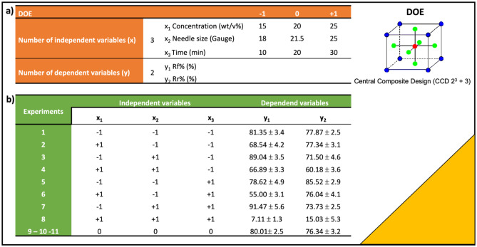
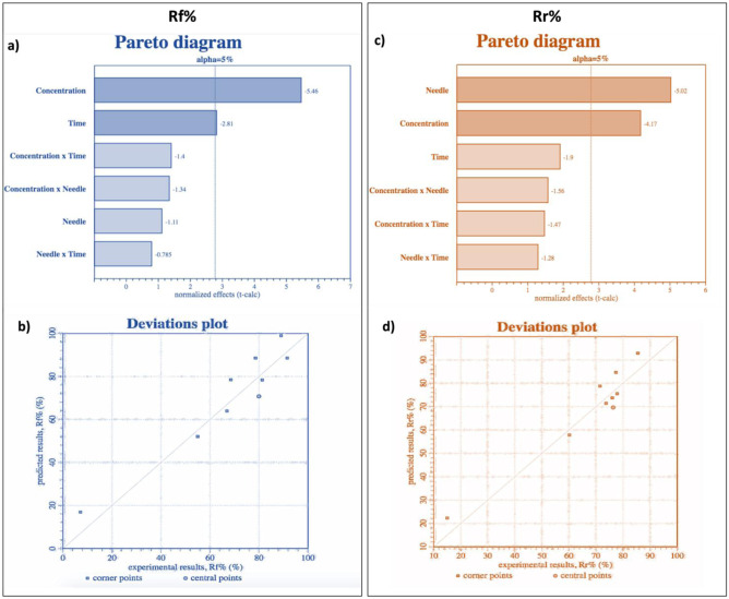
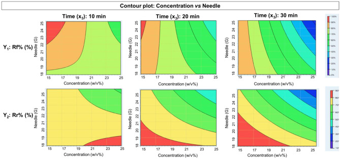
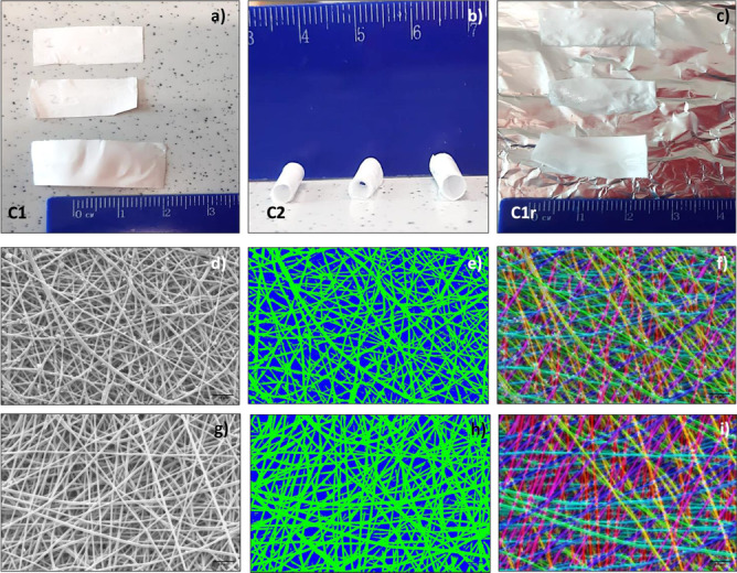
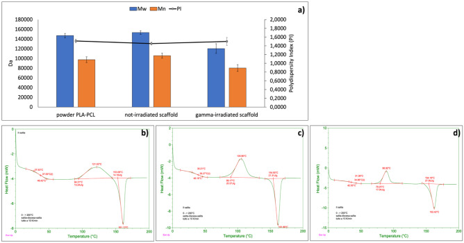
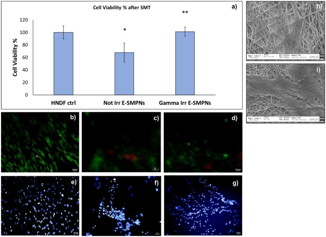
This study is a proof of concept performed to evaluate process parameters affecting shape memory effect of copolymer poly-L-lactide-co-poly-ε-caprolactone (PLA:PCL) 70:30 ratio based nanofibrous scaffolds. A design of experiment (DOE) statistical approach was used to define the interaction between independent material and process variables related to electrospun scaffold manufacturing, such as polymer solution concentration (w/v%), spinning time (min), and needle size (Gauge), and their influence on Rf% (ability of the scaffold to maintain the induced temporary shape) and Rr% (ability of the scaffold to recover its original shape) outputs. A mathematical model was obtained from DOE useful to predict scaffold Rf% and Rr% values. PLA-PCL 15% w/v, 22G needle, and 20-min spinning time were selected to confirm the data obtained from theoretical model. Subsequent morphological (SEM), chemical-physical (GPC and DSC), mechanical (uniaxial tensile tests), and biological (cell viability and adhesion) characterizations were performed.

Keywords: Design of experiment; Electrospinning; Nanofibers; Poly-L-lactide-co-poly-ε-caprolactone; Shape memory polymer.


Conflict of interest statement

SP, IG, RD, BC, and MB declare that the E-SMPN is under Italian patent # 102021000019256 filed on July 20, 2021. TM and GB declare no competing interests.

Figures

Fig. 1
Fig. 1
a DOE (CCD 23 + 3) set up and minimum (− 1), maximum (+ 1) and intermediate (0) values of independent variables (x) concentration (x1), needle size (x2), and spinning time (x3). b Experimental design and results obtained for dependent variables y1 (Rf%) and y2 (Rr%)
Fig. 2
Fig. 2
a Pareto diagram for Rf% (y1) dependent variable (p value < 0.0001). b Deviation plot for Rf% (y1) dependent variable (R2: 87.58%). c Pareto diagram for Rr% (y2) dependent variable (p value < 0.0001). d Deviation plot for Rr% (y2) dependent variable (R2: 89.89%)
Fig. 3
Fig. 3
Contour plot concentration vs needle at three different times for dependent variable y1 Rf% and y2 Rr%
Fig. 4
Fig. 4
Shape memory treatment (SMT). a Scaffold configuration before treatment, C1. b After first heating treatment (T1 > Tg°) and cooling bath (T2 < Tg°), scaffolds maintained rolled shape (configuration C2) and c after temperature treatment close to Tg° (37° C), scaffolds recovered original shape (configuration C1r). Morphological characterization of 15% PLA-PCL electrospun nanofibers obtained using 22 G needle for 20 min spinning time before SMT. d SEM image. e Fiber porosity analysis. f Fiber orientation analysis. Morphological characterization after SMT. g SEM image. h Fiber porosity analysis. i Fiber orientation analysis
Fig. 5
Fig. 5
a Results of GPC analyses reporting Mw, Mn, and PI of PLA-PCL powder and non-irradiated and gamma-irradiated PLA-PCL electrospun scaffolds. bd Results of DSC analysis performed on b PLA-PCL powder; c non-irradiated PLA-PCL electrospun nanofibers; and d gamma-irradiated PLA-PCL electrospun nanofibers
Fig. 6
Fig. 6
a Results of cell viability % determined by MTT assay on NHDF, incubated with non-irradiated and gamma-irradiated PLA-PCL electrospun scaffold after 7 days of incubation and 6 h after SMT. bg Merge of LIVE (green)/DEAD (red) staining of Human Dermal Fibroblast after 7 days of incubation: b control; c non-gamma-irradiated shape memory electrospun scaffold; d gamma-irradiated shape memory electrospun scaffold. DAPI staining of NHDF after 7 days of incubation: e control; f non-gamma-irradiated shape memory electrospun scaffold; g gamma-irradiated shape memory electrospun scaffold. SEM images of Human Dermal Fibroblast after 7 days of incubation on h non-gamma-irradiated shape memory electrospun scaffold and i gamma-irradiated shape memory electrospun scaffold

References

    1. Leng J, Lan X, Liu Y, Du S. Shape-memory polymers and their composites: Stimulus methods and applications. Prog Mater Sci. 2011;56(7):1077–1135. doi: 10.1016/j.pmatsci.2011.03.001. - DOI
    1. Zare M, Davoodi P, Ramakrishna S. Electrospun shape memory polymer micro-/nanofibers and tailoring their roles for biomedical applications. Nanomaterials. 2021;11(4). - PMC - PubMed
    1. Zhou Y, Huang WM. Shape memory effect in polymeric materials: mechanisms and optimization. Procedia IUTAM. 2015;12:83–92. doi: 10.1016/j.piutam.2014.12.010. - DOI
    1. Small IVW, Singhal P, Wilson TS, Maitland DJ. Biomedical applications of thermally activated shape memory polymers. J Mater Chem. 2010;20(17):3356–3366. doi: 10.1039/b923717h. - DOI - PMC - PubMed
    1. Hu J, Zhu Y, Huang H, Lu J. Recent advances in shape–memory polymers: structure, mechanism, functionality, modeling and applications. Prog Polym Sci. 2012;37(12):1720–1763. doi: 10.1016/j.progpolymsci.2012.06.001. - DOI

LinkOut - more resources