. 2023 Jan;91(1):91-98.
doi: 10.1002/prot.26412. Epub 2022 Aug 24.

Crystal structure of Methanococcus jannaschii dihydroorotase

Affiliations

Crystal structure of Methanococcus jannaschii dihydroorotase

Jacqueline Vitali et al. Proteins. 2023 Jan.

Abstract

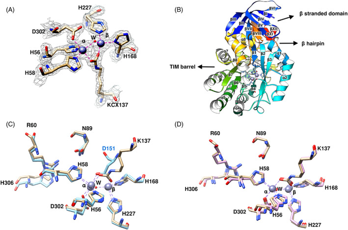
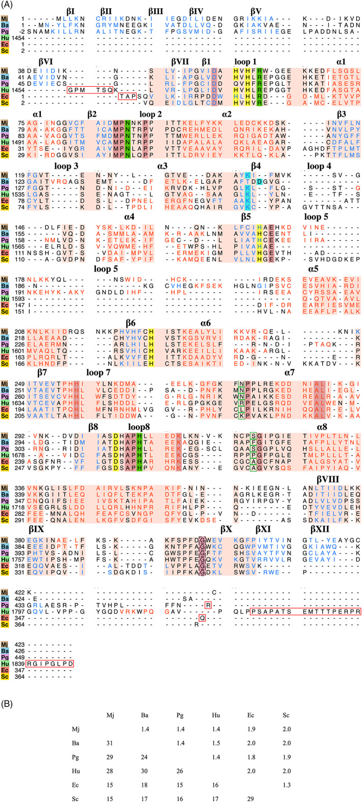
In this paper, we report the structural analysis of dihydroorotase (DHOase) from the hyperthermophilic and barophilic archaeon Methanococcus jannaschii. DHOase catalyzes the reversible cyclization of N-carbamoyl-l-aspartate to l-dihydroorotate in the third step of de novo pyrimidine biosynthesis. DHOases form a very diverse family of enzymes and have been classified into types and subtypes with structural similarities and differences among them. This is the first archaeal DHOase studied by x-ray diffraction. Its structure and comparison with known representatives of the other subtypes help define the structural features of the archaeal subtype. The M. jannaschii DHOase is found here to have traits from all subtypes. Contrary to expectations, it has a carboxylated lysine bridging the two Zn ions in the active site, and a long catalytic loop. It is a monomeric protein with a large β sandwich domain adjacent to the TIM barrel. Loop 5 is similar to bacterial type III and the C-terminal extension is long.

Keywords: Methanococcus jannaschii; crystal structure; dihydroorotase; pyrimidine biosynthesis.


Conflict of interest statement

The authors declare no potential conflict of interest.

Figures

FIGURE 1
FIGURE 1
(A) The Zn‐binding site of MjDHOase superimposed on a 2mFo‐DFc electron density map contoured at 1.7σ. Zinc ions are presented as gray spheres, water molecules (W) are represented as red spheres, and coordination bonds are shown as magenta dashed lines. This figure was drawn with UCSF Chimera. The volume viewer tool was used for the electron density map. (B) Schematic ribbons diagram of MjDHOase. The protein is shown with rainbow colors from blue at the N‐terminus to red at the C‐terminus. The TIM barrel, its adjacent β stranded domain and the β hairpin are indicated with arrows. Most of the β hairpin (in orange color) is concealed in the back of the drawing as well as strands βIX and βX. The two active‐site Zn ions and the residues that coordinate them are also included in this figure. (C) Superposition of the active site of MjDHOase (wheat carbon atoms) and BaDHOase (cyan carbon atoms, PDB: 3MPG, chain A). (D) Superposition of the active site of MjDHOase (wheat carbon atoms) and EcDHOase (pink carbon atoms, PDB: 1XGE, chain A). The active site is highly conserved, and it is composed of the Zn‐binding residues H56, H58, H168, H227, D302, KCX137, and a water W molecule. The water in the active site of BaDHOase was not determined. In addition, the substrate binding residues N89, R60, H306 are shown but the main chain of residues 275 and 320 that also interact with the substrate are not. KCX137 in MjDHOase corresponds to KCX102 of EcDHOase as seen in (D) and its carboxylate group overlaps the carboxylate of D151 in BaDHOase as seen in (C). Coordinate bonds are drawn in purple dashed lines in (C) and (D)
FIGURE 2
FIGURE 2
(A) Structure‐based multiple sequence alignment of MjDHOase with representatives of the different DHOase types: Ba (Bacillus anthracis, PDB: 3MPG chain A, bacterial type I), Pg (Porphyromonas gingivalis, PDB: 2GWN, bacterial type III), hu (PDB: 4C6C, human CAD), Ec (Escherichia coli, PDB: 1XGE chain A, bacterial type II), and Sc (Saccharomyces cerevisiae, PDB: 6L0J chain A, active fungal). The secondary structural elements are shown with blue lettering for β strands, red lettering for helices, and black lettering for loops. The secondary structural elements of the TIM barrel are labeled β1 to α8. The β strands in the adjacent domain are labeled as βI to βXII. Helices and β strands within loops are not labeled. Invariant residues involved in zinc binding are highlighted in bright yellow. Invariant residues involved in substrate binding are highlighted in dark green and invariant positions that interact with the substrate via their main chain (but not invariant in sequence) are shown within green border. Other residues that show 100% identity in all sequences are highlighted in purple. The carboxylated lysine which is invariant in archaeal, bacterial type II and III, human and active fungal is highlighted in cyan color. The invariant aspartate in bacterial type I is also highlighted in cyan color. Regions depicting missing structure are enclosed within red border. The salmon background corresponds to the fully populated columns. This figure was drawn with UCSF Chimera. (B) An identity matrix giving the sequence identity and RMSD between corresponding Cα's in the 265 fully populated columns between any two of the structures in (A). % sequence identity is below the diagonal on the bottom left and RMSD (Å) is above the diagonal on the top right. The diagonal (100% for sequence identity and 0.0 Å for RMSD) is not included

Similar articles

References

    1. Cooper C, Wilson DW. Biosynthesis of pyrimidines. Fed Proc. 1954;13:194.
    1. Lieberman I, Kornberg A. Enzymatic synthesis and breakdown of a pyrimidine, orotic acid. I. Dihydroorotic acid, ureidosuccinic acid, and 5‐carboxymethylhydantoin. J Biol Chem. 1954;207:911‐924. - PubMed
    1. Holm L, Sander C. An evolutionary treasure: unification of a broad set of amidohydrolases related to urease. Proteins. 1997;28:72‐82. - PubMed
    1. Fields C, Brichta D, Shepherdson M, Farinha M, O'Donovan G. Phylogenetic analysis and classification of dihydroorotases: a complex history for a complex enzyme. Paths Pyrimidines. 1999;7:49‐63.
    1. Thoden JB, Phillips GN Jr, Neal TM, Raushel FM, Holden HM. Molecular structure of dihydroorotase: a paradigm for catalysis through the use of a binuclear metal center. Biochemistry. 2001;40:6989‐6997. - PubMed

Publication types

LinkOut - more resources