. 2022 Jul 20;12(14):e4471.
doi: 10.21769/BioProtoc.4471.

Isolation and ex vivo Expansion of Limbal Mesenchymal Stromal Cells

Affiliations

Isolation and ex vivo Expansion of Limbal Mesenchymal Stromal Cells

Naresh Polisetti et al. Bio Protoc. .

Abstract

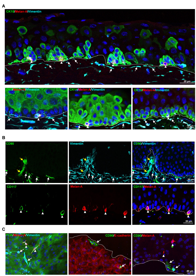
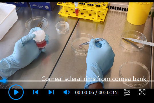
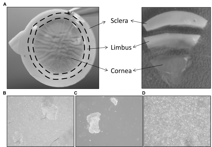
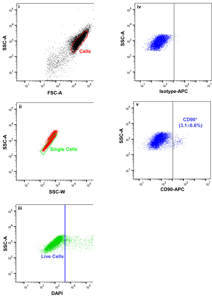
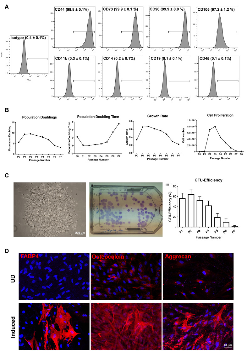
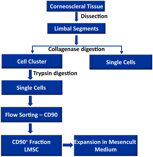
Limbal mesenchymal stromal cells (LMSC), a cellular component of the limbal stem cell niche, have the capability of determining the fate of limbal epithelial progenitor cells (LEPC), which are responsible for the homeostasis of corneal epithelium. However, the isolation of these LMSC has proven to be difficult due to the small fraction of LMSC in the total limbal population, and primary cultures are always hampered by contamination with other cell types. We recently published the efficient isolation and functional characterization of LMSC from the human corneal limbus using CD90 as a selective marker. We observed that flow sorting yielded a pure population of LMSC with superior self-renewal capacity and transdifferentiation potential, and supported the maintenance of the LEPC phenotype. Here, we describe an optimized protocol for the isolation of LMSC from cadaveric corneal limbal tissue by combined collagenase digestion and flow sorting with expansion of LMSC on plastic. Graphical abstract.

Keywords: Cornea; Expansion; Isolation; Limbal mesenchymal stromal cells; Limbal niche cells; Limbal stem cells.


Conflict of interest statement

Competing interests The authors declare that they have no competing interests.

Figures

Figure 1.
Figure 1.. Localization of limbal niche cells in situ .
A . Triple immunostaining analysis of limbal tissue sections showing melanocytes [melan-A + (red) vimentin + (cyan) cells, arrow heads] in close contact with clusters of cytokeratin (CK)15 + , CK14 + , CK19 + (green) limbal epithelial progenitor cells (LEPC), whereas sub-epithelial stromal cells [vimentin + cells (cyan), arrows] were in close association with basal limbal epithelial cells, and not with more superficial CK3 + cells (green). Dashed line represents the basement membrane (BM), and nuclear counterstaining was done with 4′,6-diamidino-2-phenylindole (DAPI, blue). B . Double immunostaining of limbal sections showing the co-localization of CD90 (green) and vimentin (cyan) in the sub-epithelial stromal cells (white arrows), which were in close association with basal layers of limbal epithelium (dotted line represents the BM), as well as blood vessels of the limbal stroma (yellow arrows). The limbal sections also show the co-localization of CD117 (green) and melan A (red) in the melanocytes (arrow heads) at the basal layer of the limbal epithelium. Nuclear counterstaining with DAPI (blue). C . Immunofluorescence analysis of cultured limbal clusters showing the expression of keratins (PCK, green) and vimentin (cyan) in epithelial cells, melan-A (red) and vimentin (cyan) expression in melanocytes (arrow heads), and only vimentin expression in stromal cells (arrows). Double immunostaining of cultured limbal clusters showing the CD90 + stromal cells (green, arrow) at the edge of clusters and also in between E-cadherin + epithelial cells (red, dashed line represents the edge of the cluster), whereas melan-A + melanocytes were located between the cells (red, arrow heads). Nuclear counterstaining with DAPI (blue). Reprinted from Polisetti et al. (2022) , licensed under a CC BY 4.0.
Video 1.
Video 1.. Limbal cell isolation
Figure 2.
Figure 2.. Isolation of limbal cluster cells.
A. The corneal scleral rim (left) was cut into sectors, and each sector was trimmed off 1 mm before and after the limbal region (right). Reprinted from Polisetti et al. (2019), licensed under a CC BY 4.0. B. Different sizes of limbal clusters and single cells (left) formed after overnight incubation of limbal segments in collagenase solution (x40 magnification). C. Limbal clusters separated from single cells after filtration. D. Single cell suspension of limbal cells after digestion of limbal clusters with trypsin-EDTA (x40 magnification).
Figure 3.
Figure 3.. Fluorescence activated cells sorting (FACS) images demonstrating the gating strategy used to isolate limbal mesenchymal stromal cells.
Forward scatter (FSC-A) vs. side scatter (SSC-A) graph showing the cells of interest selected on the basis of size and granularity (i). Side scatter area vs. width graph showing the selection of single cells by excluding doublets or clumps, (ii) followed by dead cell exclusion using 4′,6-diamidino-2-phenylindole DAPI (iii). The isotype control graph shows the set of gates (iv) used to select the CD90 + cells (iv). Percentages (%) of positive cells are expressed as the means ± SEM.
Figure 4.
Figure 4.. Phase contrast images showing the spindle shaped, elongated with prominent nucleolus of CD90 + cells after day 3, 5, and 10 of seeding [×40 magnification (upper row); ×100 magnification (bottom row)].
Figure 5.
Figure 5.. Phenotypic profile and functional characterization of CD90 + (LMSC) cells.
A. Flow cytometry analysis showing the expression of CD markers. Percentage of cells expressed as mean ± SEM of four individual experiments. B. Graphs showing the population doublings [log 10 (y/x)/log10 2, where y is the final density of the cells, and x is the initial seeding density of the cells], population doubling time [(t − t 0 )log2/(logy – logx), where t, t 0 represents the time at cell counting, y equals the number of cells at time t, and x equals the number of cells at time t 0 ], growth rate [ln(N t /N o )/t, where N t represents final cell number, N o represents the initial cell number, and t equals the number of days in culture], and proliferation potential of LMSC over the passages. Data are expressed as means of five individual experiments. C. Phase contrast micrograph showing the LMSC colony (i), and T75 flask showing crystal violet stained colonies of LMSC (ii). The graph represents the colony forming efficiency of LMSC over the passages. Percentage of colonies expressed as means ± standard deviation (n = 5). D. Immunostaining analysis showing the expression of fatty acid binding protein 4 (FABP4), osteocalcin, and aggrecan in adipogenic, osteogenic, and chondrogenic induced cells, respectively. No staining has been seen for FABP4 in undifferentiated (UD) controls, but weak staining was observed for osteocalcin and aggrecan in UD controls. Nuclear counterstaining with DAPI (blue).
None

Similar articles

Cited by

References

    1. Al-Jaibaji O. , Swioklo S. and Connon C. J. ( 2019 . ). Mesenchymal stromal cells for ocular surface repair . Expert Opin Biol Ther 19 ( 7 ): 643 - 653 . - PubMed
    1. Chen S. Y. , Hayashida Y. , Chen M. Y. , Xie H. T. and Tseng S. C. ( 2011 . ). A new isolation method of human limbal progenitor cells by maintaining close association with their niche cells . Tissue Eng Part C Methods 17 ( 5 ): 537 - 548 . - PMC - PubMed
    1. Chen S. Y. , Han B. , Zhu Y. T. , Mahabole M. , Huang J. , Beebe D. C. and Tseng S. C. ( 2015 . ). HC-HA/PTX3 Purified From Amniotic Membrane Promotes BMP Signaling in Limbal Niche Cells to Maintain Quiescence of Limbal Epithelial Progenitor/Stem Cells . Stem Cells 33 ( 11 ): 3341 - 3355 . - PubMed
    1. Dziasko M. A. , Armer H. E. , Levis H. J. , Shortt A. J. , Tuft S. and Daniels J. T. ( 2014 . ). Localisation of epithelial cells capable of holoclone formation in vitro and direct interaction with stromal cells in the native human limbal crypt . PLoS One 9 ( 4 ): e94283 . - PMC - PubMed
    1. Funderburgh J. L. , Funderburgh M. L. and Du Y. ( 2016 . ). Stem Cells in the Limbal Stroma . Ocul Surf 14 ( 2 ): 113 - 120 . - PMC - PubMed

LinkOut - more resources