

Follicular lymphoma presenting like marginal zone lymphoma: A case report
- PMID: 35979114
- PMCID: PMC9258390
- DOI: 10.12998/wjcc.v10.i17.5877
Follicular lymphoma presenting like marginal zone lymphoma: A case report
Abstract
Background: Follicular lymphoma (FL), a common type of indolent lymphoma, carries markers of the germinal center, and the rearrangement of the BCL-2 gene is regarded as an initiating event and a hallmark of the neoplasm. When FL has marginal zone differentiation, some marginal zone features are carried by the neoplasm.
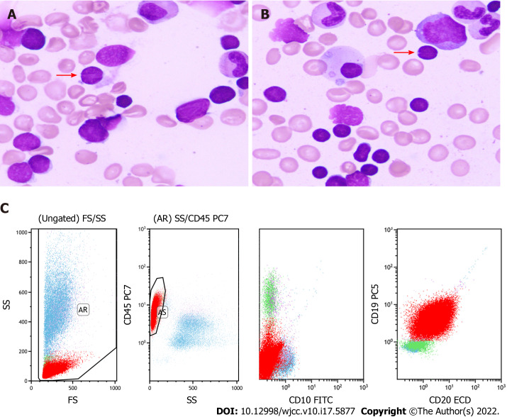
Case summary: A 54-year-old male with lymphadenopathy, splenomegaly and hyperlymphocytosis was diagnosed with FL with marginal zone differentiation. The tumor demonstrated different features in the bone marrow (BM) compared with the follicle of the lymph node (LN). Some component of the neoplasm mimicked marginal zone lymphoma, such as infiltrating the marginal zone of the LN, displaying a monocytoid shape and lacking the expression of CD10 in the BM. The diagnosis of FL was made due to the concurrent detection of BCL-2 rearrangement in the LN and BM.
Conclusion: Discordant pathological features in LN and BM could mislead diagnosis. When clinical and pathological manifestations are confusing in diagnosis, typical genetic abnormalities are decisive.
Keywords: Case report; Discordant immunophenotypes; Follicular lymphoma; Gene rearrangement; Marginal zone differentiation.
©The Author(s) 2022. Published by Baishideng Publishing Group Inc. All rights reserved.
Conflict of interest statement
Conflict-of-interest statement: The authors have no conflicts of interest to declare.
Figures


References
-
- Küppers R. Mechanisms of B-cell lymphoma pathogenesis. Nat Rev Cancer. 2005;5:251–262. - PubMed
-
- Chapman JR, Alvarez JP, White K, Sanchez S, Khanlari M, Algashaamy K, Cassidy D, Peng JH, Fan YS, Alencar A, Alderuccio JP, Lossos IS, Vega F. Unusual Variants of Follicular Lymphoma: Case-based Review. Am J Surg Pathol. 2020;44:329–339. - PubMed
-
- Abou-Elella A, Shafer MT, Wan XY, Velanker M, Weisenburger DD, Nathwani BN, Gascoyne RD, Greiner TC, Chan WC. Lymphomas with follicular and monocytoid B-cell components. Evidence for a common clonal origin from follicle center cells. Am J Clin Pathol. 2000;114:516–522. - PubMed
-
- Schmid U, Cogliatti SB, Diss TC, Isaacson PG. Monocytoid/marginal zone B-cell differentiation in follicle centre cell lymphoma. Histopathology. 1996;29:201–208. - PubMed
Publication types
LinkOut - more resources
Full Text Sources
