

Chondromyxoid fibroma of the cervical spine: A case report
- PMID: 35979139
- PMCID: PMC9258375
- DOI: 10.12998/wjcc.v10.i17.5748
Chondromyxoid fibroma of the cervical spine: A case report
Abstract
Background: Chondromyxoid fibroma (CMF) is an unusual benign tumour of cartilaginous tissues that may be confused with other malignant tumours. It is rarely seen in the cervical spine.
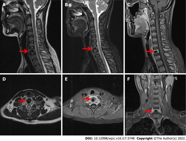
Case summary: A 24-year-old young woman was admitted to the hospital because of neck and shoulder pain. Computed tomography, magnetic resonance imaging, X-ray and other imaging examinations of the cervical spine and laboratory-related indicators combined with intraoperative pathology revealed that the patient had cervical CMF. We performed total resection of the vertebral body and intervertebral disc, and internal fixation was performed to simultaneously maintain the stability of the entire spine. The clinical results from extensive resection were satisfactory. At the 2-year follow-up, the patient's symptoms had not recurred.
Conclusion: CMF is a benign primary bone tumour that is rarely located in the vertebral bone. Accurate initial diagnosis of these tumours is important for appropriate treatment. En bloc surgical resection of the tumour is the cornerstone of treatment.
Keywords: Benign cartilaginous tumour; Case report; Cervical tumour; Chondromyxoid fibroma.
©The Author(s) 2022. Published by Baishideng Publishing Group Inc. All rights reserved.
Conflict of interest statement
Conflict-of-interest statement: The authors declare that there is no conflict of interest.
Figures


Similar articles
-
Chondromyxoid fibroma of frontal bone: a case report and review of the literature.Turk Neurosurg. 2008 Jul;18(3):249-53. Turk Neurosurg. 2008. PMID: 18814113 Review.
-
Diagnostic challenge in chondromyxoid fibroma clinically mimicking neuroma.Contemp Oncol (Pozn). 2024;28(3):267-269. doi: 10.5114/wo.2024.144081. Epub 2024 Oct 15. Contemp Oncol (Pozn). 2024. PMID: 39512533 Free PMC article.
-
Chondromyxoid fibroma of the temporal bone: A case report and review of the literature.World J Clin Cases. 2018 Dec 26;6(16):1210-1216. doi: 10.12998/wjcc.v6.i16.1210. World J Clin Cases. 2018. PMID: 30613685 Free PMC article.
-
Chondromyxoid fibroma of the lumbar spine: case report and literature review.Eur Spine J. 2012 Jun;21 Suppl 4(Suppl 4):S458-62. doi: 10.1007/s00586-011-2078-x. Epub 2011 Nov 18. Eur Spine J. 2012. PMID: 22094389 Free PMC article. Review.
-
Chondromyxoid Fibroma of the Rib: A Rare Benign Tumor With Potential for Local Recurrence.Cureus. 2021 Oct 31;13(10):e19172. doi: 10.7759/cureus.19172. eCollection 2021 Oct. Cureus. 2021. PMID: 34873515 Free PMC article.
Cited by
-
Chondromyxoid fibroma of distal phalanx of great toe: a rare case report with literature review.Int J Burns Trauma. 2024 Dec 15;14(6):142-147. doi: 10.62347/MHUS7790. eCollection 2024. Int J Burns Trauma. 2024. PMID: 39850785 Free PMC article.
References
-
- JAFFE HL, LICHTENSTEIN L. Chondromyxoid fibroma of bone; a distinctive benign tumor likely to be mistaken especially for chondrosarcoma. Arch Pathol (Chic) 1948;45:541–551. - PubMed
-
- Douis H, Saifuddin A. The imaging of cartilaginous bone tumours. II. Chondrosarcoma. Skeletal Radiol. 2013;42:611–626. - PubMed
Publication types
LinkOut - more resources
Full Text Sources
Research Materials
