Case report: Bilateral central serous chorioretinopathy-like abnormalities in a man with pulmonary arterial hypertension
- PMID: 35979218
- PMCID: PMC9376321
- DOI: 10.3389/fmed.2022.983548
Case report: Bilateral central serous chorioretinopathy-like abnormalities in a man with pulmonary arterial hypertension
Abstract
Background: Pulmonary arterial hypertension (PAH) leads to progressive increases in pulmonary vascular resistance, right heart failure, and death if left untreated. Ocular complications secondary to PAH were less reported. In this study, we reported a case of bilateral visual loss and metamorphopsia in a patient with PAH, who developed central serous chorioretinopathy (CSCR)-like abnormalities and optic disc atrophy.
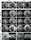
Case summary: A 45-year-old man presented with decreasing central vision and metamorphopsia in both eyes. He had a history of PAH and 6-year history of low-dose oral sildenafil treatment. Slit-lamp examination revealed prominent dilated and tortuous episcleral and conjunctival vessels. Ultrawide-field color picture showed retinal pigment epithelial mottling and atrophy in ring-like configurations. Ultrawide-field autofluorescence showed multiple irregular hyper-autofluorescence with a constellation-like pattern surrounding the optic nerve head and macular region. Optical coherence tomography angiography (OCTA) b-scan demonstrated CSCR-like changes. Swept-source optical coherence tomography (SS-OCT) analysis showed optic nerve atrophy with enlarged cup/disc ratio in right eye, which was confirmed with perimetry. Fluorescein angiography (FA) showed marked leakage of macula and optic nerve head with time, cystoid macular edema, early blocking with late staining of the flecks as shown in the backgrounds of infrared and autofluorescence, and mild leakage in peripheral retina. Indocyanine green angiography (ICGA) showed dilation, tortuosity and congestion of all vortex veins without obvious leakage.
Conclusion: Undertreated PAH may cause the congestion of the choroid and induce CSCR-like abnormalities.
Keywords: case report; central serous chorioretinopathy; fundus abnormalities; hypertension - complications; macular edema; pulmonary arterial hypertension; vascular disease.
Copyright © 2022 Zhou, Zhang, Gu, Zhou and Zhang.
Conflict of interest statement
The authors declare that the research was conducted in the absence of any commercial or financial relationships that could be construed as a potential conflict of interest.
Figures


Similar articles
-
En face enhanced-depth swept-source optical coherence tomography features of chronic central serous chorioretinopathy.Ophthalmology. 2014 Mar;121(3):719-26. doi: 10.1016/j.ophtha.2013.10.014. Epub 2013 Nov 26. Ophthalmology. 2014. PMID: 24289918 Free PMC article.
-
VISION LOSS IN A PATIENT WITH PRIMARY PULMONARY HYPERTENSION AND LONG-TERM USE OF SILDENAFIL.Retin Cases Brief Rep. 2017 Fall;11(4):325-328. doi: 10.1097/ICB.0000000000000355. Retin Cases Brief Rep. 2017. PMID: 27355186
-
Findings of OCT-angiography compared to fluorescein and indocyanine green angiography in central serous chorioretinopathy.Lasers Surg Med. 2018 Dec;50(10):987-993. doi: 10.1002/lsm.22952. Epub 2018 Jun 12. Lasers Surg Med. 2018. PMID: 29896889
-
[A new approach for studying the retinal and choroidal circulation].Nippon Ganka Gakkai Zasshi. 2004 Dec;108(12):836-61; discussion 862. Nippon Ganka Gakkai Zasshi. 2004. PMID: 15656089 Review. Japanese.
-
Biomarkers for central serous chorioretinopathy.Ther Adv Ophthalmol. 2020 Aug 24;12:2515841420950846. doi: 10.1177/2515841420950846. eCollection 2020 Jan-Dec. Ther Adv Ophthalmol. 2020. PMID: 32923941 Free PMC article. Review.
References
-
- Izbicki G, Rosengarten D, Picard E. Sildenafil citrate therapy for pulmonary arterial hypertension. N Engl J Med. (2006) 354:1091–3. - PubMed
Publication types
LinkOut - more resources
Full Text Sources

