

Tumor-to-tumor metastasis of clear cell renal cell carcinoma to contralateral synchronous pheochromocytoma: A case report
- PMID: 35979292
- PMCID: PMC9294876
- DOI: 10.12998/wjcc.v10.i19.6750
Tumor-to-tumor metastasis of clear cell renal cell carcinoma to contralateral synchronous pheochromocytoma: A case report
Abstract
Background: Tumor-to-tumor metastasis (TTM) is an uncommon condition. Only a few cases of renal cell carcinoma (RCC) as donor tumor of TTM have been reported in literature, and none of these studies have described RCC metastasizing to synchronous pheochromocytoma (PCC).
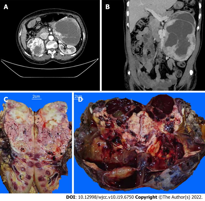
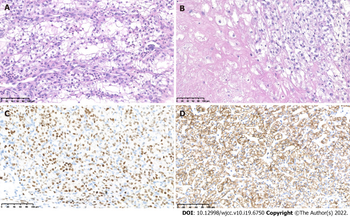
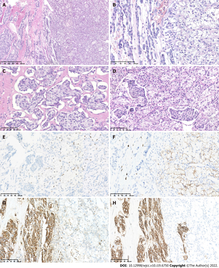
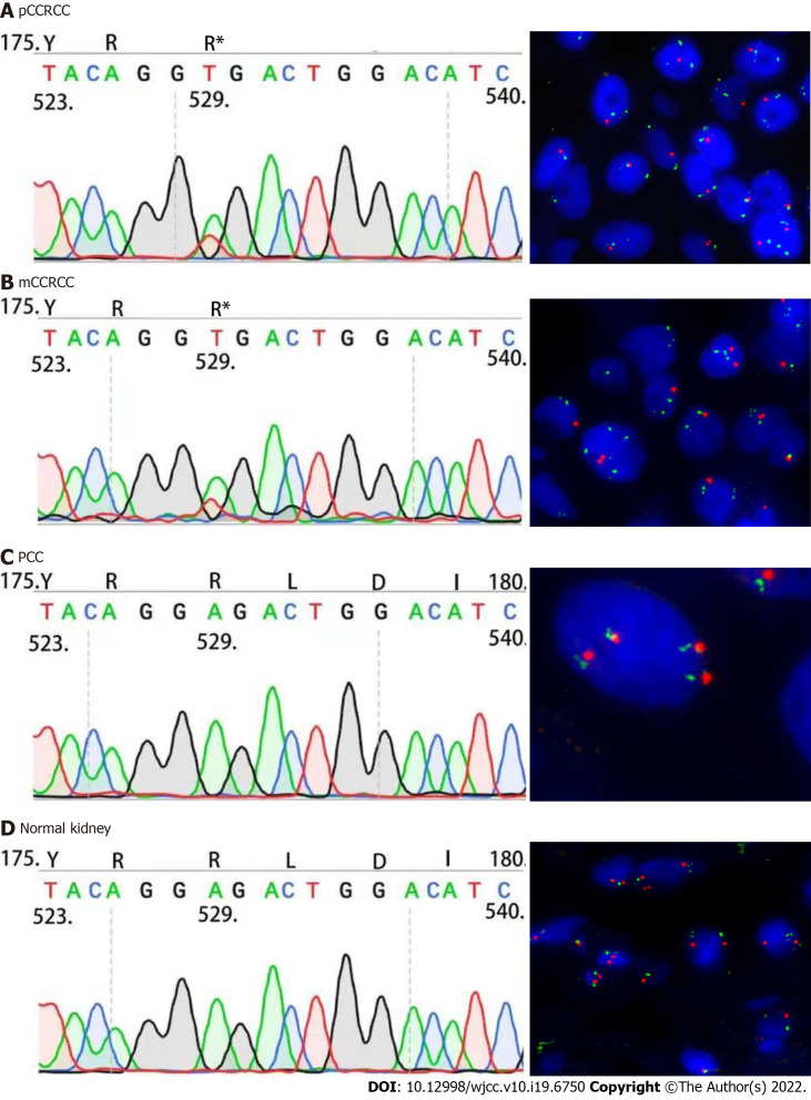
Case summary: The patient was a 54-year-old woman who presented with recurrent dull abdominal pain for six months, which was further aggravated for one more month. Enhanced computed tomography revealed a tumor mass in the right kidney and another mass in the left retroperitoneum/adrenal gland. Histopathology and immunochemistry of resected specimens confirmed the diagnosis of clear cell renal cell carcinoma (CCRCC) of the right kidney, and the left retroperitoneum revealed a typical PCC with CCRCC metastasis. Whole exome sequencing revealed the presence of a c.529A>T somatic mutation of the Von Hippel Lindau (VHL) gene in the metastasized CCRCC, which was also present in the primary right kidney CCRCC, as confirmed by Sanger sequencing. No VHL mutation was detected in the PCC or in normal right kidney tissue. Fluorescence in situ hybridization revealed loss of chromosome 3p in both the primary right kidney CCRCC and CCRCC metastasized to PCC in the left kidney.
Conclusion: This is the first case showing metastasis of CCRCC to PCC, thus leading to tumor-to-tumor metastasis.
Keywords: Case report; Clear cell renal cell carcinoma; Pheochromocytoma; Tumor-to-tumor metastasis; Von Hippel Lindau somatic mutation.
©The Author(s) 2022. Published by Baishideng Publishing Group Inc. All rights reserved.
Conflict of interest statement
Conflict-of-interest statement: The authors declare no competing interests.
Figures
References
-
- Bukowski RM. Natural history and therapy of metastatic renal cell carcinoma: the role of interleukin-2. Cancer . 1997;80:1198–1220. - PubMed
-
- von Knobloch R, Schrader AJ, Walthers EM, Hofmann R. Simultaneous adrenalectomy during radical nephrectomy for renal cell carcinoma will not cure patients with adrenal metastasis. Urology . 2009;73:333–336. - PubMed
-
- Bartoš V. Tumor-to-Tumor Metastasis - a Unique Case of Clear Cell Renal Cell Carcinoma Harboring Metastasis of Adenocarcinoma of Unknown Origin. Klin Onkol . 31:366–370. - PubMed
-
- Campbell LV Jr, Gilbert E, Chamberlain CR Jr, Watne AL. Metastases of cancer to cancer. Cancer . 1968;22:635–643. - PubMed
-
- Wimmer JL, Coffey DM, Kaplan AL, Ayala AG, Ro JY. Tumor-to-tumor metastasis with endometrial carcinoma metastatic to squamous cell carcinoma of vulva: the first reported case. Arch Pathol Lab Med . 2013;137:1825–1828. - PubMed
Publication types
LinkOut - more resources
Full Text Sources
