

Genetic association of apolipoprotein E genotype with EEG alpha rhythm slowing and functional brain network alterations during normal aging
- PMID: 35979332
- PMCID: PMC9376365
- DOI: 10.3389/fnins.2022.931173
Genetic association of apolipoprotein E genotype with EEG alpha rhythm slowing and functional brain network alterations during normal aging
Abstract
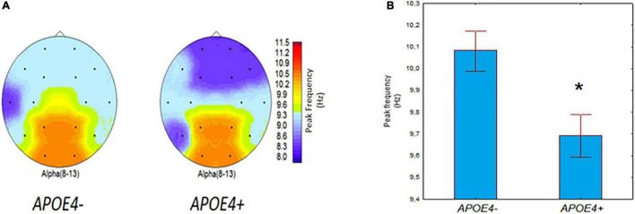
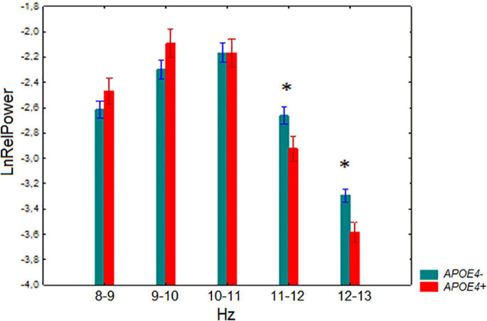
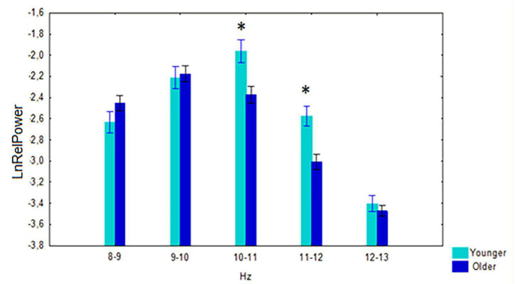
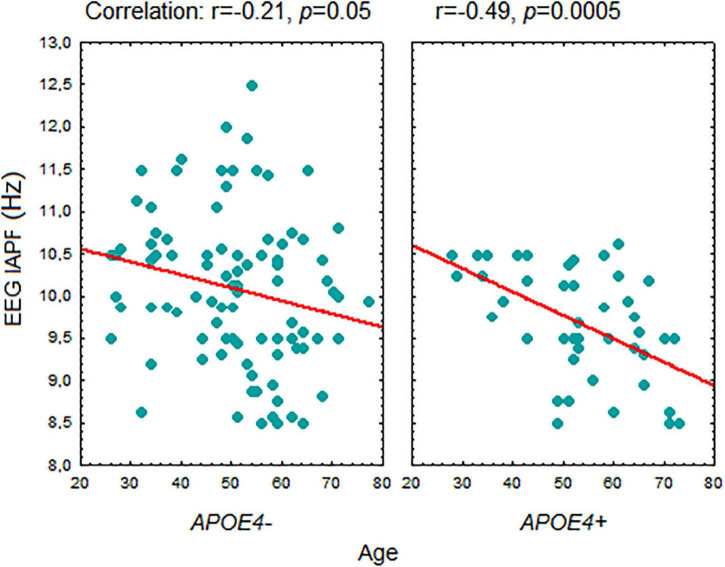
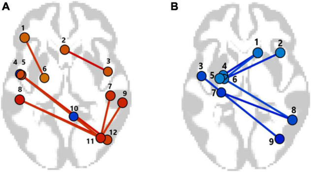
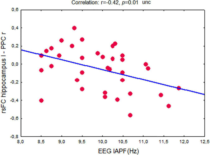
The ε4 allele of the apolipoprotein E (APOE4+) genotype is a major genetic risk factor for Alzheimer's disease (AD), but the mechanisms underlying its influence remain incompletely understood. The study aimed to investigate the possible effect of the APOE genotype on spontaneous electroencephalogram (EEG) alpha characteristics, resting-state functional MRI (fMRI) connectivity (rsFC) in large brain networks and the interrelation of alpha rhythm and rsFC characteristics in non-demented adults during aging. We examined the EEG alpha subband's relative power, individual alpha peak frequency (IAPF), and fMRI rsFC in non-demented volunteers (age range 26-79 years) stratified by the APOE genotype. The presence of the APOE4+ genotype was associated with lower IAPF and lower relative power of the 11-13 Hz alpha subbands. The age related decrease in EEG IAPF was more pronounced in the APOE4+ carriers than in the APOE4+ non-carriers (APOE4-). The APOE4+ carriers had a stronger fMRI positive rsFC of the interhemispheric regions of the frontoparietal, lateral visual and salience networks than the APOE4- individuals. In contrast, the negative rsFC in the network between the left hippocampus and the right posterior parietal cortex was reduced in the APOE4+ carriers compared to the non-carriers. Alpha rhythm slowing was associated with the dysfunction of hippocampal networks. Our results show that in adults without dementia APOE4+ genotype is associated with alpha rhythm slowing and that this slowing is age-dependent. Our data suggest predominant alterations of inhibitory processes in large-scale brain network of non-demented APOE4+ carriers. Moreover, dysfunction of large-scale hippocampal network can influence APOE-related alpha rhythm vulnerability.
Keywords: APOE genotype; Alzheimer’s disease; aging; alpha rhythm; brain networks; functional MRI; functional connectivity; genetic predisposition.
Copyright © 2022 Ponomareva, Andreeva, Protasova, Konovalov, Krotenkova, Kolesnikova, Malina, Kanavets, Mitrofanov, Fokin, Illarioshkin and Rogaev.
Conflict of interest statement
The authors declare that the research was conducted in the absence of any commercial or financial relationships that could be construed as a potential conflict of interest.
Figures
Similar articles
-
Abnormal Temporal Slowing on EEG Findings in Preclinical Alzheimer's Disease Patients With the ApoE4 Allele: A Pilot Study.Cureus. 2023 Oct 28;15(10):e47852. doi: 10.7759/cureus.47852. eCollection 2023 Oct. Cureus. 2023. PMID: 38021568 Free PMC article.
-
Modulation of APOE and SORL1 genes on hippocampal functional connectivity in healthy young adults.Brain Struct Funct. 2017 Aug;222(6):2877-2889. doi: 10.1007/s00429-017-1377-3. Epub 2017 Feb 22. Brain Struct Funct. 2017. PMID: 28229235 Free PMC article.
-
Differences in resting state functional connectivity underlie visuomotor performance declines in older adults with a genetic risk (APOE ε4) for Alzheimer's disease.Front Aging Neurosci. 2022 Dec 1;14:1054523. doi: 10.3389/fnagi.2022.1054523. eCollection 2022. Front Aging Neurosci. 2022. PMID: 36533177 Free PMC article.
-
Neuroimaging biomarkers for Alzheimer's disease in asymptomatic APOE4 carriers.Rev Neurol (Paris). 2013 Oct;169(10):729-36. doi: 10.1016/j.neurol.2013.07.025. Epub 2013 Sep 6. Rev Neurol (Paris). 2013. PMID: 24016463 Review.
-
APOE-related biomarker profiles in non-pathological aging and early phases of Alzheimer's disease.Neurosci Biobehav Rev. 2013 Sep;37(8):1322-35. doi: 10.1016/j.neubiorev.2013.05.006. Epub 2013 May 20. Neurosci Biobehav Rev. 2013. PMID: 23701948 Review.
Cited by
-
Neurophysiological hallmarks of Huntington's disease progression: an EEG and fMRI connectivity study.Front Aging Neurosci. 2023 Dec 15;15:1270226. doi: 10.3389/fnagi.2023.1270226. eCollection 2023. Front Aging Neurosci. 2023. PMID: 38161585 Free PMC article.
-
Neuronal Hyperactivation in EEG Data during Cognitive Tasks Is Related to the Apolipoprotein J/Clusterin Genotype in Nondemented Adults.Int J Mol Sci. 2023 Apr 5;24(7):6790. doi: 10.3390/ijms24076790. Int J Mol Sci. 2023. PMID: 37047762 Free PMC article.
-
Transcranial direct current stimulation and neuronal functional connectivity in MCI: role of individual factors associated to AD.Front Psychiatry. 2024 Aug 19;15:1428535. doi: 10.3389/fpsyt.2024.1428535. eCollection 2024. Front Psychiatry. 2024. PMID: 39224475 Free PMC article.
-
Disrupted theta oscillation propagation in healthy elderly individuals with apolipoprotein E epsilon 4 allele.Front Neurosci. 2025 Aug 6;19:1579329. doi: 10.3389/fnins.2025.1579329. eCollection 2025. Front Neurosci. 2025. PMID: 40842730 Free PMC article.
-
Abnormal Temporal Slowing on EEG Findings in Preclinical Alzheimer's Disease Patients With the ApoE4 Allele: A Pilot Study.Cureus. 2023 Oct 28;15(10):e47852. doi: 10.7759/cureus.47852. eCollection 2023 Oct. Cureus. 2023. PMID: 38021568 Free PMC article.
References
-
- Alzheimer’s Association (2017). 2017 Alzheimer’s disease facts and figures. Alzheimers Dement. 13 325–373. 10.1016/j.jalz.2017.02.001 - DOI
-
- Babiloni C., Carducci F., Lizio R., Vecchio F., Baglieri A., Bernardini S., et al. (2013). Resting state cortical electroencephalographic rhythms are related to gray matter volume in subjects with mild cognitive impairment and Alzheimer’s disease. Hum. Brain Mapp. 34 1427–1446. 10.1002/hbm.22005 - DOI - PMC - PubMed
-
- Babiloni C., Del Percio C., Lizio R., Noce G., Lopez S., Soricelli A., et al. (2018a). Abnormalities of resting-state functional cortical connectivity in patients with dementia due to Alzheimer’s and Lewy body diseases: an EEG study. Neurobiol. Aging 65 18–40. 10.1016/j.neurobiolaging.2017.12.023 - DOI - PubMed
-
- Babiloni C., Del Percio C., Lizio R., Noce G., Lopez S., Soricelli A., et al. (2018b). Functional cortical source connectivity of resting state electroencephalographic alpha rhythms shows similar abnormalities in patients with mild cognitive impairment due to Alzheimer’s and Parkinson’s diseases. Clin. Neurophysiol. 129 766–782. 10.1016/j.clinph.2018.01.009 - DOI - PubMed
LinkOut - more resources
Full Text Sources
Miscellaneous