Herpesvirus entry mediator on T cells as a protective factor for myasthenia gravis: A Mendelian randomization study
- PMID: 35979348
- PMCID: PMC9376372
- DOI: 10.3389/fimmu.2022.931821
Herpesvirus entry mediator on T cells as a protective factor for myasthenia gravis: A Mendelian randomization study
Abstract
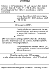
Background and objectives: Myasthenia gravis (MG) is a T cell-driven, autoantibody-mediated disorder affecting transmission in neuromuscular junctions. The associations between the peripheral T cells and MG have been extensively studied. However, they are mainly of observational nature, thus limiting our understanding of the effect of inflammatory biomarkers on MG risk. With large data sets now available, we used Mendelian randomization (MR) analysis to investigate whether the biomarkers on T cells are causally associated with MG and further validate the relationships.
Methods: We performed a two-sample MR analysis using genetic data from one genome-wide association study (GWAS) for 210 extensive T-cell traits in 3,757 general population individuals and the largest GWAS for MG currently available (1,873 patients versus 36,370 age/gender-matched controls) from US and Italy. Then the biomarkers of interest were validated separately in two GWASs for MG in FIN biobank (232 patients versus 217,056 controls) and UK biobank (152 patients versus 386,631 controls).
Results: In the first analysis, three T-cell traits were identified to be causally protective for MG risk: 1) CD8 on terminally differentiated CD8+ T cells (OR [95% CI] = 0.71 [0.59, 0.86], P = 5.62e-04, adjusted P =2.81e-02); 2) CD4+ regulatory T proportion in T cells (OR [95% CI] = 0.44 [0.26, 0.72], P = 1.30e-03, adjusted P =2.81e-02); 3) HVEM expression on total T cells (OR [95% CI] = 0.67 [0.52, 0.86], P = 1.61e-03, adjusted P =2.81e-02) and other eight T-cell subtypes (e.g., naïve CD4+ T cells). In particular, HVEM is a novel immune checkpoint on T cells that has never been linked to MG before. The SNPs on the TNFRSF14 per se further support a more direct link between the HVEM and MG. The validation analysis replicated these results in both FIN and UK biobanks. Both datasets showed a concordant protective trend supporting the findings, albeit not significant.
Conclusion: This study highlighted the role of HVEM on T cells as a novel molecular-modified factor for MG risk and validated the causality between T cells and MG. These findings may advance our understanding of MG's immunopathology and facilitate the future development of predictive disease-relevant biomarkers.
Keywords: GWAS; HVEM; Mendelian randomization; T cell; myasthenia gravis.
Copyright © 2022 Zhong, Jiao, Huan, Zhao, Su, Goh, Zheng, Zhou, Luo and Zhao.
Conflict of interest statement
The authors declare that the research was conducted in the absence of any commercial or financial relationships that could be construed as a potential conflict of interest.
Figures


Similar articles
-
Mendelian randomization analyses of known and suspected risk factors and biomarkers for myasthenia gravis overall and by subtypes.BMC Neurol. 2024 Jan 18;24(1):33. doi: 10.1186/s12883-024-03529-y. BMC Neurol. 2024. PMID: 38238684 Free PMC article.
-
Serum Inflammatory Factors Levels and Risk of Myasthenia Gravis: A Bidirectional Mendelian Randomization Study.Mol Neurobiol. 2025 Jun;62(6):7738-7746. doi: 10.1007/s12035-025-04744-5. Epub 2025 Feb 11. Mol Neurobiol. 2025. PMID: 39932515
-
Decoding the causal association between immune cells and three chronic respiratory diseases: Insights from a bi-directional Mendelian randomization study.BMC Pulm Med. 2025 Apr 15;25(1):183. doi: 10.1186/s12890-025-03641-w. BMC Pulm Med. 2025. PMID: 40234829 Free PMC article.
-
Causal effects of gut microbiota, metabolites, immune cells, liposomes, and inflammatory proteins on anorexia nervosa: A mediation joint multi-omics Mendelian randomization analysis.J Affect Disord. 2025 Jan 1;368:343-358. doi: 10.1016/j.jad.2024.09.115. Epub 2024 Sep 17. J Affect Disord. 2025. PMID: 39299582
-
Causal role of the pyrimidine deoxyribonucleoside degradation superpathway mediation in Guillain-Barré Syndrome via the HVEM on CD4 + and CD8 + T cells.Sci Rep. 2024 Nov 9;14(1):27418. doi: 10.1038/s41598-024-78996-x. Sci Rep. 2024. PMID: 39521826 Free PMC article.
Cited by
-
Genetically predicted effects of physical activity and sedentary behavior on myasthenia gravis: evidence from mendelian randomization study.BMC Neurol. 2023 Aug 11;23(1):299. doi: 10.1186/s12883-023-03343-y. BMC Neurol. 2023. PMID: 37568096 Free PMC article.
-
Mendelian randomization analyses of known and suspected risk factors and biomarkers for myasthenia gravis overall and by subtypes.BMC Neurol. 2024 Jan 18;24(1):33. doi: 10.1186/s12883-024-03529-y. BMC Neurol. 2024. PMID: 38238684 Free PMC article.
-
An angel or a devil? Current view on the role of CD8+ T cells in the pathogenesis of myasthenia gravis.J Transl Med. 2024 Feb 20;22(1):183. doi: 10.1186/s12967-024-04965-7. J Transl Med. 2024. PMID: 38378668 Free PMC article. Review.
-
Association between COVID-19 and myasthenia gravis (MG): A genetic correlation and Mendelian randomization study.Brain Behav. 2023 Nov;13(11):e3239. doi: 10.1002/brb3.3239. Epub 2023 Aug 28. Brain Behav. 2023. PMID: 37638499 Free PMC article.
References
-
- Wolfe GI, Kaminski HJ, Aban IB, Minisman G, Kuo H-C, Marx A, et al. . Long-term effect of thymectomy plus prednisone versus prednisone alone in patients with non-thymomatous myasthenia gravis: 2-year extension of the MGTX randomised trial. Lancet Neurol (2019) 18:259–68. doi: 10.1016/S1474-4422(18)30392-2 - DOI - PMC - PubMed
Publication types
MeSH terms
Substances
Grants and funding
LinkOut - more resources
Full Text Sources
Medical
Research Materials

