. 2022 Aug 1:13:884362.
doi: 10.3389/fimmu.2022.884362. eCollection 2022.

YAP1 alleviates sepsis-induced acute lung injury via inhibiting ferritinophagy-mediated ferroptosis

Affiliations

YAP1 alleviates sepsis-induced acute lung injury via inhibiting ferritinophagy-mediated ferroptosis

Jing Zhang et al. Front Immunol. .

Abstract

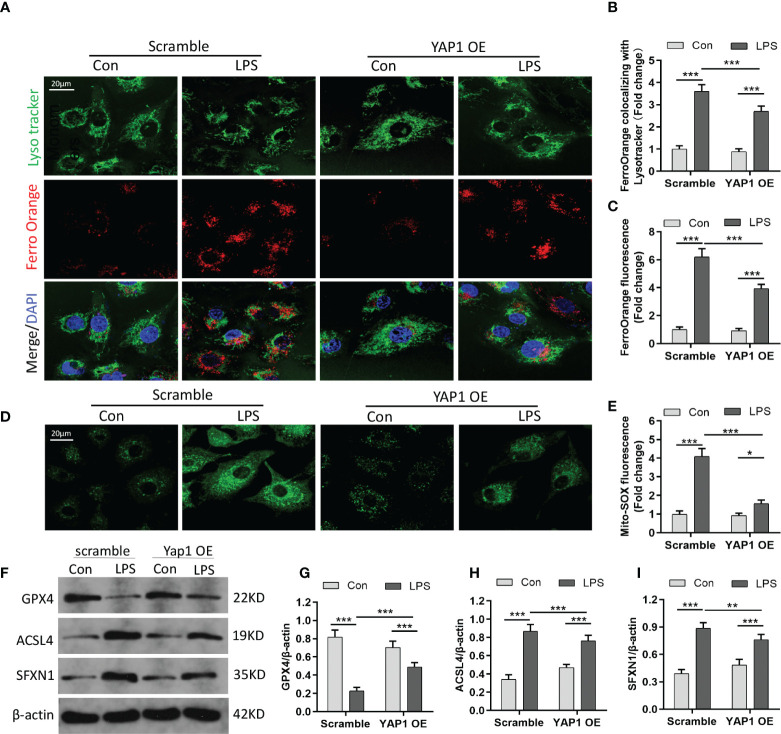
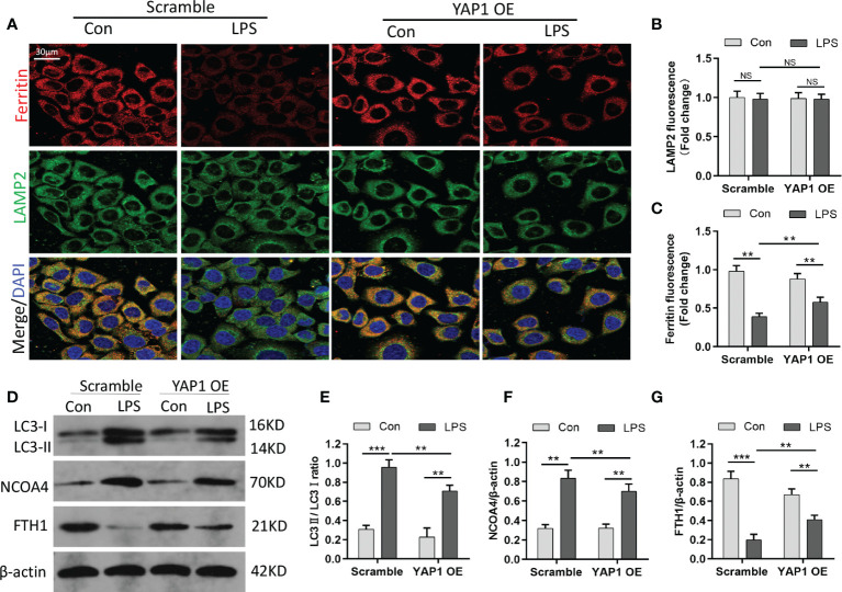
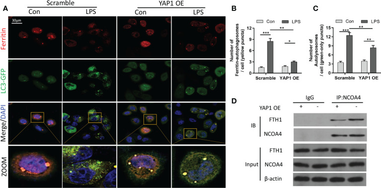
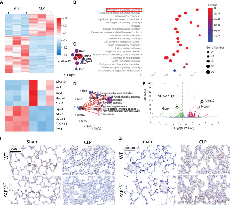
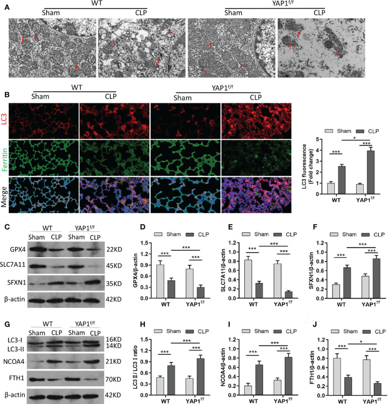
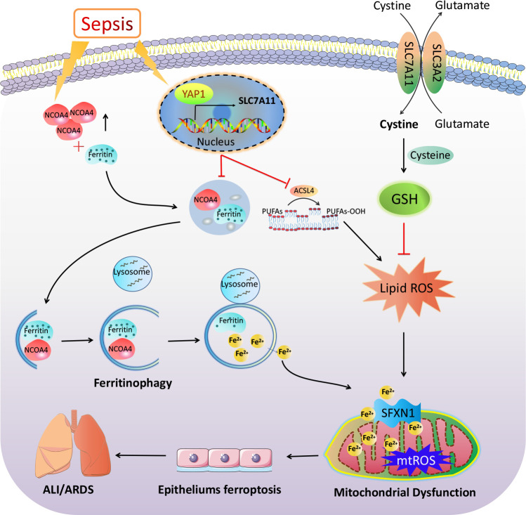
Ferroptosis is a phospholipid peroxidation-mediated and iron-dependent cell death form, involved in sepsis-induced organ injury and other lung diseases. Yes-associated protein 1 (YAP1), a key regulator of the Hippo signaling pathway, could target multiple ferroptosis regulators. Herein, this study aimed to explore the involvement of ferroptosis in the etiopathogenesis of sepsis-induced acute lung injury (ALI) and demonstrate that YAP1 could disrupt ferritinophagy and moderate sepsis-induced ALI. Cecal ligation and puncture (CLP) models were constructed in wild-type (WT) and pulmonary epithelium-conditional knockout (YAP1f/f) mice to induce ALI, while MLE-12 cells with or without YAP1 overexpression were stimulated by lipopolysaccharide (LPS) in vitro. In-vivo modes showed that YAP1 knockout aggravated CLP-induced ALI and also accelerated pulmonary ferroptosis, as presented by the downregulated expression of GPX4, FTH1, and SLC7A11, along with the upregulated expression of SFXN1 and NCOA4. Transcriptome research identified these key genes and ferroptosis pathways involved in sepsis-induced ALI. In-vitro modes consistently verified that YAP1 deficiency boosted the ferrous iron accumulation and mitochondrial dysfunction in response to LPS. Furthermore, the co-IP assay revealed that YAP1 overexpression could prevent the degradation of ferritin to a mass of Fe2+ (ferritinophagy) via disrupting the NCOA4-FTH1 interaction, which blocked the transport of cytoplasmic Fe2+ into the mitochondria via the mitochondrial membrane protein (SFXN1), further reducing the generation of mitochondrial ROS. Therefore, these findings revealed that YAP1 could inhibit ferroptosis in a ferritinophagy-mediated manner, thus alleviating sepsis-induced ALI, which may provide a new approach to the therapeutic orientation for sepsis-induced ALI.

Keywords: NCOA4; YAP1; ferritinophagy; ferroptosis; sepsis-induced acute lung injury.


Conflict of interest statement

The authors declare that the research was conducted in the absence of any commercial or financial relationships that could be construed as a potential conflict of interest.

Figures

Figure 1
Figure 1
Yes-associated protein 1 (YAP1) alleviated reactive oxygen species (ROS) accumulation and lipid peroxidation in LPS-induced MLE-12 cells. (A) Representative images of fluorescence probes (DCFH-DA) for intracellular ROS production and (B) quantification of ROS fluorescence intensity. (C) Lipid ROS generation was analyzed by using BODIPY 581/591 and determined by flow cytometry in MLE-12 cells treated with the contextual stimuli and (D) quantification of lipid ROS. (E) Cell viability was evaluated by the cell counting kit-8. The contents of MDA (F), GSH (G), and Fe2+ (H) were determined using the indicated commercial kits. Data are expressed as mean ± SD. n = 3; *p < 0.05, **p < 0.01, ***p < 0.001. LPS, lipopolysaccharide; Con, control group; Scramble, negative control; YAP1 OE, YAP1 overexpression.
Figure 2
Figure 2
YAP1 activation inhibited ferroptosis by decreasing the amount of intracellular free ferrous iron. (A) LysoTracker (green) and FerroOrange (red) were shown by confocal imaging in MLE-12 cells stimulated with LPS. The images exhibited the localization of ferrous iron in survival cells. The lysosomes were stained by LysoTracker Green fluorescence. Nuclei were stained with DAPI (blue). (B) Quantification of the fluorescence intensity of FerroOrange colocalized with LysoTracker (green). (C) Quantification of the FerroOrange fluorescence intensity. (D) Representative images of mitochondrial ROS stained with MitoSOX (green). (E) Quantitative results of mitochondrial ROS. (F–I) The expression levels of GPX4 (G), ACSL4 (H), and SFXN1 (I) were shown by WB. Data are expressed as mean ± SD. n = 3; *p < 0.05, **p < 0.01, ***p < 0.001.
Figure 3
Figure 3
YAP1 restrained the degradation of ferritin in lysosomes to suppress ferritinophagy. (A) Confocal images of ferritin (red) and LAMP2 (green) were shown in LPS-treated MLE-12 cells, while lysosomes were stained with LAMP2 (green). (B) Quantification measurement of lysosome fluorescence intensity. (C) Quantification measurement of ferritin fluorescence intensity. (D–G) Data showed the expression contents of LC3II/LC3I (E), NCOA4 (F), and FTH1 (G). Data are expressed as mean ± SD. n = 3; *p < 0.05, **p < 0.01, ***p < 0.001. NS: no significance, P>0.05.
Figure 4
Figure 4
YAP1 inhibited ferritinophagy by disrupting the NCOA4−FTH1 interaction. (A) Cells in the indicated groups were transfected with the LC3-GFP plasmid, and the autophagosome in cells presented green fluorescence puncta. Representative confocal images of cells exhibited fluorescent colocalization (yellow puncta) of ferritin (red fluorescence) with LC3-GFP (green puncta). Cells were treated with LPS for 24 (h) (B) Fluorescence quantification of ferritin colocalizing with autophagosomes (LC3-GFP). The colocalization puncta per cell were calculated. (C) Fluorescence quantification of autophagosomes (LC3-GFP). The autophagosome puncta per cell were calculated. (D) Immunoprecipitation analysis of NCOA4 and FTH1 interaction in MLE-12 cells. IgG was used for control. Data are expressed as mean ± SD. n = 3; *p < 0.05, **p < 0.01, ***p < 0.001.
Figure 5
Figure 5
YAP1 deficiency aggravated oxidative stress-mediated lung injury in CLP mice. (A) Histological images of lung samples exhibited by HE staining in mice following CLP, YAP1f/f, and CLP+YAP1f/f treatment. (B) The pathological lesion scores. (C) The content of BALF protein. (D) Representative fluorescent images of ROS staining and the quantification of ROS fluorescence intensity in lung tissue. (E) The certification of YAP1 conditional knockout in mice pulmonary epithelial cells. (F–G) Pulmonary content of MDA (F) and GSH (G) was assessed by the corresponding commercial kit. Data are expressed as mean ± SD. n = 6; *p < 0.05, **p < 0.01, ***p < 0.001.
Figure 6
Figure 6
Gene expression analysis of lung tissue in WT+CLP mice and WT+Sham mice. (A) Hierarchical clustered heatmap of differentially expressed genes (DEGs) in lung tissues between WT+CLP mice and WT+Sham mice, n = 3. (B) The Kyoto Encyclopedia of Genes and Genomes enrichment analysis (KEGG pathway) was adopted to identify the most significantly altered signaling pathways in lung tissues of CLP mice. The differentially expressed genes enriched in the ferroptosis-related pathway, indicating that the ferroptosis signaling pathway was obviously activated, n = 3. (C) Construction of protein–protein interaction (PPI) regulatory network based on DEGs. (D) Network of KEGG pathway based on similarity of their gene expression profiles. These enrichment genes were related to several signaling pathways. (E) The volcano plot shows DEGs between CLP mice and WT mice. Genes colored in red represented dramatically upregulated genes, genes colored in green displayed remarkably downregulated genes, while insignificantly altered genes are colored in gray, log2 FC > 1, Q value < 0.05. (F) SLC7A11 of mouse lung parenchyma was stained by immunohistochemical (IHC) staining. (G) NCOA4 of mouse lung parenchyma was stained by IHC staining.
Figure 7
Figure 7
YAP1 deficiency aggravated CLP-induced ferroptosis and ferritinophagy in lung tissues. (A) Representative images shown by TEM. The red arrow indicates representative mitochondria in WT and YAP1f/f mice lungs treated by CLP or not (n = 3 mice/group). (B) Fluorescence analysis showed representative images of ferritin colocalization (green) with tissue expression of LC3 (red) and quantification fluorescence intensity of LC3. Nuclei were counterstained by DAPI (blue). Light pink fluorescence represented the colocalization of ferritin and LC3. (C–F) Immunoblotting detection of the expression contents of GPX4 (D), SLC7A11 (E), and SFXN1 (F) in mice with indicated treatment. (G–J) Immunoblotting determination of the expression levels of LC3II/LC3I (H), NCOA4 (I), and FTH1 (J) in mice with equivalent treatment. Data are expressed as mean ± SD. n = 6; *p < 0.05, **p < 0.01, ***p < 0.001.
Figure 8
Figure 8
The mechanism illustration on the involvement of YAP1 in ferritinophagy and ferroptosis in sepsis-induced acute lung injury. Sepsis induces increased intracellular NCOA4 expression, which then has an interaction with ferritin and brings about the autophagic degradation of ferritin. YAP1 can suppress the degradation of ferritin into autophagosomes via inhibiting NCOA4-mediated ferritinophagy, then prevent ferrous iron from transporting into the mitochondria. YAP1 attenuates the accumulation of mitochondrial reactive oxygen species and lipid peroxidation and contributes to protecting mitochondrial function and repressing ferroptosis. Therefore, we conclude that YAP1 is involved in sepsis-induced acute lung injury by regulating the process of ferritinophagy-mediated ferroptosis.

Similar articles

Cited by

References

    1. Singer M, Deutschman CS, Seymour CW, Shankar-Hari M, Annane D, Bauer M, et al. . The third international consensus definitions for sepsis and septic shock (sepsis-3). JAMA (2016) 315(8):801–10. doi: 10.1001/jama.2016.0287 - DOI - PMC - PubMed
    1. Angus DC, van der Poll T. Severe sepsis and septic shock. N Engl J Med (2013) 369(9):840–51. doi: 10.1056/NEJMra1208623 - DOI - PubMed
    1. Torio CM, Moore BJ. National inpatient hospital costs: The most expensive conditions by payer, 2013 Stat Brief. In: Healthcare Cost and Utilization Project (HCUP) Statistical Briefs (2006) 204 Agency for Healthcare Research and Quality (US):Rockville (MD). - PubMed
    1. Hall MJ, Levant S, DeFrances CJ. Trends in inpatient hospital deaths: National hospital discharge survey, 2000-2010 NCHS Data Brief. US Department of Health and Human Services, Centers for Disease Control and Prevention (2013) 118):1–8. National Center for Health Statistics. - PubMed
    1. Sessler CN, Bloomfield GL, Fowler AA, 3rd. Current concepts of sepsis and acute lung injury. Clin Chest Med (1996) 17(2):213–35. doi: 10.1016/S0272-5231(05)70310-5 - DOI - PubMed